. 2015 Feb;185(2):446-61.
doi: 10.1016/j.ajpath.2014.10.005. Epub 2014 Dec 4.

Type I interferon contributes to noncanonical inflammasome activation, mediates immunopathology, and impairs protective immunity during fatal infection with lipopolysaccharide-negative ehrlichiae

Affiliations

Type I interferon contributes to noncanonical inflammasome activation, mediates immunopathology, and impairs protective immunity during fatal infection with lipopolysaccharide-negative ehrlichiae

Qin Yang et al. Am J Pathol. 2015 Feb.

Abstract

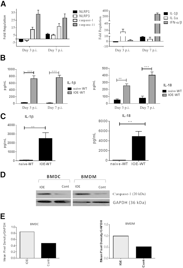
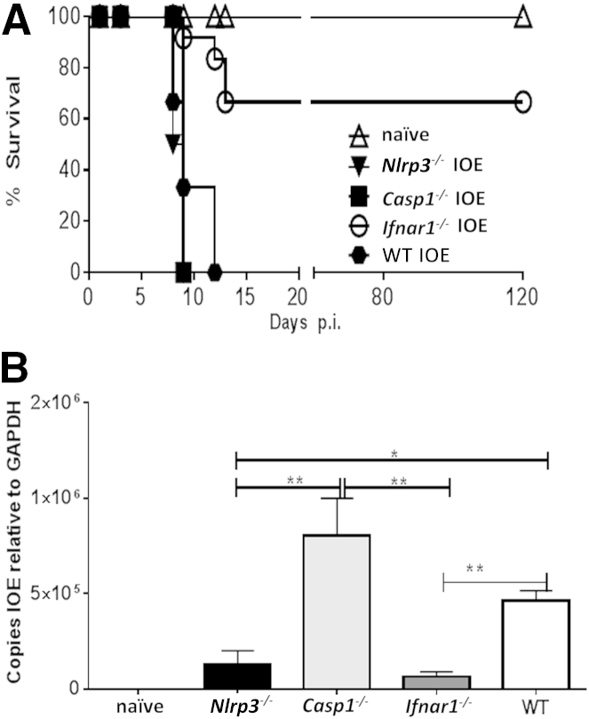
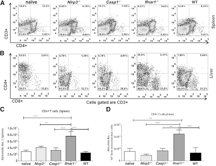
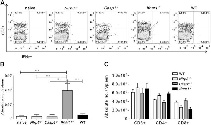
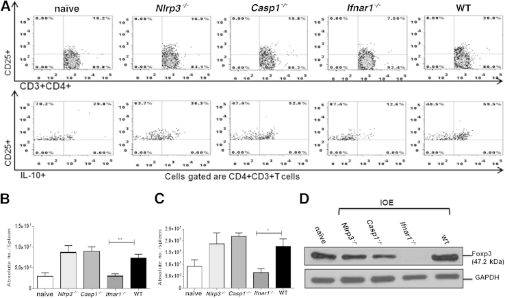
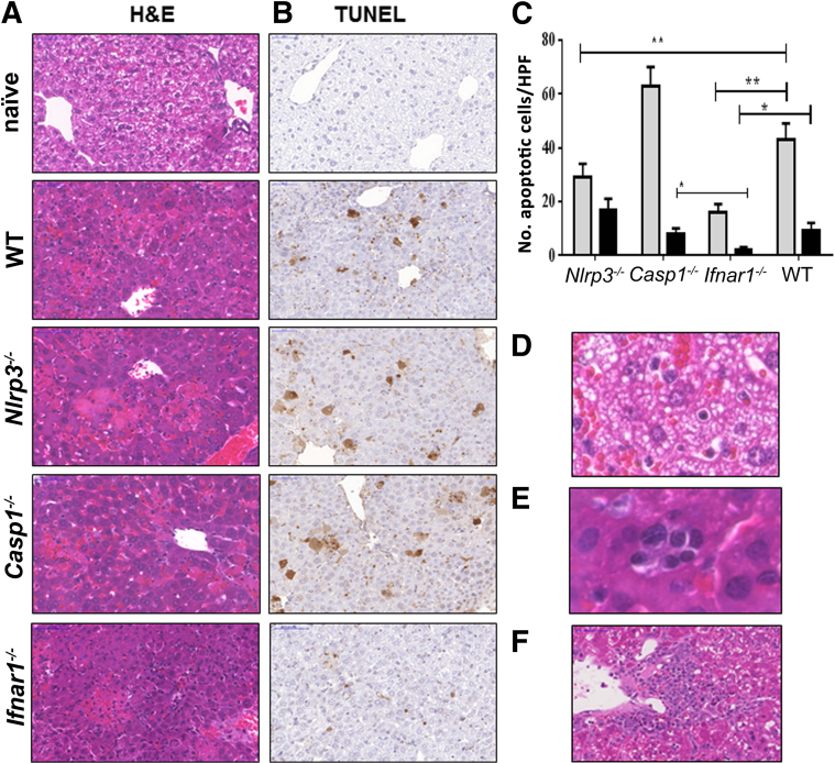
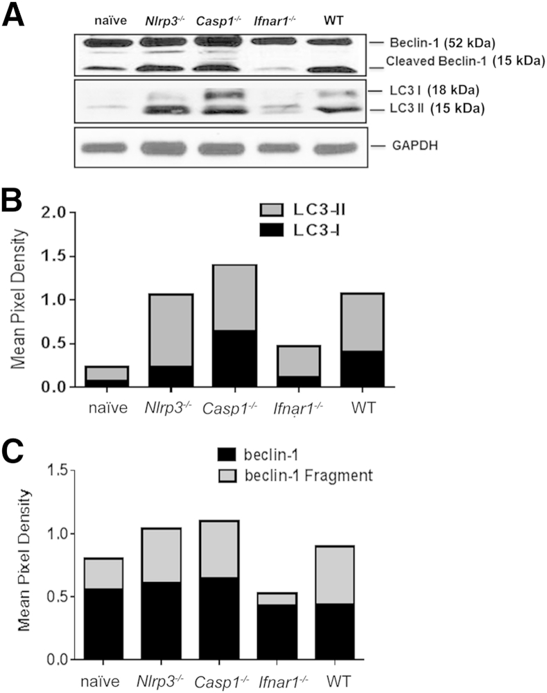
Ehrlichia species are intracellular bacteria that cause fatal ehrlichiosis, mimicking toxic shock syndrome in humans and mice. Virulent ehrlichiae induce inflammasome activation leading to caspase-1 cleavage and IL-18 secretion, which contribute to development of fatal ehrlichiosis. We show that fatal infection triggers expression of inflammasome components, activates caspase-1 and caspase-11, and induces host-cell death and secretion of IL-1β, IL-1α, and type I interferon (IFN-I). Wild-type and Casp1(-/-) mice were highly susceptible to fatal ehrlichiosis, had overwhelming infection, and developed extensive tissue injury. Nlrp3(-/-) mice effectively cleared ehrlichiae, but displayed acute mortality and developed liver injury similar to wild-type mice. By contrast, Ifnar1(-/-) mice were highly resistant to fatal disease and had lower bacterial burden, attenuated pathology, and prolonged survival. Ifnar1(-/-) mice also had improved protective immune responses mediated by IFN-γ and CD4(+) Th1 and natural killer T cells, with lower IL-10 secretion by T cells. Importantly, heightened resistance of Ifnar1(-/-) mice correlated with improved autophagosome processing, and attenuated noncanonical inflammasome activation indicated by decreased activation of caspase-11 and decreased IL-1β, compared with other groups. Our findings demonstrate that IFN-I signaling promotes host susceptibility to fatal ehrlichiosis, because it mediates ehrlichia-induced immunopathology and supports bacterial replication, perhaps via activation of noncanonical inflammasomes, reduced autophagy, and suppression of protective CD4(+) T cells and natural killer T-cell responses against ehrlichiae.


Figures

Figure 1
Figure 1
Lethal ehrlichial infection differentially activates inflammasomes, compared with uninfected control. A: Mice infected with the lethal dose of Ixodes ovatus Ehrlichia (IOE) had a higher mRNA expression of inflammasome components NLRP1, NLRP3, caspase-1, caspase-11, IL-1α, IL-β, and cytokine IFN-α/β in liver at different time points after infection, compared with control. Results were normalized to housekeeping genes and expressed as fold regulation, compared with gene expression in naïve mice. B: Levels of IL-1β and IL-18 increased in spleen of lethally IOE-infected WT mice, compared with uninfected sham controls on days 3 and 7 after infection (p.i.). C: Levels of IL-1β and IL-18 increased in serum of lethally IOE-infected WT mice, compared with uninfected sham controls on day 7 p.i. D: Western blot analysis indicated higher expression of active caspase-1 (p20) in bone marrow–derived dendritic cells (BMDCs) and bone marrow derived–macrophages (BMDMs), either infected in vitro with IOE or left uninfected (control). E: Mean pixel density of Western blotting bands for active caspase-1 was determined by ImageJ software. Data are expressed as means ± SD. n = 9 mice per group (A–C), pooled from two independent experiments. Western blot results (D and E) represent one of three independent experiments with similar results. ∗∗P ≤ 0.01, ∗∗∗P ≤ 0.001. Cont, control.
Figure 2
Figure 2
Enhanced resistance of Ifnar1−/− mice to lethal ehrlichiosis. A: Survival curves for IOE-infected mice show long-term survival of Ifnar1−/−. B: Bacterial burden in liver of IOE-infected mice and uninfected controls was determined by quantitative real-time PCR on day 7 p.i. IOE-infected Ifnar1−/− and Nlrp3−/− mice had significantly lower bacterial burden, compared with WT and Casp1−/− mice. Data are expressed as means ± SD. n = 12 mice per group (A); n = 9 mice per group, pooled from three independent experiments (B). P ≤ 0.05, ∗∗P ≤ 0.01.
Figure 3
Figure 3
IFN-I impairs the expansion of NKT cells in liver during fatal ehrlichial infection. A: For flow cytometry, liver mononuclear cells prepared from the liver of naïve and IOE-infected WT, Nlrp3−/−, Casp1−/−, and Ifnar1−/− mice. B: Liver mononuclear cells were then analyzed for expression of TCRβ and CD1d using specific anti-TCRβ monoclonal antibody and CD1d tetramer. C: TCRβ+CD1dtet+ cells were then gated and reanalyzed for B220 expression. NKT cells were defined as TCRβ+CD1dtet+B220 cells. D: Absolute numbers of NKT cells in liver of naïve mice or IOE-infected WT Nlrp3−/−, Casp1−/−, and Ifnar1−/− mice were determined on day 7 p.i. Data are expressed as means ± SD and are representative of three independent experiments with similar results. n = 3 mice per group. ∗∗P ≤ 0.01, ∗∗∗P ≤ 0.001.
Figure 4
Figure 4
Expansion of the CD4+ T-cell subset in Ifnar1−/− mice after lethal IOE infection. A and B: Flow cytometry analysis of the percentage of CD4+ T cells in spleen (A) and liver (B) of mice on day 7 p.i. with lethal Ixodes ovatus Ehrlichia infection. C and D: Absolute numbers of CD4+ T cells in spleen (C) and liver (D) of the same mice. Data are expressed as means ± SD, and are representative of three independent experiments with similar results. n = 9 mice per group. ∗∗P ≤ 0.01, ∗∗∗P ≤ 0.001.
Figure 5
Figure 5
Enhanced protective type I immune responses in IOE-infected Ifnar1−/− mice. A and B: Flow cytometry analysis of the percentage (A) and absolute number (B) of intracellular IFN-γ–expressing CD3+ splenocytes from IOE-infected WT, Ifnar1−/−, Nlrp3−/−, and Casp1−/− mice on day 7 p.i. C: Flow cytometry analysis of the absolute numbers of CD3+, CD4+, and CD8+ T cells shows similar number of these T-cell subsets in naïve mice of different backgrounds (WT, Ifnar1−/−, Nlrp3−/−, and Casp1−/−). Data are expressed as means ± SD and are representative of three independent experiments (N = 9 mice). ∗∗∗P ≤ 0.001.
Figure 6
Figure 6
IFNAR1 inhibits splenic and systemic IFN-γ production while enhancing IL-10 secretion during fatal ehrlichial infection. A: Levels of IFN-γ and IL-10 and the ratio of IL-10 to IFN-γ in serum from all infected groups of mice at day 7 p.i. B: Levels of IFN-γ and IL-10 and the IL-10/ IFN-γ ratio in culture supernatants from spleen harvested from all infected groups of mice on day 7 p.i. Data are expressed as means ± SD and are representative of three independent experiments with similar results. n = 9 mice per group. P ≤ 0.05, ∗∗P ≤ 0.01, and ∗∗∗P ≤ 0.001.
Figure 7
Figure 7
IFNAR1 signaling increased the numbers of IL-10–producing-CD3+CD4+CD25+ T cells and the number of FOXP3-expressing Tregs in liver during fatal ehrlichial infection. A: Flow cytometry analysis of the percentage of CD3+CD4+CD25+ and IL-10+CD3+CD4+CD25+ T cells in spleen at day 7 p.i. B and C: Absolute number of CD3+CD4+CD25+ T cells (B) and IL-10+CD3+CD4+CD25+ T cells (C) in spleen at day 7 p.i. D: FOXP3 protein expression in liver was determined via Western blot analysis of liver lysate. Data are expressed as means ± SD and are representative of three independent experiments with similar results. n = 9 mice per group. P ≤ 0.05, ∗∗P ≤ 0.01.
Figure 8
Figure 8
IFNAR1 signaling promotes expansion of FOXP3+ Treg cells in spleen during fatal ehrlichial infection. A: Flow cytometry analysis of the percentage of CD3+CD4+CD25+FOXP3+ Tregs. B and C: Absolute number of CD4+CD25+FOXP3+ (B) and of CD3+CD4+CD25FOXP3+ (C) Tregs in spleen of the different groups, including uninfected control, at day 7 p.i. Data are expressed as means ± SD and are representative of two independent experiments with similar results. n = 9 mice per group. P ≤ 0.05, ∗∗P ≤ 0.01, and ∗∗∗P ≤ 0.001.
Figure 9
Figure 9
IOE-infected Ifnar1−/− mice have altered hepatic pathology, with increased cellular infiltration and less apoptosis, compared with WT mice. A and B: Livers were harvested on day 7 p.i., and liver sections were stained by hematoxylin and eosin (H&E) (A) or by TUNEL (B). C: Tunel assay revealed significantly lower numbers of apoptotic Kupffer cells (gray bars, P < 0.01) and hepatocytes (black bars, P < 0.01) in IFNaR1−/− mice, compared with WT and casp1−/− mice. The number of apoptotic Kupffer cells, but not hepatocytes, in NLRP3−/− mice was also significantly lower than that detected in WT and casp1−/− mice. The number of apoptotic cells was counted within 10 high-power fields (×40 magnification). D: Liver from IOE-infected WT mice, but not Ifnar1−/− mice, exhibited microvesicular steatosis. E: Compared with the other groups, liver of IOE-infected Ifnar1−/− mice had more inflammatory cell infiltration (composed primarily of lymphocytes) and pronounced regenerative changes, including extramedullary hematopoiesis with aggregates of erythrocyte precursors observed within sinusoids. F: By day 10 p.i., when most WT mice had died of infection, Ifnar1−/− mice exhibited prominent perivascular granuloma-like lymphohistiocytic infiltrates. Data are expressed as means ± SD and are representative of three independent experiments with similar results. n = 9 mice per group. P ≤ 0.05, ∗∗P ≤ 0.01. Original magnification, ×40. HPF, high-power field.
Figure 10
Figure 10
IFNAR1 regulates IL-1β secretion during fatal ehrlichial infection. A: Liver lysate from the different groups of mice was analyzed for expression of IL-1β (p17) and active caspase-11 (p30) by Western blotting on day 7 p.i. GAPDH expression was used as loading control. B: Serum levels of IL-1β were measured using enzyme-linked immunosorbent assay on day 7 p.i. C and D: BMDMs from IOE-infected WT and Ifnar1−/− mice express pro–caspase-11 (p45). Expression of pro–caspase-11 was normalized to GAPDH. Results in A, C, and D are from one mouse in each group and are representative of three independent experiments with similar results (n = 9 mice per group). Data in B are expressed as means ± SD and are representative of three independent experiments with similar results (n = 9 mice per group). P ≤ 0.05.
Figure 11
Figure 11
IFN-I blocks degradation of autophagy during fatal ehrlichial infection. Liver lysate from the different groups of mice was analyzed for expression of autophagy markers LC3I and LC3II (A and B) and beclin-1 (A and C) by Western blotting on day 7 p.i. GAPDH expression was used as loading control. Band density was quantified using ImageJ software and was normalized to GAPDH. Results are from one mouse in each group and are representative of three independent experiments with similar results. n = 3 mice per group.

Similar articles

Cited by

References

    1. Olano J.P., Walker D.H. Human ehrlichioses. Med Clin North Am. 2002;86:375–392. - PubMed
    1. Walker D.H., Dumler J.S. Human monocytic and granulocytic ehrlichioses. Discovery and diagnosis of emerging tick-borne infections and the critical role of the pathologist. Arch Pathol Lab Med. 1997;121:785–791. - PubMed
    1. Fichtenbaum C.J., Peterson L.R., Weil G.J. Ehrlichiosis presenting as a life-threatening illness with features of the toxic shock syndrome. Am J Med. 1993;95:351–357. - PubMed
    1. Wakeel A., den Dulk-Ras A., Hooykaas P.J., McBride J.W. Ehrlichia chaffeensis tandem repeat proteins and Ank200 are type 1 secretion system substrates related to the repeats-in-toxin exoprotein family. Front Cell Infect Microbiol. 2011;1:22. - PMC - PubMed
    1. Ismail N., Crossley E.C., Stevenson H.L., Walker D.H. Relative importance of T-cell subsets in monocytotropic ehrlichiosis: a novel effector mechanism involved in Ehrlichia-induced immunopathology in murine ehrlichiosis. Infect Immun. 2007;75:4608–4620. - PMC - PubMed

Publication types

MeSH terms