. 2014 Dec 3;34(49):16369-84.
doi: 10.1523/JNEUROSCI.1309-14.2014.

Entrapment via synaptic-like connections between NG2 proteoglycan+ cells and dystrophic axons in the lesion plays a role in regeneration failure after spinal cord injury

Affiliations

Entrapment via synaptic-like connections between NG2 proteoglycan+ cells and dystrophic axons in the lesion plays a role in regeneration failure after spinal cord injury

Angela R Filous et al. J Neurosci. .

Abstract

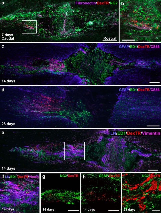
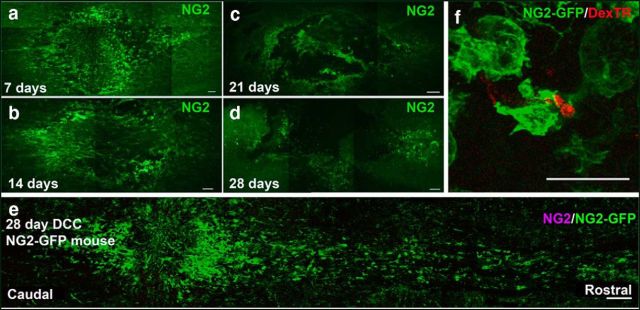
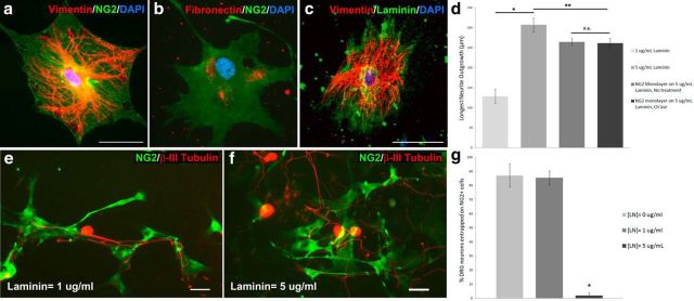
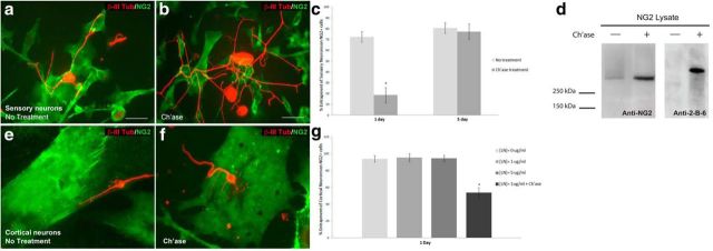
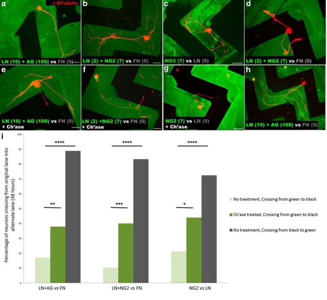
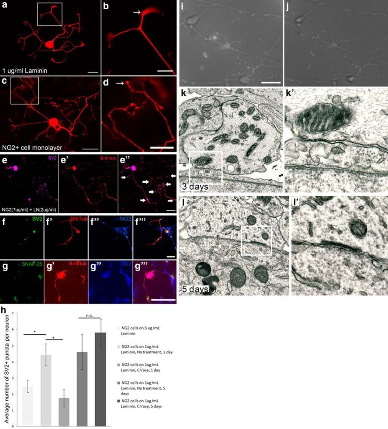
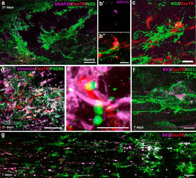
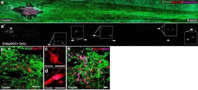
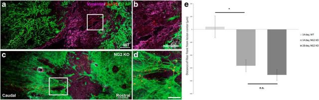
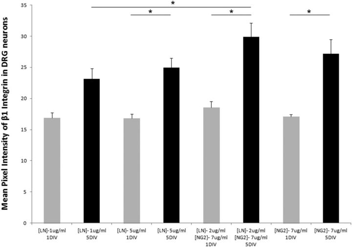
NG2 is purportedly one of the most growth-inhibitory chondroitin sulfate proteoglycans (CSPGs) produced after spinal cord injury. Nonetheless, once the severed axon tips dieback from the lesion core into the penumbra they closely associate with NG2+ cells. We asked if proteoglycans play a role in this tight cell-cell interaction and whether overadhesion upon these cells might participate in regeneration failure in rodents. Studies using varying ratios of CSPGs and adhesion molecules along with chondroitinase ABC, as well as purified adult cord-derived NG2 glia, demonstrate that CSPGs are involved in entrapping neurons. Once dystrophic axons become stabilized upon NG2+ cells, they form synaptic-like connections both in vitro and in vivo. In NG2 knock-out mice, sensory axons in the dorsal columns dieback further than their control counterparts. When axons are double conditioned to enhance their growth potential, some traverse the lesion core and express reduced amounts of synaptic proteins. Our studies suggest that proteoglycan-mediated entrapment upon NG2+ cells is an additional obstacle to CNS axon regeneration.

Keywords: NG2 proteoglycan; chondroitinase; dystrophic axons; glial scar; oligodendrocyte progenitor cells; spinal cord injury.


Figures

Figure 1.
Figure 1.
Injured fibers remain associated with NG2+ cells for several weeks after injury. a, Confocal montage of a longitudinal section through the injured section of rat cord, 7 d after injury. At 7 d after injury, Dextran-Texas Red (DexTR)-labeled fibers of dystrophic axons (red) can be found caudal to the lesion, in an area rich in NG2 (green) and fibronectin (purple). The slice is oriented with the rostral end of tissue to the right and caudal to the left throughout the paper, unless otherwise noted. b, High magnification of the area outlined in a. c, Dextran-Texas Red-labeled fibers remain in the penumbra of the lesion, in an area rich in CS56, with some GFAP and ED1 expression. The fibers are associated most closely with CS56 expression. d, At 28 d after injury, the fibers remain in this area, even as ED1 expression is reduced. e, Fourteen days after injury these fibers are also found in an area rich in laminin and vimentin. f, High magnification of the area outlined in e. g, At 14 d after injury, the fibers are still associated with NG2+ cells (green) in the lesion, but not with astrocytes labeled with GFAP (green) at the caudal end of the lesion (h), where the majority of the fibers have stabilized. i, Close association with NG2+ cells persists at 21 d after injury. Scale bars: a, c–e, g, h, 200 μm; b, i, 10 μm; f, 100 μm.
Figure 2.
Figure 2.
NG2 expression declines over time, but formerly expressing NG2-cells remain in the lesion. a–d, Level of NG2 expression at 7 (a), 14 (b), 21 (c), and 28 d (d) after injury in rat. e, A montage of the lesion in an NG2-lck-eGFP mouse 28 d after injury. Even small amounts of NG2 promoter activity are able to drive large amounts of GFP expression. f, A z-stack projection of a fiber enwrapped by a GFP+ cell at the caudal end of the lesion, 28 d after injury, from a different slice than that imaged in e. Scale bars: e, 200 μm; a–d, 100 μm; f, 25 μm. DexTR, Dextran-Texas Red
Figure 3.
Figure 3.
NG2+ cells provide a growth-permissive substrate. a, NG2+ cells also express vimentin (red), a progenitor marker. b, NG2+ cells express growth-permissive extracellular matrix molecules such as laminin (red) and fibronectin (red, c). d, Neurite outgrowth on the low-laminin substrate (1 μg/ml) was significantly lower than on the high-laminin substrate (5 μg/ml) or NG2+ cell monolayer. Outgrowth on the NG2+ cell monolayer was significantly less than on a high-laminin substrate. No significant difference in outgrowth was observed on the NG2+ cell monolayer with or without ch'ase treatment. Kruskal–Wallis test, Mann–Whitney U test; n = 33 (1 μg/ml laminin), 97 (5 μg/ml laminin), 176 (NG2 monolayer, no treatment), 174 (NG2 monolayer, ch'ase); *p < 0.0005, **p < 0.015. H(3) = 43.57. e, Neurons whose cell bodies begin on the surface of an NG2+ cell remain on the NG2+ cell surface and do not extend processes off onto a low-laminin (1 μg/ml) substrate. f, DRG neurons beginning on an NG2+ cell surface can extend neurites off the NG2+ cell onto a high-laminin (5 μg/ml) substrate. g, The percentage of DRG neurons per coverslip that began on the surface of an NG2+ cell and did not extend neurons onto the laminin background at different laminin concentrations was calculated. Neurons remain entrapped on the surface of the NG2+ cell when laminin concentrations are at 1 μg/ml or below. One-way ANOVA, Tukey post hoc test, n = 6 coverslips per condition, *p < 0.02. F(9) = 36.41. Scale bar: 50 μm.
Figure 4.
Figure 4.
Chondroitinase treatment reverses entrapment. a, When NG2+ cells and DRG neurons are cocultured on low-laminin (1 μg/ml) substrates, the neurons usually do not extend processes off the surface of the NG2+ cell. b, Chondroitinase treatment (0.1 U/ml) at the time of DRG plating allows neurites to extend onto the laminin substrate more frequently. c, The percentage of neurons that begin on the NG2+ cell surface and are unable to extend processes onto the laminin substrate was significantly reduced when the cocultures were treated with ch'ase (0.1 U/ml) before DRG plating when cocultured for 1 DIV. At 5 DIV, neurites are unable to extend off the NG2+ cell surface, even after ch'ase treatment. Two-proportions test, n = 106 (1 d, no treatment), 160 (1 d, ch'ase), 90 (5 d, no treatment), 71 (5 d, ch'ase); *p < 0.0005, p = 0.828, z = 11.16, 0.22. d, A Western blot using protein from NG2+ cell lysate reveals a high molecular weight smear ∼300 kDa in lane 2. Lane 1 contains a protein ladder with a band at 250 kDa. Lane 2 contains lysate from NG2+ cells without treatment and lane 3 contains lysate from NG2+ cells pretreated with ch'ase for 1 h, which removes the smear. e–g, Cortical neurons (stained with β-III-tubulin, red) cocultured with NG2+ cells for 24 h become entrapped, just as the adult DRG neurons in a. e, A cortical neuron whose cell body begins on the surface on an NG2+ cells (green) becomes entrapped on the NG2+ cell surface and does not extend processes onto the low-laminin substrate (1 μg/ml). f, When cocultures are treated with ch'ase at the time of neuronal plating, neurites extend off the NG2+ cell surface onto low laminin. g, Quantification of the percentage of neurons per coverslip that become entrapped on the NG2+ cell. Ch'ase treatment at 1 DIV allows neurites to extend onto laminin. Units are percentage entrapment per coverslip and data are presented as mean + SEM; *p value < 0.05. Scale bars: a, b, 50 μm.
Figure 5.
Figure 5.
Proteoglycans play a role in entrapping neurons. DRG neurons were plated on striped substrates, given the choice between a laminin-proteoglycan lane (green) and a fibronectin (FN) or laminin-only background (black). a, b, Neurons prefer to grow in the laminin-proteoglycan (a, aggrecan, 100 μg/ml; b, NG2, 7 μg/ml) lane, rather than the fibronectin-only lane (5 μg/ml). c, Neurons preferentially grow in lanes containing only NG2 (7 μg/ml), rather than laminin-only (5 μg/ml). d, When the cell bodies of neurons begin in the lane containing fibronectin only, they freely extend into and out of the various lanes. e–g, When cultures were ch'ase treated (0.1 U/ml) at the time of plating DRG neurons, neurons extend processes onto the fibronectin (e, f) or laminin (g) background. h, When a neuron beginning on laminin only projects for an extended period in the CSPG-containing lane, it becomes entrapped. i, The percentage of neurons beginning in a green lane and extending neurites from the green to the black substrate were quantified. The same was done for those crossing from black to green when cell bodies began in the black lane. Two-proportions test, n = 85 (LN/AG vs FN, no treatment), 53 (LN/AG vs FN, ch'ase), 9 (LN/AG vs FN, no treatment, crossing black to green) 39 (LN/NG2 vs FN, no treatment), 25 (LN/NG2 vs FN, ch'ase), 12 (LN/NG2 vs FN, no treatment crossing black to green), 80 (NG2 vs LN, no treatment), 25 (NG2 vs LN, ch'ase), 40 (NG2 vs LN, no treatment, crossing black to green). Two-proportions test, *p = 0.037,**p = 0.026, ***p = 0.005, and ****p < 0.0005. z= −2.23, −4.08, −2.81, −4.96, 2.08, −6.09. Scale bar, 50 μm. AG, aggrecan.
Figure 6.
Figure 6.
NG2+ cells and adult DRG neurons form synaptic-like connections in vitro. a, Dissociated neurons growing on a uniform laminin substrate extend many neurites with lamellipodia at the growth cones. b, Inset from a, showing a growth cone. c, Neurons have a different morphology when growing on an NG2+ cell monolayer. Neurons appear to have en passant synapses along their neurites. d, Inset from c, showing bulb endings. e, An adult DRG neuron growing on a uniform substrate of NG2 (7 μg/ml) and laminin (2 μg/ml) has SV2 accumulation in puncta along its processes. f, DRG neurons growing on sparsely plated NG2+ cells for 2 d begin to express SV2+ puncta along their processes where the neurites contact NG2+ cell processes. g, An adult DRG neuron expresses the presynaptic marker, SNAP-25, when it contacts an NG2+ cell in culture. h, Quantification of the number of SV2+ puncta in a DRG neuron in a defined area when grown on an NG2+ cell monolayer for 1 and 5 d. One-way ANOVA, Tukey post hoc test, n = 40 (NG2 cells on 5 μg/ml LN, 1 d),18 (NG2 cells on 1 μg/ml LN, 1 d, no treatment), 29 (NG2 cells on 1 μg/ml LN, 1 d, ch'ase),18 (NG2 cells on 1 μg/ml LN, 5 d, no treatment), 23 (NG2 cells on 1 μg/ml LN, 1 d, ch'ase); *p < 0.0005 F(4) = 6.20. i, DRG neuron growing on an NG2+ cell monolayer for 5 DIV after high K+ stimulation in the presence of FM dye. j, The same neuron imaged in i after another high K+ stimulation without dye, showing a reduction in fluorescence. k, Electron micrograph image of a neuron in close association with an NG2+ cell after 3 d in coculture. k′, Higher magnification of the area boxed in k. l, Electron micrograph of a neuron in close association with an NG2+ cell after 5 d in coculture. L′, Boxed area in l, showing a subsurface cistern in the neuron. Scale bars: a, c, e, f, g, 50 μm; b, d, i, 25 μm.
Figure 7.
Figure 7.
NG2+ cells and dystrophic axons colocalize with synaptic markers in vivo. Dystrophic axons stabilize on NG2+ cells at the caudal end of the lesion after spinal cord injury and begin to form synapses. a, Montage of confocal images of a longitudinal section of the lesion 21 d after injury. Endballs of retrogradely labeled fibers are found associated with NG2+ cells at 21 d after a dorsal column crush. b, SNAP-25 accumulates in the endings of Dextran-Texas Red (DexTR)-labeled fibers. c, Projection of z-stack confocal images depicting an NG2+ cell enwrapping a Dextran-Texas Red-labeled fiber. d, Vimentin expression is highest adjacent to the lesion. PSD-95 expression is observed in vimentin+ cells where labeled fibers stabilize. e, Higher magnification, showing PSD-95 puncta accumulating in vimentin+ cells adjacent to stabilized fibers. f, Z-stack projection of a Dextran-Texas Red-labeled fiber expressing SV2 and enwrapped by an NG2+ cell. g, As early as 7 d after injury, SV2 accumulates in the endings of labeled fibers, caudal to the lesion. Scale bars: a,100 μm; d, g, 50 μm; b, 25 μm; c, e, f, 10 μm.
Figure 8.
Figure 8.
Conditioned fibers beyond the lesion still associate with NG2+ cells. Double conditioning lesion (2xCL) of the sciatic nerve allows several injured fibers to grow beyond the lesion center 21 d after SCI. a, GFAP expression (green) shows the glial scar surrounding the lesion. Vimentin (purple) is expressed within the lesion center and the penumbra. Arrows show fibers that have grown rostral to the lesion. a′, Dextran-Texas Red (DexTR)-labeled fibers from image a. The boxes illustrate select fiber endings at higher magnification. b, Higher magnification of the area near the asterisk in a′ from a serial slice, demonstrating that the majority of Dextran-Texas Red-labeled fibers remain caudal to the lesion in association with NG2+ cells. NG2 (green) expression is found in the lesion center and throughout the tissue. The brightness in a and b differ, so that NG2 expression could be visualized. SV2 expression is found accumulated in the endings of fibers caudal to the lesion but not those that have crossed rostrally. c, Fiber rostral to the lesion without SV2 (purple) expression. d, Fiber from b caudal to the lesion, with SV2 expression in its ending. e, Z-stack projection of a Dextran-Texas Red-labeled fiber found rostral to lesion core associated with NG2 expression. Scale bars: a,100 μm; b, 50 μm; c, d,10 μm; e, 20 μm.
Figure 9.
Figure 9.
Removal of NG2 causes increased dieback of sensory fibers after SCI. NG2 may be necessary to prevent further dieback after DCC. a, Dextran-Texas Red (DexTR)-labeled fibers are found within the lesion core of WT mice 14 d after DCC. b, Inset from a, showing where the majority of fibers stabilize in WT mice. c, DexTR-labeled fibers are found more caudal to the lesion in NG2-KO mice littermates. d, Inset from c, showing the majority of fibers in the KO animals are found caudal to the lesion, rather than within the lesion core. e, Quantification of the distance of the fiber front away from the lesion center. Two-sample t test, *p = 0.035, p = 0.302. t(4) = 3.13, t(7) = 1.11. Scale bars: a, c,100 μm; b, d,50 μm.
Figure 10.
Figure 10.
DRG neurons plated on an NG2 substrate upregulated integrin β1 after 5 DIV. At 1 DIV, there was no difference in integrin expression within the DRG neuron on any substrate. By 5 DIV, integrin expression significantly increased on high laminin, a laminin-NG2 mixture, or an NG2 only substrate. Units are mean pixel intensity per region. All data are presented as group mean + SEM, *p value < 0.05.

Similar articles

Cited by

References

    1. Asher RA, Morgenstern DA, Shearer MC, Adcock KH, Pesheva P, Fawcett JW. Versican is upregulated in CNS injury and is a product of oligodendrocyte lineage cells. J Neurosci. 2002;22:2225–2236. - PMC - PubMed
    1. Bai L, Hecker J, Kerstetter A, Miller RH. Myelin repair and functional recovery mediated by neural cell transplantation in a mouse model of multiple sclerosis. Neurosci Bull. 2013;29:239–250. doi: 10.1007/s12264-013-1312-4. - DOI - PMC - PubMed
    1. Bergles DE, Roberts JD, Somogyi P, Jahr CE. Glutamatergic synapses on oligodendrocyte precursor cells in the hippocampus. Nature. 2000;405:187–191. doi: 10.1038/35012083. - DOI - PubMed
    1. Bradbury EJ, Moon LD, Popat RJ, King VR, Bennett GS, Patel PN, Fawcett JW, McMahon SB. Chondroitinase ABC promotes functional recovery after spinal cord injury. Nature. 2002;416:636–640. doi: 10.1038/416636a. - DOI - PubMed
    1. Burden-Gulley SM, Payne HR, Lemmon V. Growth cones are actively influenced by substrate-bound adhesion molecules. J Neurosci. 1995;15:4370–4381. - PMC - PubMed

Publication types

MeSH terms