. 2014 Dec 3;34(49):16180-93.
doi: 10.1523/JNEUROSCI.3020-14.2014.

Impairment of glymphatic pathway function promotes tau pathology after traumatic brain injury

Affiliations

Impairment of glymphatic pathway function promotes tau pathology after traumatic brain injury

Jeffrey J Iliff et al. J Neurosci. .

Abstract

Traumatic brain injury (TBI) is an established risk factor for the early development of dementia, including Alzheimer's disease, and the post-traumatic brain frequently exhibits neurofibrillary tangles comprised of aggregates of the protein tau. We have recently defined a brain-wide network of paravascular channels, termed the "glymphatic" pathway, along which CSF moves into and through the brain parenchyma, facilitating the clearance of interstitial solutes, including amyloid-β, from the brain. Here we demonstrate in mice that extracellular tau is cleared from the brain along these paravascular pathways. After TBI, glymphatic pathway function was reduced by ∼60%, with this impairment persisting for at least 1 month post injury. Genetic knock-out of the gene encoding the astroglial water channel aquaporin-4, which is importantly involved in paravascular interstitial solute clearance, exacerbated glymphatic pathway dysfunction after TBI and promoted the development of neurofibrillary pathology and neurodegeneration in the post-traumatic brain. These findings suggest that chronic impairment of glymphatic pathway function after TBI may be a key factor that renders the post-traumatic brain vulnerable to tau aggregation and the onset of neurodegeneration.

Keywords: AQP4; aquaporin-4; cerebrospinal fluid; neurodegeneration; tauopathy; traumatic brain injury.


Figures

Figure 1.
Figure 1.
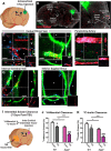
TBI causes loss of perivascular AQP4 polarization in astrocytic end feet. A, Schematic depicting Hit & Run model of moderate TBI (Ren et al., 2013). Mouse is briefly anesthetized with isoflurane, suspended by its incisors from a string, and a calibrated temporal impact is delivered by a pneumatic-controlled, cortical impactor device. The animal falls to an underlying pad and rapidly awakens from anesthesia. B–C, Twenty-eight days after TBI, the traumatic lesion is bounded by a dense glial scar and surrounded by a wide field of reactive astrogliosis. Changes in AQP4 expression and localization between 3 and 28 d after TBI have been extensively characterized previously (Ren et al., 2013). Within the control cortex (D), AQP4 localization is polarized to perivascular astrocytic end feet surrounding the cerebral microcirculation (arrows). Inset depicts GFAP immunoreactivity alone. Twenty-eight days after, AQP4 immunoreactivity in the contralateral cortex (E) appears normal. In regions of reactive astrogliosis (inset shows GFAP immunoreactivity alone) surrounding the traumatic lesion in the ipsilateral cortex (F), perivascular AQP4 polarization is persistently lost, as AQP4 immunoreactivity increases in nonperivascular structures (arrows identify the location of perivascular astrocytic end feet. Scale bars: 20 μm.
Figure 2.
Figure 2.
AQP4 immunoreactivity is absent in Aqp4−/− mice. To ensure the specificity of the anti-AQP4 primary antibody used in the present study, we conducted immunolabeling of control and TBI-treated brains from Aqp4−/− mice perfused 7 d after injury. Although GFAP expression was readily detectable in control (A), contralateral TBI (B), and ipsilateral TBI (C) cortex, no AQP4 immunoreactivity was detectable. Scale bar, 20 μm.
Figure 3.
Figure 3.
Paravascular CSF–ISF exchange is chronically impaired after TBI. A, B, Paravascular CSF–ISF exchange was evaluated by intracisternal injection of CSF tracer (ovalbumin-conjugated Alexa 555; OA-45) 1, 3, 7, and 28 d after TBI. C–F, Ex vivo whole-slice fluorescence imaging shows that paravascular CSF influx evaluated 30 min post injection was dramatically reduced 7 d after TBI. Interestingly, reduced glymphatic influx was observed bilaterally despite the unilateral traumatic injury. Quantification of tracer influx into the cortex (G) demonstrates that the effect of TBI upon CSF influx peaks at 7 d post injury; however, a significant impairment of glymphatic function remains 28 d after injury (***p < 0.001 vs Control; ###p < 0.001 vs contralateral structure; two-way ANOVA with Tukey's post hoc test for multiple comparisons, n = 5–12 animals per group). Although impaired CSF influx is observed bilaterally in both cortex and striatum, the impairment is greatest in the ipsilateral cortex.
Figure 4.
Figure 4.
Interstitial tau is cleared along the paravascular glymphatic pathway. A, Recombinant hTau was injected into the cortex of Tie2-GFP:NG2-DsRed double-transgenic mice, which express GFP in the vascular endothelium and DsRed fluorescent protein in cerebral vascular smooth muscle and pericytes. B–H, Movement of interstitial hTau through the intact brain was evaluated 30 min post injection by immunofluorescence. hTau moved diffusely from the injection site (B, C), moving along subcortical white matter tracks and perivascular spaces, to exit the brain along large draining veins. High-power micrographs of the intraparenchymal segment (D) and the exit site from the brain (E) of the caudal rhinal vein show hTau accumulation (arrowheads) in paravascular spaces surrounding this vein. This includes the wall of the large surface venous structure present at the brain surface (arrow). Insets depict X-Z and Y-Z orthogonal views of confocal stack. Inset at bottom right depicts vascular reporter proteins without hTau fluorescence channel. Asterisk denotes vessel lumen. F, The absence of hTau along a nearby penetrating artery shows that para-arterial spaces are not long-range pathways for interstitial hTau clearance. Intense hTau labeling of the distant medial internal cerebral veins (G) and the inferior sagittal sinus (H) shows that these venous structures are major outflow routes for interstitial hTau. Scale bars: 20 μm. I, The effect of TBI upon interstitial solute clearance from the cortex was evaluated 7 d post injury. The clearance of radiolabeled 3H-mannitol (MW 182 Da; J) and 14C-inulin (MW∼5 kDa; K) was measured 60 min after infusion into contralateral frontal cortex. In wild-type mice, TBI significantly slowed the clearance of both 3H-mannitol and 14C-inulin (#p < 0.05, ###p < 0.001 TBI vs sham; two-way ANOVA with Tukey's post hoc test for multiple comparisons; n = 6 animals per group). Clearance studies conducted in Aqp4−/− mice demonstrated that impairment of solute clearance after TBI was exacerbated by Aqp4 gene deletion (*p < 0.05, ***p < 0.001 wild-type vs Aqp4−/− mice; two-way ANOVA with Tukey's post hoc test for multiple comparisons, n = 6 animals per group).
Figure 5.
Figure 5.
Aqp4 gene deletion does not exacerbate chronic traumatic lesion development. The effect of Aqp4 gene deletion upon traumatic lesion volume was evaluated in brains harvested 28 d after TBI. A, B, Brains were serially sliced and brain structure was evaluated by H&E staining. Red arrow indicates site of traumatic impact and area of greatest cortical damage. Ipsilateral and contralateral cortical areas were measured for each slice, then integrated through serial slices to derive a cortical volume. Ipsilateral cortical volume was expressed as a ratio to the contralateral volume (C). No significant difference was observed in ipsilateral lesion volume between wild-type and Aqp4−/− mice (unpaired t test).
Figure 6.
Figure 6.
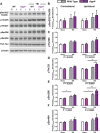
Post-traumatic P-tau accumulation is exacerbated by Aqp4 gene deletion. The effect of impairing glymphatic pathway function by Aqp4 gene deletion upon the development of tauopathy after TBI was evaluated. A, Wild-type and Aqp4−/− brains were harvested 28 d post injury and probed for the presence of P-tau epitopes. Representative blots are presented showing the effect of mouse genotype (wild-type vs Aqp4−/−), injury status (sham vs TBI), and hemisphere [contralateral (C) vs ipsilateral (I)] upon labeling by various P-tau monoclonal antibodies targeting different tau phosphorylation epitopes. Total tau was also measured (Pan-tau) and all P-tau levels were normalized to P-tau levels within each biological sample. B–F, Across all epitopes, TBI tended to increase P-tau labeling, particularly in the ipsilateral hemisphere. This effect of TBI on P-tau labeling was statistically significant when antibodies targeting the pThr205 (C), pThr231 (D), and pSer396 (E) were used (two-way ANOVA, n = 3–4 per group). Post hoc analysis revealed that Aqp4 gene deletion significantly increased pThr231 labeling after TBI (*p < 0.05, **p < 0.01, ***p < 0.001; two-way ANOVA with Tukey's post hoc test for multiple comparisons, n = 3–4 per group).
Figure 7.
Figure 7.
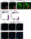
Neuronal P-tau accumulation and axonal degeneration after TBI in Aqp4−/− mice. Wild-type and Aqp4−/− mice were subjected to TBI and the presence of P-tau was evaluated by immunofluorescence 28 d post injury. Double labeling with the AT8 P-tau antibody (specific for pSer202/pThr205 epitopes) and the neuronal marker NeuN showed that in the wild-type cortex, P-tau immunoreactivity was not observed under control conditions (A). After TBI, P-tau immunoreactivity was diffusely increased, but at a low level (B). In the Aqp4−/− cortex (C), marked P-tau labeling was observed after TBI, both within neuronal soma (arrows) and in surrounding neurites (arrowheads). Quantification of P-tau immunoreactivity in cortical (D) and striatal (E) regions proximal to the traumatic lesion showed that P-tau accumulation was dramatically increased in the Aqp4−/− brain after TBI. F–K, Axonal degeneration was evaluated in the ipsilateral cortex and underlying corpus callosum 28 d after TBI by staining for phosphorylated neurofilament with the SMI34 monoclonal antibody. In the control cortex (F) and corpus callosum (I), no axonal spheroids or varicosities were evident. In the wild-type cortex after TBI (G), SMI34-immunoreactive varicosities were sparsely present (arrows). In the Aqp4−/− cortex (H) and corpus callosum (K), SMI34-positive spheroids and varicosities were readily detectable (arrows).
Figure 8.
Figure 8.
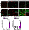
Aqp4 gene deletion promotes neuroinflammation after TBI. The effect of Aqp4 gene deletion upon the persistence of neuroinflammation after TBI was evaluated. A–F, Wild-type and Aqp4−/− mice were subjected to TBI and reactive astrogliosis (GFAP expression), and microgliosis (Iba1 expression) in the contralateral and ipsilateral cortex was evaluated by immunofluorescence 28 d post injury. Compared with wild-type mice after TBI, markedly elevated GFAP- and Iba1-immunoreactivity were observed in the ipsilateral cortex of Aqp4−/− mice after TBI. G, Quantification of GFAP labeling demonstrated significantly increased reactive astrogliosis in the ipsilateral cortex of Aqp4−/− mice (###p < 0.001, Ipsilateral vs Contralateral Aqp4−/−; two-way ANOVA with Tukey's post hoc test for multiple comparisons; n = 4 animals per group), which was not present in wild-type mice (**p < 0.01, Aqp4−/− Ipsilateral vs WT Ipsilateral). H, Quantification of Iba1 labeling showed that microglial activation was similarly increased in the ipsilateral cortex of Aqp4−/− mice (#p < 0.05, Ipsilateral vs Contralateral Aqp4−/−).
Figure 9.
Figure 9.
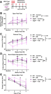
Aqp4 gene deletion exacerbates post-traumatic cognitive impairment. A, The effect of impairing the glymphatic pathway upon post-traumatic cognitive deficits was evaluated in wild-type and Aqp4−/− mice subjected to TBI. Animals underwent baseline behavioral testing 2 d before injury, then weekly after TBI. B, Gross motor behavior was assessed by the open field test. TBI did not significantly alter performance in the open field test in wild-type mice; however, performance in the open field test was significantly impaired after TBI in the Aqp4−/− mice (#p < 0.05 Aqp4−/− Control vs Aqp4−/− TBI; two-way repeated-measures ANOVA; n = 9–14 per group). C, Motor coordination was evaluated by the rotarod test. While TBI did not significantly impair rotarod performance in wild-type animals, in Aqp4−/− mice TBI significantly impaired test performance (#p < 0.05 Aqp4−/− Control vs Aqp4−/− TBI; two-way repeated-measures ANOVA with Sidak's post hoc test for multiple comparisons, n = 9–14 animals per group). Cognitive function was evaluated with the novel-object recognition test (D) and the Barnes maze test (E). In both tests, TBI impaired cognitive performance among both wild-type and Aqp4−/− mice [*p < 0.05, **p < 0.01 WT Control vs WT TBI; #p < 0.05, ##p < 0.05 Aqp4−/− Control vs Aqp4−/− TBI; two-way repeated-measures ANOVA with Sidak's post hoc test for multiple comparisons; n = 9–14 animals per group (Novel-Object Recognition), n = 5 per group (Barnes Maze)]. Post-traumatic cognitive impairment was exacerbated in Aqp4−/− mice compared with wild-type animals (p < 0.05 WT TBI vs Aqp4−/− TBI).
Figure 10.
Figure 10.
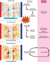
Post-traumatic impairment of glymphatic pathway function promotes tau aggregation. Schematic representation of proposed relationship between perivascular AQP4 polarization, glymphatic pathway function, and interstitial tau clearance after TBI. We propose that chronic loss of perivascular AQP4 polarization after TBI impairs paravascular clearance of interstitial tau, promoting tau aggregation, neurodegeneration, and persistent neuroinflammation in the post-traumatic brain.

Similar articles

Cited by

References

    1. Auguste KI, Jin S, Uchida K, Yan D, Manley GT, Papadopoulos MC, Verkman AS. Greatly impaired migration of implanted aquaporin-4-deficient astroglial cells in mouse brain toward a site of injury. FASEB J. 2007;21:108–116. doi: 10.1096/fj.06-6848com. - DOI - PubMed
    1. Badaut J, Ashwal S, Obenaus A. Aquaporins in cerebrovascular disease: a target for treatment of brain edema? Cerebrovasc Dis. 2011;31:521–531. doi: 10.1159/000324328. - DOI - PMC - PubMed
    1. Chesser AS, Pritchard SM, Johnson GV. Tau clearance mechanisms and their possible role in the pathogenesis of Alzheimer Disease. Front Neurol. 2013;4:122. doi: 10.3389/fneur.2013.00122. - DOI - PMC - PubMed
    1. Cirrito JR, Deane R, Fagan AM, Spinner ML, Parsadanian M, Finn MB, Jiang H, Prior JL, Sagare A, Bales KR, Paul SM, Zlokovic BV, Piwnica-Worms D, Holtzman DM. P-glycoprotein deficiency at the blood-brain barrier increases amyloid-beta deposition in an Alzheimer disease mouse model. J Clin Invest. 2005;115:3285–3290. doi: 10.1172/JCI25247. - DOI - PMC - PubMed
    1. Crane JM, Bennett JL, Verkman AS. Live cell analysis of aquaporin-4 m1/m23 interactions and regulated orthogonal array assembly in glial cells. J Biol Chem. 2009;284:35850–35860. doi: 10.1074/jbc.M109.071670. - DOI - PMC - PubMed

Publication types

MeSH terms

LinkOut - more resources