. 2015 Feb 19;518(7539):409-12.
doi: 10.1038/nature13975. Epub 2014 Dec 1.

Towards a therapy for Angelman syndrome by targeting a long non-coding RNA

Affiliations

Towards a therapy for Angelman syndrome by targeting a long non-coding RNA

Linyan Meng et al. Nature. .

Abstract

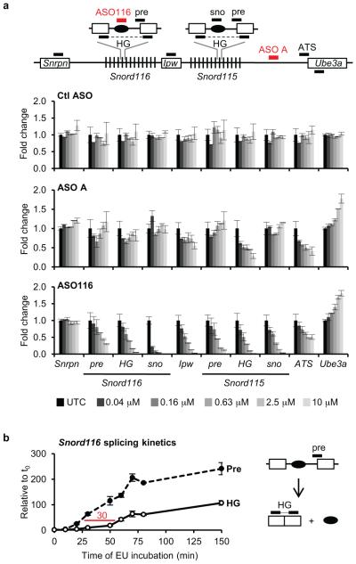
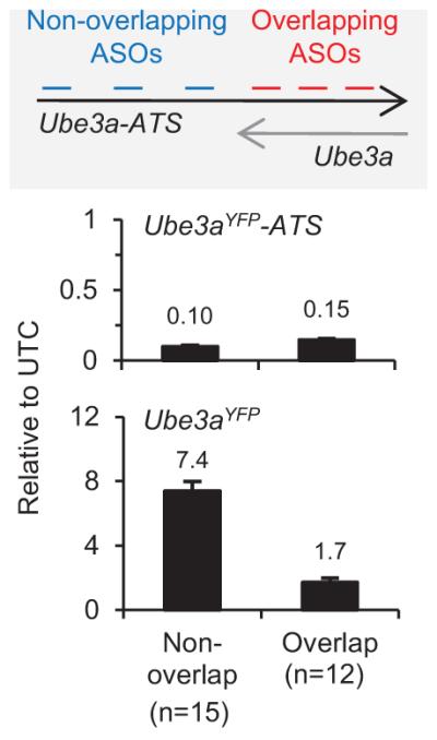
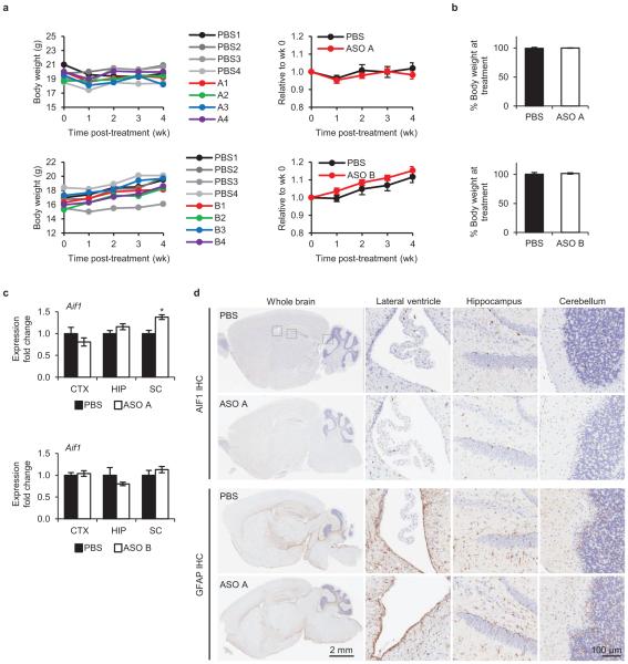
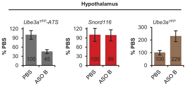
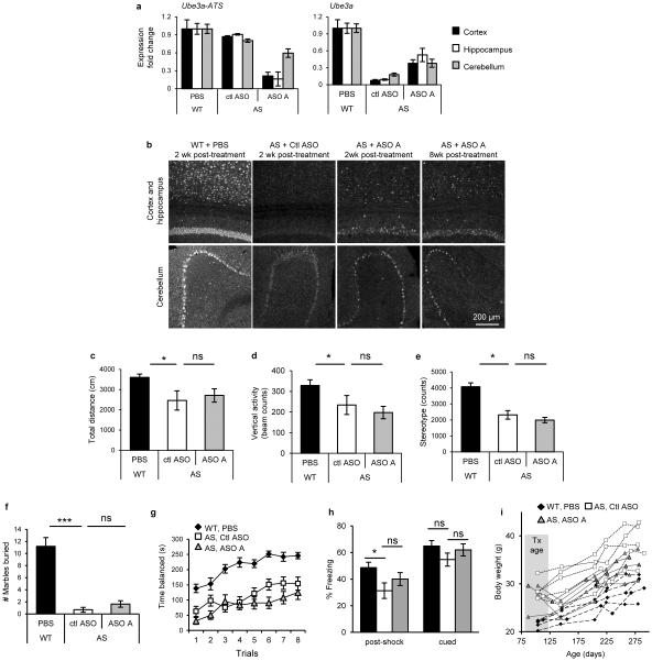
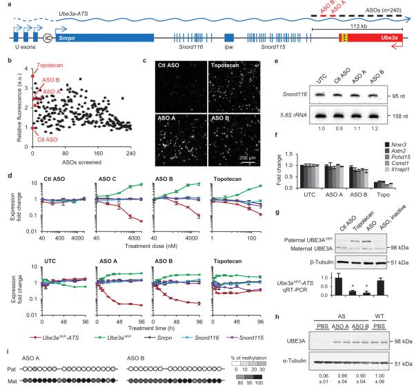
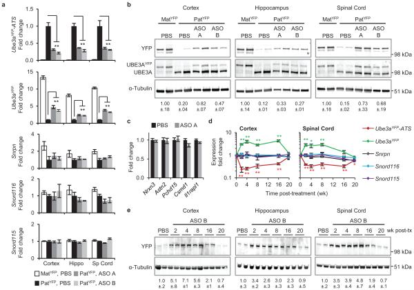
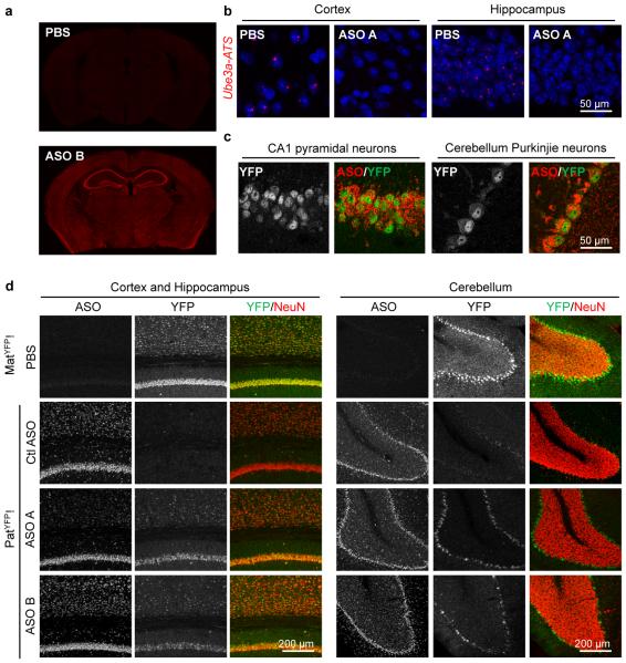
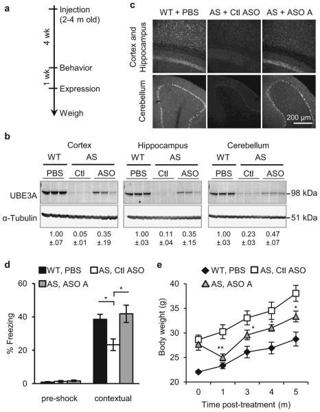
Angelman syndrome is a single-gene disorder characterized by intellectual disability, developmental delay, behavioural uniqueness, speech impairment, seizures and ataxia. It is caused by maternal deficiency of the imprinted gene UBE3A, encoding an E3 ubiquitin ligase. All patients carry at least one copy of paternal UBE3A, which is intact but silenced by a nuclear-localized long non-coding RNA, UBE3A antisense transcript (UBE3A-ATS). Murine Ube3a-ATS reduction by either transcription termination or topoisomerase I inhibition has been shown to increase paternal Ube3a expression. Despite a clear understanding of the disease-causing event in Angelman syndrome and the potential to harness the intact paternal allele to correct the disease, no gene-specific treatment exists for patients. Here we developed a potential therapeutic intervention for Angelman syndrome by reducing Ube3a-ATS with antisense oligonucleotides (ASOs). ASO treatment achieved specific reduction of Ube3a-ATS and sustained unsilencing of paternal Ube3a in neurons in vitro and in vivo. Partial restoration of UBE3A protein in an Angelman syndrome mouse model ameliorated some cognitive deficits associated with the disease. Although additional studies of phenotypic correction are needed, we have developed a sequence-specific and clinically feasible method to activate expression of the paternal Ube3a allele.


Figures

Extended Data Figure 1
Extended Data Figure 1. ASOs targeting Snord116 reduced Ube3a-ATS pre-mRNA
a, Upper panel, schematic of the ASO binding sites and location of qRT-PCR primer and probe sets. Lower panel, qRT-PCR from WT primary neurons treated with ASO A or ASO116 (72 h) using primer and probe sets to the indicated regions of Ube3a-ATS pre-mRNA and mRNA. b, Nascent transcripts were isolated from WT primary neurons incubated with 5-ethynyl uridine (see Methods) for the indicated time. qRT-PCR for pre-mRNA and mature mRNA (HG, host gene) within the Snord116 region. The red line indicates the 30 min delay between the appearance of pre-mRNA and mature mRNA. Assuming a transcription elongation rate of 4 kb/min, it would take RNAPII 80 min to transcribe the 332 kb distance from the last copy of Snord116 to the ASO binding site. n=2 per group, mean ± absolute deviation.
Extended Data Figure 2
Extended Data Figure 2. ASOs complementary to two regions of Ube3a-ATS differed in their ability to unsilence paternal Ube3a
PatYFP primary neurons were treated with ASOs that bind Ube3a-ATS 5′ of Ube3a (non-overlap ASOs, n=15) or that bind to the gene body region (overlap ASOs, n=12) for 72 h. The level of Ube3aYFP-ATS reduction and Ube3aYFP up-regulation was analyzed by qRT-PCR and normalized to untreated control (UTC) neurons. Mean ± s.e.m.
Extended Data Figure 3
Extended Data Figure 3. In vivo ASO administration was well tolerated
a, Left, body weight of individual WT C57BL/6 female mice (2 months old) treated with PBS or ASO measured weekly for 4 wk post-treatment. Right, Change in body weight at each time point relative to body weight at time of treatment. n=4 per group, mean ± s.e.m. b, Percent change in body weight of PatYFP mice 4 wk post-treatment relative to pre-treatment. n=3-4, mean ± s.e.m. c, Microglial activation was measured by Aif1 qRT-PCR 4 wk post-treatment. CTX, cortex; HIP, hippocampus; SC, thoracic spinal cord. *P<0.05, two-tailed t-test, n=3-4 per group, mean ± s.e.m. d, Immunohistochemistry for AIF1 and GFAP on sagittal brain sections from WT C57BL/6 female mice treated with PBS or ASO for 2 wk.
Extended Data Figure 4
Extended Data Figure 4. Snord116 was not reduced in the hypothalamus
qRT-PCR on RNA isolated from PatYFP mice 4 wk post-treatment of PBS or ASO B.
Extended Data Figure 5
Extended Data Figure 5. UBE3A unsilencing persisted 4 months post-treatment
ASO and YFP immunofluorescence on brain sections of cortex and cerebellum in PatYFP mice 4 months post-treatment of ASO A.
Extended Data Figure 6
Extended Data Figure 6. UBE3AYFP was up-regulated throughout the brain
Whole brain image of YFP fluorescence in PatYFP mice treated with PBS or ASO 4 wk post-treatment.
Extended Data Figure 7
Extended Data Figure 7. Imaging of unsilenced UBE3AYFP in specific brain regions
Immunofluorescence for ASO, UBE3AYFP, and NeuN 4 wk post-treatment in MatYFP or PatYFP mice of the amygdala, hippocampus CA2 and CA3 layers, dentate gyrus, and striatrum (panel 1); thalamus, hypothalamus, medulla, and third ventricle (panel 2); motor cortex, somatosensory cortex, auditory cortex, and visual cortex (panel 3).
Extended Data Figure 8
Extended Data Figure 8. Intrahippocampal injection of ASO A in PatYFP mice resulted in near complete unsilencing of paternal UBE3AYFP
YFP immunofluorescence on brain sections from PatYFP mice treated with non-targeting control ASO (Ctl ASO), 100 μg ASO A via intrahippocampal injection, or 700 μg ASO A via ICV injection. A MatYFP mouse treated with PBS was included for comparison.
Extended Data Figure 9
Extended Data Figure 9. ASO treatment in AS mice up-regulated Ube3a
a, RNA levels of Ube3a-ATS and Ube3a were determined by qRT-PCR in WT mice treated with PBS and AS mice treated with non-targeting control ASO (ctl ASO), or ASO A. n=2-3 per group, mean ± s.e.m. b, UBE3A immunofluorescence on brain sections was performed 2 to 8 wk post-treatment. c-h, ASO treatment in adult AS mice did not reverse some disease-associated phenotypes. c, Total distance traveled in the open field assay. d, Vertical activity in the open field assay. e, Stereotype activity in the open field assay. f, Marble burying test. The y axis represents the number of marbles at least 50% buried. g, Accelerating rotarod test during eight trials. h, Post-shock and cued response measured during the fear conditioning assay. n=13-15 per group *P<0.05, ***P<0.001 (one way ANOVA with Newman-keuls post-hoc analysis). i, Growth curve of age-matched female mice. Each line represents weight measurements of a single mouse over a 5 month time course post-injection, n=5 per group. Tx age, age of mouse at time of treatment.
Figure 1
Figure 1. Unsilencing of the Ube3a paternal allele by Ube3a-ATS targeted ASOs in cultured mouse neurons
a, Schematic mouse Ube3a genomic locus. IC, imprinting center. b, UBE3AYFP fluorescence (arbitrary units, a.u.) in ASO-treated primary neurons relative to untreated control. Ctl ASO, non-targeting control ASO. c, YFP fluorescent imaging of treated PatYFP neurons. d, Normalized mRNA levels in PatYFP neurons treated with increasing dose (upper panel) or for increasing time (lower panel). e, Northern blot of Snord116 expression. Snord116 intensity relative to 5.8S rRNA is quantified. f, Normalized mRNA levels of long genes. g, Western blot (upper) and qRT-PCR (lower) from PatYFP neurons. ASO, inactive is a sequence-matched RNase H inactive ASO. *P<0.05, two-tailed t-test, n=2 per group, mean ± absolute deviation. h, Western blot from WT or AS primary neurons. UBE3A signal intensity was quantified relative to α-Tubulin. i, DNA methylation analysis of the PWS imprinting center. The paternal allele was distinguished by the conversion of a CpG dinucleotide (CG > AA) in CAST.Chr7 mice.
Figure 2
Figure 2. A single administration of Ube3a-ATS ASOs resulted in paternal UBE3A unsilencing for 4 months
a and b, mRNA levels (a), and UBE3AYFP protein (b) in cortex, hippocampus, and spinal cord 4 wk after ICV injection of PBS or ASO to PatYFP mice. MatYFP mice are included for comparison. c, Normalized mRNA levels of long genes in the cortex. d and e, RNA levels (d) and UBE3AYFP protein (e) in ASO-treated PatYFP mice 2 to 20 wk post-treatment. *P<0.05, **P<0.005, two-tailed t-test, n=3-4 per group, mean ± s.e.m. For Western blot quantification, YFP signal intensity was calculated relative to α-Tubulin.
Figure 3
Figure 3. Widespread distribution of paternal UBE3A unsilencing throughout the brain
Imaging of brain sections 4 wk post-treatment of non-targeting control ASO (Ctl ASO) or Ube3a-ATS ASOs (ASO A and/or B) in PatYFP mice, as labelled. a, ASO immunofluorescence on whole brain coronal sections. b, In situ hybridization of Ube3a-ATS. c, High magnification staining of UBE3AYFP and ASO. d, Immunofluorescence for ASO, UBE3AYFP, and NeuN in critical brain regions. MatYFP mice treated with PBS were included for expression comparison.
Figure 4
Figure 4. ASO administration in adult AS mice unsilenced paternal UBE3A and ameliorated abnormal phenotypes
a, Experimental schedule. b, Western blot with anti-UBE3A in brain regions of treated mice. Quantification of UBE3A normalized to α-Tubulin is indicated below the images. c, UBE3A immunofluorescence in WT or AS mice. d, Contextual fear measured during the fear conditioning assay. *P<0.05, one-way ANOVA with Newman–Keuls post-hoc, n=13-15 per group. e, Growth curve of age-matched female mice, *P<0.05, **P<0.01 (ASO A versus Ctl ASO), two-way ANOVA of repeated measurements with Newman–Keuls post-hoc), n=5 per group. Ctl or Ctl ASO, non-targeting control ASO.

Comment in

Similar articles

Cited by

References

    1. Dagli A, Buiting K, Williams CA. Molecular and Clinical Aspects of Angelman Syndrome. Molecular syndromology. 2012;2:100–112. - PMC - PubMed
    1. Williams CA, Driscoll DJ, Dagli AI. Clinical and genetic aspects of Angelman syndrome. Genetics in medicine : official journal of the American College of Medical Genetics. 2010;12:385–395. - PubMed
    1. Kishino T, Lalande M, Wagstaff J. UBE3A/E6-AP mutations cause Angelman syndrome. Nature genetics. 1997;15:70–73. - PubMed
    1. Matsuura T, et al. De novo truncating mutations in E6-AP ubiquitin-protein ligase gene (UBE3A) in Angelman syndrome. Nature genetics. 1997;15:74–77. - PubMed
    1. Albrecht U, et al. Imprinted expression of the murine Angelman syndrome gene, Ube3a, in hippocampal and Purkinje neurons. Nature genetics. 1997;17:75–78. - PubMed

Publication types

MeSH terms