. 2015 Jan 16;290(3):1485-95.
doi: 10.1074/jbc.M114.593871. Epub 2014 Nov 26.

Interferon-stimulated gene 15 (ISG15) and ISG15-linked proteins can associate with members of the selective autophagic process, histone deacetylase 6 (HDAC6) and SQSTM1/p62

Affiliations

Interferon-stimulated gene 15 (ISG15) and ISG15-linked proteins can associate with members of the selective autophagic process, histone deacetylase 6 (HDAC6) and SQSTM1/p62

Hiroshi Nakashima et al. J Biol Chem. .

Abstract

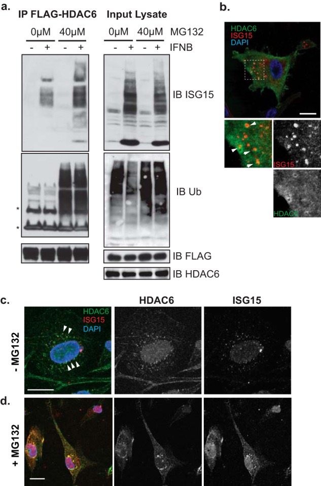
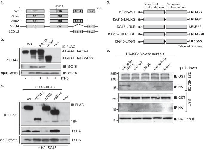
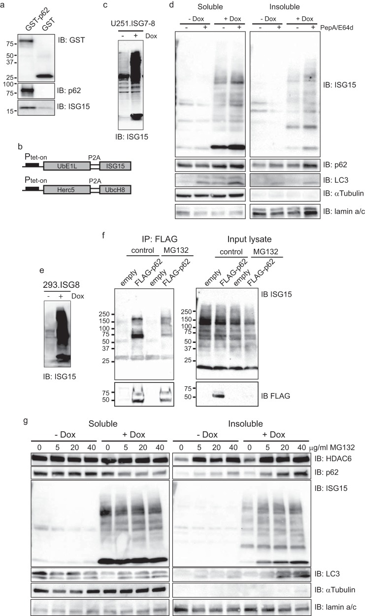
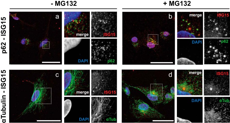
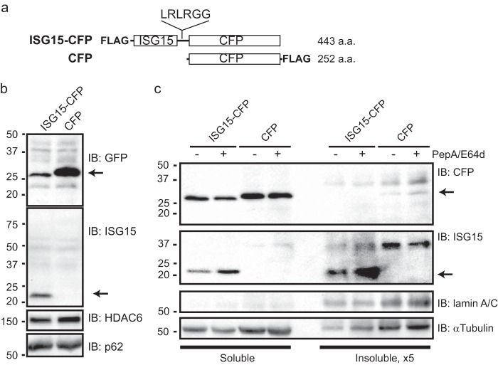
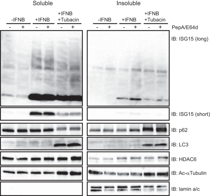
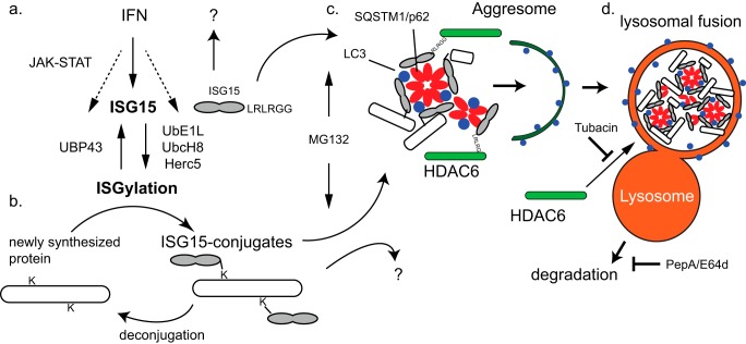
The ubiquitin-like interferon (IFN)-stimulated gene 15 (ISG15) and its specific E1, E2, and E3 enzymes are transcriptionally induced by type I IFNs. ISG15 conjugates newly synthesized proteins. ISG15 linkage to proteins appears to be an important downstream IFN signaling event that discriminates cellular and pathogenic proteins synthesized during IFN stimulation from existing proteins. This eliminates potentially pathogenic proteins as the cell attempts to return to normal homeostasis after IFN "stressed" conditions. However, the molecular events that occur in this process are not well known. Here, we show that the C-terminal LRLRGG of ISG15 interacts with the binder of ubiquitin zinc finger (BUZ) domain of histone deacetylase 6 (HDAC6). Because HDAC6 is involved in the autophagic clearance of ubiquitinated aggregates during which SQSTM1/p62 plays a major role as a cargo adapter, we also were able to confirm that p62 binds to ISG15 protein and its conjugated proteins upon forced expression. Both HDAC6 and p62 co-localized with ISG15 in an insoluble fraction of the cytosol, and this co-localization was magnified by the proteasome inhibitor MG132. In addition, ISG15 was degraded via the lysosome. Overexpression of ISG15, which leads to an increased conjugation level of the cellular proteome, enhanced autophagic degradation independently of IFN signaling transduction. These results thus indicate that ISG15 conjugation marks proteins for interaction with HDAC6 and p62 upon forced stressful conditions likely as a step toward autophagic clearance.

Keywords: Autophagy; Histone Deacetylase 6 (HDAC6); ISG15; Infection; Lysosome; Post-translational Modification (PTM); SQSTM1; Ubiquitin; Virus; p62.


Figures

FIGURE 1.
FIGURE 1.
HDAC6 interacts with ISG15-conjugated cellular proteins. a, U251 expressing FLAG-HDAC6 WT (full length) were treated with IFNβ (1,000 units/ml; 16 h) with and without MG132 (40 μm; 4 h). Anti-FLAG-immunoprecipitated (IP) proteins and input cell lysates were immunoblotted (IB) with anti-FLAG, anti-ISG15, and anti-Ub antibodies. b–d, U251 cells expressing FLAG-HDAC6 WT were treated with IFNβ (1,000 units/ml; 16 h) in the absence (b and c) or the presence (d) of MG132 (40 μm; 5 h). Cytoskeletal insoluble fractions were visible in cells prepermeabilized with Triton X-100 (c and d). Fluorescence signals for Alexa Fluor 488 (green) for FLAG, Alexa Fluor 555 (red) for ISG15, and DAPI (blue) were visualized using confocal microscopy. High magnification views highlighted by broken line boxes are shown in the bottom panels of the image (b). Scale bars, 20 μm.
FIGURE 2.
FIGURE 2.
Mapping the interacting domains of HDAC6 and ISG15. a, schematic of FLAG-tagged HDAC6 and its deletion mutants. The two catalytic domains (CD1 and CD2), SE14 domain, and BUZ domain are shown as closed boxes. An asterisk (*) represents the H611A point mutation in WT. b, U251 cells that express FLAG-HDAC6 WT, mutant without an active site histidine (H611A), an SE14-BUZ-truncated mutant (ΔCter), or empty vector (Vec) were treated with IFNβ (1,000 units/ml; 16 h). Lysates were subjected to co-immunoprecipitation (IP) with antibody against FLAG and analysis using antibodies against FLAG and ISG15. c, 293 cells were transiently co-transfected with expression vectors for FLAG-HDAC6 WT, a C-terminal truncation (ΔCter) mutant, an N-terminal truncation (ΔCD1/2) mutant, a BUZ domain truncation (ΔBUZ) mutant, an SE14 domain truncation (ΔSE14) mutant, or an empty vector (Vec) along with an HA-tagged ISG15 expression vector. Lysates and immunoprecipitates were analyzed with antibodies against FLAG and HA. d, schematic of HA-tagged ISG15 with wild type or mutants. The two Ub-like domains are shown as closed boxes. Peptides were tagged with HA at the N terminus, and amino acids were deleted from the LRLRGG peptide sequences at the C-terminal end (c-end) as indicated by asterisks (*). e, 293A cells were transiently transfected with plasmids carrying HA-tagged ISG15 vectors with mutants. In a pulldown assay, cell lysates were incubated with purified recombinant GST-tagged HDAC6 or control GST, bound to glutathione-Sepharose beads, and subjected to analysis with antibodies against HA and GST. IB, immunoblotting.
FIGURE 3.
FIGURE 3.
ISG15 up-regulation of aggregate formation of p62. a, purified recombinant ISG15 proteins were pulled down with glutathione-Sepharose bead-bound purified recombinant GST tag-p62 fusion or control GST and analyzed as indicated. b, schematic inducible co-expression vectors of ISG15 and the core enzyme genes ISG15, E1 UbE1L, E2 UbcH8, and E3 Herc5 using self-cleavage peptide sequences (P2A) under the control of the Tet-On promoter (Ptet-on). The details of the vector constructs are described under “Experimental Procedures.” c, U251.ISG7–8 exhibited overexpression of ISG15 and ISG15 conjugation to cellular proteins in the presence of Dox (100 ng/ml; 24 h) prior to immunoblotting (IB) with anti-ISG15 antibody. d, immunoblots of detergent-soluble (Soluble) and -insoluble (Insoluble) fractions of U251.ISG7–8 cells with and without Dox treatment (100 ng/ml; 24 h) in the presence or absence of PepA/E64d (each 20 μg/ml; 4 h) were probed for the indicated proteins. e, 293.ISG8 exhibited overexpression of ISG15 and conjugation in the presence of Dox (200 ng/ml; 24 h). f, 293-ISG8 cells were transiently transfected with FLAG-tagged p62 or control empty vector and treated with MG132 (40 μm; 6 h). Anti-FLAG-immunoprecipitated (IP) proteins and input cell lysates were immunoblotted (IB) with anti-FLAG and anti-ISG15 antibodies. g, immunoblots of 0.5% Triton X-100 detergent-soluble (Soluble) and -insoluble (Insoluble) lysate fractions of 293.ISG8 cells treated with Dox (200 ng/ml; 24 h) in the presence or absence of MG132 (6 h) at the indicated doses prior to collecting the cells were probed for the indicated proteins.
FIGURE 4.
FIGURE 4.
ISG15 and p62 co-localization in insoluble fraction. a–d, U251 cells were treated with IFNβ (1,000 units/ml; 16 h) before treatment with MG132 (40 μm; 5 h) (b and d) or without it (a and c). Prepermeabilized cells were fixed with 4% paraformaldehyde and subjected to fluorescence microscopy analysis using Alexa Fluor 488 (green) against p62 and α-tubulin, Alexa Fluor 555 (red) against ISG15, and DAPI (blue). High magnification rescans of the broken line box regions of interest are shown in the panels on the right of each sample image. Scale bars, 50 μm.
FIGURE 5.
FIGURE 5.
Lysosomal degradation of cleaved ISG15. a, schematic of ISG15-CFP and CFP-expressing cDNA vector and its product protein (in amino acids (a.a.)). The LRLRGG C-terminal end of FLAG-tagged human ISG15 is linked to the N terminus of mTurquoise2 (referred to as CFP). Control CFP is tagged with FLAG at the C-terminal end. b, cell lysates were prepared in radioimmune precipitation assay buffer 3 days after transient transfection of the vectors as indicated and immunoblotted using antibodies against GFP, ISG15, HDAC6, and p62. c, immunoblots (IB) of detergent-soluble (Soluble) and -insoluble (Insoluble) fractions of U251 with 24-h transient transfection of the indicated vectors in the presence or absence of PepA/E64d (each 20 μg/ml; 6 h) were probed for the indicated proteins. Volumes of insoluble fractions are 5-fold (×5) higher concentration than those of the soluble fractions. Numbers shown on the left indicate protein sizes (kDa). Arrows indicate the bands of cleaved ISG15 or CFP protein.
FIGURE 6.
FIGURE 6.
The HDAC6 deacetylase inhibitor, Tubacin, suppresses ISG15 degradation. U251 cells were treated with IFNβ (1,000 units/ml; 12 h) and tubacin (5 μm; 13 h) before treatment with PepA/E64d (20 μg/ml; 6 h). Cell lysates prepared as the soluble and insoluble fractions were immunoblotted (IB) using antibodies against the indicated proteins. An immunoblot against ISG15 shows both a short exposure (short) and a long exposure (long) time to compare the levels in non-conjugated ISG15.
FIGURE 7.
FIGURE 7.
Schematic model of ISG15-mediated degradation. a, IFNs are known to stimulate the JAK-STAT pathway to induce a number of interferon-stimulated genes including ISG15, UbE1L, UbcH8, and Herc5 that lead to the ISGylation process and UBP43 that deconjugates ISGylated proteins. b, the Lys63-linked di-Ub-like protein ISG15 conjugates to lysine (K) residues of newly synthesized proteins at the LRLRGG-terminal end (2). Some ISG15 conjugates are also deconjugated to remove ISG15. c, our findings indicate that HDAC6 and SQSTM1/p62 can independently bind ISG15 to recognize ISG15 and ISG15 conjugates directly or indirectly. ISG15 aggregates or aggresomes would be formed through oligomerization and recruitment of autophagosome-bound LC3 by p62. The proteasome inhibitor MG132 induces and enhances this aggregate formation and accumulation in the insoluble fraction. d, the deacetylase activity of HDAC6 can mediate lysosomal fusion of ISG15-containing aggregates for clearance, and this is inhibited by the specific HDAC6 inhibitor tubacin. PepA/E64d inhibit lysosomal digestion of peptide contents.

Similar articles

Cited by

References

    1. Sadler A. J., Williams B. R. (2008) Interferon-inducible antiviral effectors. Nat. Rev. Immunol. 8, 559–568 - PMC - PubMed
    1. Durfee L. A., Lyon N., Seo K., Huibregtse J. M. (2010) The ISG15 conjugation system broadly targets newly synthesized proteins: implications for the antiviral function of ISG15. Mol. Cell 38, 722–732 - PMC - PubMed
    1. Malakhov M. P., Malakhova O. A., Kim K. I., Ritchie K. J., Zhang D. E. (2002) UBP43 (USP18) specifically removes ISG15 from conjugated proteins. J. Biol. Chem. 277, 9976–9981 - PubMed
    1. Morales D. J., Lenschow D. J. (2013) The antiviral activities of ISG15. J. Mol. Biol. 425, 4995–5008 - PMC - PubMed
    1. Romijn R. A., Westein E., Bouma B., Schiphorst M. E., Sixma J. J., Lenting P. J., Huizinga E. G. (2003) Mapping the collagen-binding site in the von Willebrand factor-A3 domain. J. Biol. Chem. 278, 15035–15039 - PubMed

Publication types

MeSH terms