. 2015 Jan 1;194(1):334-48.
doi: 10.4049/jimmunol.1401548. Epub 2014 Nov 24.

Myeloid cell TRAF3 regulates immune responses and inhibits inflammation and tumor development in mice

Affiliations

Myeloid cell TRAF3 regulates immune responses and inhibits inflammation and tumor development in mice

Almin I Lalani et al. J Immunol. .

Abstract

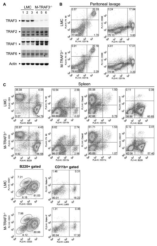
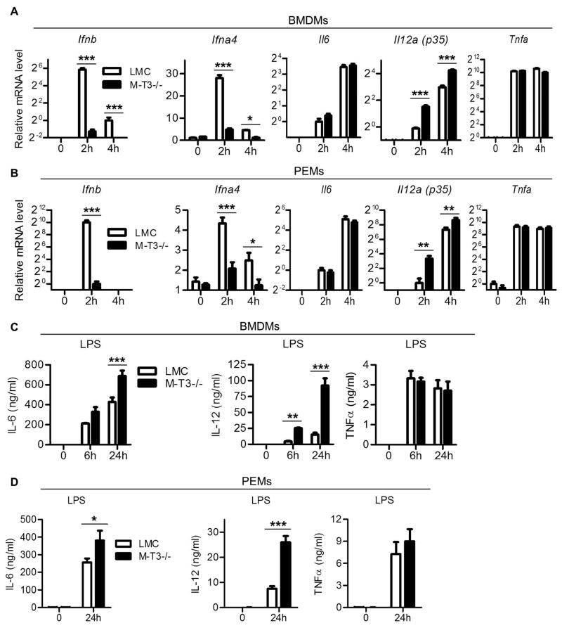
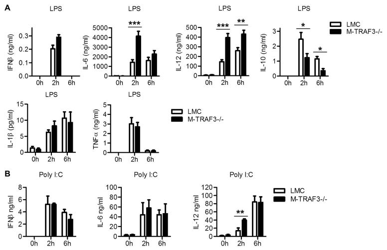
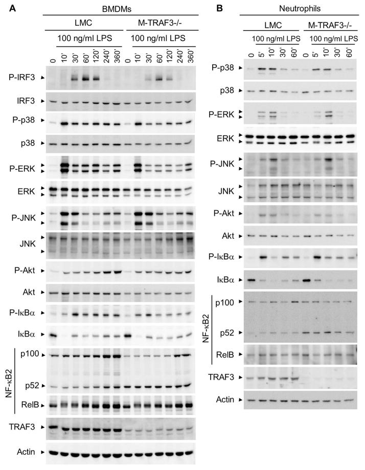
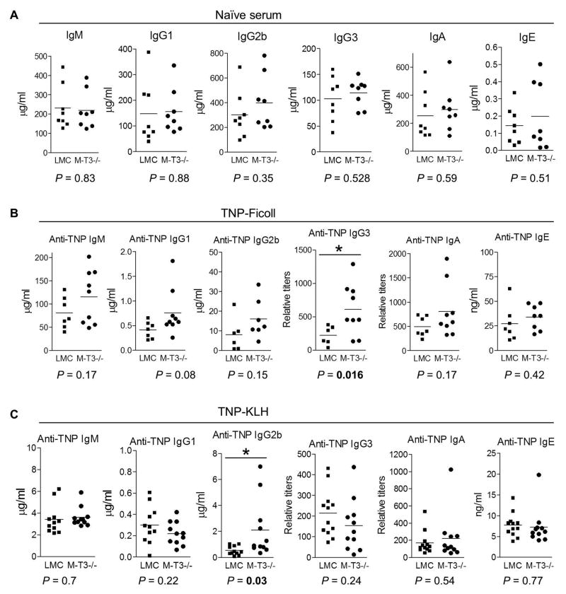
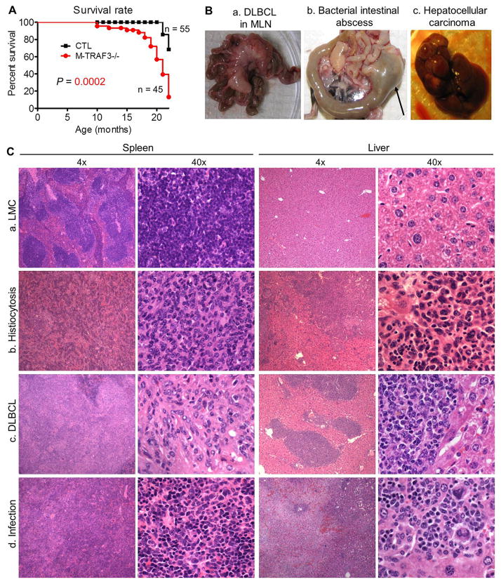
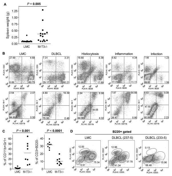
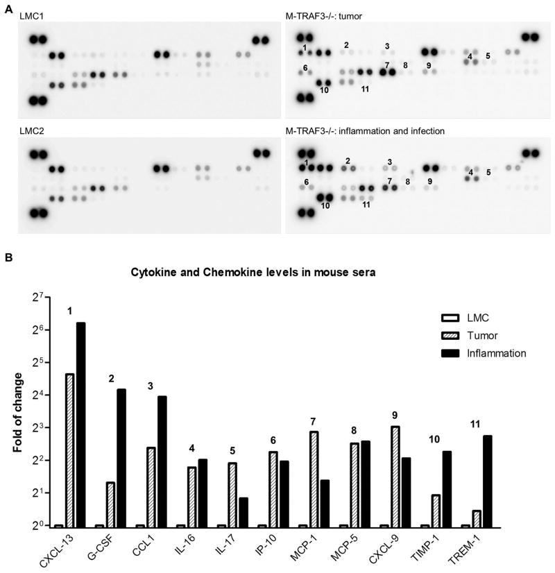
Myeloid cells, including granulocytes, monocytes, macrophages, and dendritic cells, are crucial players in innate immunity and inflammation. These cells constitutively or inducibly express a number of receptors of the TNFR and TLR families, whose signals are transduced by TNFR-associated factor (TRAF) molecules. In vitro studies showed that TRAF3 is required for TLR-induced type I IFN production, but the in vivo function of TRAF3 in myeloid cells remains unknown. In this article, we report the generation and characterization of myeloid cell-specific TRAF3-deficient (M-TRAF3(-/-)) mice, which allowed us to gain insights into the in vivo functions of TRAF3 in myeloid cells. We found that TRAF3 ablation did not affect the maturation or homeostasis of myeloid cells in young adult mice, even though TRAF3-deficient macrophages and neutrophils exhibited constitutive NF-κB2 activation. However, in response to injections with LPS (a bacterial mimic) or polyinosinic-polycytidylic acid (a viral mimic), M-TRAF3(-/-) mice exhibited an altered profile of cytokine production. M-TRAF3(-/-) mice immunized with T cell-independent and -dependent Ags displayed elevated T cell-independent IgG3 and T cell-dependent IgG2b responses. Interestingly, 15- to 22-mo-old M-TRAF3(-/-) mice spontaneously developed chronic inflammation or tumors, often affecting multiple organs. Taken together, our findings indicate that TRAF3 expressed in myeloid cells regulates immune responses in myeloid cells and acts to inhibit inflammation and tumor development in mice.


Conflict of interest statement

Competing interests statement

The authors declare that they have no competing financial interests.

Figures

Figure 1
Figure 1. Normal lymphocyte and myeloid cell populations in young adult M-TRAF3−/− mice
TRAF3flox/flox littermate control (LMC) and M-TRAF3−/− mice analyzed were 8 to 12 weeks old. (A) Verification of TRAF3 deletion in BMDMs by Western blot analysis. Total cellular proteins were prepared from BMDMs derived from 3 pairs of LMC and M-TRAF3−/− mice. Proteins were immunoblotted for TRAF3, followed by TRAF2, TRAF1, TRAF6 and actin. (B) Representative FACS profiles of peritoneal lavage prepared from LMC and M-TRAF3−/− mice. Cells were stained with fluorochrome-conjugated antibodies specific for CD11b, F4/80, and CD68, and then analyzed by a FACSCalibur. Peritoneal macrophages are identified as CD11b+F4/80+CD68+. (C) Representative FACS profiles of splenocytes prepared from LMC and M-TRAF3−/− mice. Cells were stained with fluorochrome-conjugated antibodies specific for B220, CD3, CD11b, F4/80, CD11c, MHC ClassII, CD21, CD23, Ly6G, Siglec-F, and CD317 (PDCA-1), and then analyzed by a FACSCalibur. FACS profiles were scatter-gated on live cells. Gated cell populations include: B cells, B220+CD3−; T cells, CD3+B220−; red pulp macrophages, CD11b+F4/80+; cDCs, CD11c+MHC ClassII+; follicular (FO) B cells, B220+CD21IntCD23+; marginal zone (MZ) B cells, B220+CD21+CD23Int; neutrophils, CD11b+Ly6G+Siglec-F-; pDCs, B220+CD317 (PDCA-1)+. Similar results were observed in 4 additional experiments.
Figure 2
Figure 2. Impaired type I IFN but enhanced IL-6 and IL-12 production in TRAF3−/− macrophages in response to LPS stimulation
(A and B) LPS-induced mRNA expression of cytokines in macrophages. BMDMs (A) or PEMs (B) derived from M-TRAF3−/− and LMC mice (7–10 week-old) were cultured in the absence or presence of 100 ng/ml of LPS for 2 or 4 hours. RNA was extracted, and real-time PCR was performed using TaqMan primers and probes specific for Ifnb, Ifna4, Il6, Il12a (p35), and Tnfa. Each reaction also included the probe (VIC-labeled) and primers for b-actin mRNA, which served as an endogenous control. Relative mRNA expression levels of each cytokine were analyzed using the Sequence Detection Software (Applied Biosystems) and the comparative Ct method. Graphs depict the results of three independent experiments with duplicate reactions in each experiment (mean ± S.D.). (C and D) LPS-induced secretion of cytokines in macrophages. BMDMs (C) or PEMs (D) derived from M-TRAF3−/− and LMC mice (7–10 week-old) were cultured in the absence or presence of 100 ng/ml of LPS for indicated time periods. The levels of IL-6, IL-12 and TNFα in culture supernatants were analyzed by ELISA. Graphs depict the results of three independent experiments with duplicate samples in each experiment (mean ± S.D.). *, significantly different between LMC and M-TRAF3−/− (t test, p < 0.05); **, very significantly different between LMC and M-TRAF3−/− (t test, p < 0.01); ***, highly significantly different between LMC and M-TRAF3−/− (t test, p < 0.001).
Figure 3
Figure 3. In vivo cytokine production in response to injection with LPS or polyI:C
Gender and age-matched M-TRAF3−/− and LMC mice (8–12 week-old) were i.p. injected with LPS (300 μg LPS/20g body weight, n=9 for each group, A) or polyI:C (200 μg polyI:C/20g body weight, n=4 for each group, B), and sera were collected at 2 and 6 h post-injection. Serum levels of IFN-β, IL-6, IL-12, IL-10, IL-1β, and TNFα were measured by ELISA. *, significantly different between LMC and M-TRAF3−/− (t test, p < 0.05); **, very significantly different between LMC and M-TRAF3−/− (t test, p < 0.01); ***, highly significantly different between LMC and M-TRAF3−/− (t test, p < 0.001).
Figure 4
Figure 4. Altered signaling events in TRAF3−/− BMDMs and neutrophils in response to LPS stimulation
BMDMs (A) or thioglycollate elicited peritoneal neutrophils (B) prepared from M-TRAF3−/− and LMC mice (8–12 week-old) were stimulated with 100 ng/ml of LPS for indicated time periods. Total protein lysates were prepared, and signaling events of TLR4 were analyzed by immunoblot analysis. Proteins were immunoblotted for phosphorylated (P-) or total IRF-3, p38, ERK, JNK, Akt, IκBα, followed by NF-κB2 (p100 and p52), RelB, TRAF3 and actin. Results shown are representative of 3 independent experiments.
Figure 5
Figure 5. Altered antibody responses in M-TRAF3−/− mice
LMC and M-TRAF3−/− mice analyzed were 8 to 12 weeks old. (A) Basal serum titers of Ig isotypes. Sera from naïve LMC and M-TRAF3−/− mice (n=8 for each group) were tested for IgM, IgG1, IgG2b, IgG3, IgA, and IgE levels by ELISA. (B) TI antibody responses. LMC and M-TRAF3−/− mice (n=9 for each group) were immunized with the TI Ag TNP-Ficoll, and sera were collected on day 7 after immunization. Serum titers of anti-TNP IgM, IgG1, IgG2b, IgG3, IgA, and IgE were analyzed by ELISA. (C) TD antibody responses. LMC and M-TRAF3−/− mice (n=12 for each group) were immunized with the TD Ag TNP-KLH in Alum. Sera were collected on day 7 after immunization. Serum titers of anti-TNP IgM, IgG1, IgG2b, IgG3, IgA, and IgE were measured by ELISA. Multiple serial dilutions of each serum sample were tested to ensure that the readout is within the linear range of the assay. *, significantly different from LMC (t-test, P < 0.05).
Figure 6
Figure 6. Gross and histopathological features of M-TRAF3−/− mice with spontaneous tumor, inflammation or infection
(A) Accelerated mortality of M-TRAF3−/− mice. Survival curves of LMC and M-TRAF3−/− mice were generated using the Kaplan-Meier method. P<0.001 as determined by the Mantel-Cox log-rank test. (B) Representative images of affected organs in diseased M-TRAF3−/− mice. a, Massively enlarged mesenteric lymph node (MLN) in a mouse with DLBCL (mouse ID: 237-2). b, GI tract and MLN of a mouse with bacterial infection and inflammation (mouse ID: 228-5). Arrow indicates the large bacterial intestinal abscess. c, Liver of a mouse with hepatocellular adenoma (mouse ID: 237-4). (C) Representative micrographs of the spleen and liver of diseased M-TRAF3−/− mice. Sections of the spleen and liver were stained with hematoxylin and eosin, and representative micrographs of LMC and M-TRAF3−/− mice are shown for comparison. a, Normal tissues from a LMC mouse. b, Mouse (ID: 237-5) with extensive histiocytosis and increased erythroid activity in the spleen and areas of pure histiocytosis in the liver. c, Mouse (ID: 233-5) with DLBCL showing marked enlargement of the splenic white pulp by tumor cells and large perivascular infiltrates with tumor cells in the liver. d, Mouse (ID: 274-12) with infection showing inflammation and necrotic areas in the liver and marked red pulp hyperplasia of myeloid and erythroid elements in the spleen.
Figure 7
Figure 7. Abnormal lymphocyte and myeloid cell populations in the spleen of diseased M-TRAF3−/− mice
(A) Enlarged spleens of M-TRAF3−/− mice (age: 15–22 month-old). The graph depicts spleen weights of age-matched LMC and M-TRAF3−/− mice (n=15 for each group of mice). (B) Representative FACS profiles of splenocytes of age-matched LMC and diseased M-TRAF3−/− mice. Splenocytes were stained with fluorochrome-conjugated B220, CD3, CD11b, Gr-1, CD21 and CD23 Abs, and then analyzed by a FACSCalibur. FACS profiles were scatter-gated on live cells. Gated cell populations include: B cells, B220+CD3−; T cells, CD3+B220−; myeloid cells, CD11b+Gr-1+; a distinct population of histiocytes (gated with a circle in the 3rd and 5th panel): CD11blowGr-1low. M-TRAF3−/− mice shown include: 237-2, DLBCL; 237-5, histiocytosis; 237-1, inflammation; 274-12, infection and inflammation. FACS profiles shown are representative of 2 cases of DLBCL, 2 cases of histiocytosis, 2 cases of inflammation, and 2 cases of infection, respectively. (C) Increased percentage of CD11b+Gr-1+ myeloid cells and decreased percentage of CD3+B220− T cells in diseased M-TRAF3−/− mice. The graphs depict the percentages of CD11b+Gr-1+ myeloid cells or CD3+B220− T cells of age-matched LMC and diseased M-TRAF3−/− mice (n=8 for each group of mice) analyzed by FACS. P values (t-test) between LMC and M-TRAF3−/− mice are shown. (D) Representative FACS profiles of B220+ splenic B cells of M-TRAF3−/− mice with DLBCL. Follicular (FO) B cells are identified as B220+CD21IntCD23+, and marginal zone (MZ) B cells are identified as B220+CD21+CD23Int. The small population (6.91%) of B220+CD21−CD23− observed in LMC are immature and activated B cells, and the major population (41.34% in mouse ID 237-5 and 68.48% in mouse ID 233-5) of B220+CD21−CD23− observed in M-TRAF3−/− mice are predominantly DLBCL cells.
Figure 8
Figure 8. Aberrant serum cytokine and chemokine levels of diseased M-TRAF3−/− mice
(A) Cytokine and chemokine protein array blots of age-matched LMC and diseased M-TRAF3−/− mice. Cytokines and chemokines in mouse sera were detected using the Mouse Cytokine Array Assay kit (R&D) following the manufacturer’s protocol. In each blot, combined sera of three mice (70 μl serum of each mouse) were used for the cytokine array assay. Sera of M-TRAF3−/− mice with tumor examined include 237-5 (histiocytosis), 228-7 (DLBCL), and 274-11 (FL). Sera of M-TRAF3−/− mice with inflammation and infection examined include 228-5, 237-1, and 274-12. (B) Quantification of cytokine and chemokine levels of diseased M-TRAF3−/− mice measured by the cytokine protein array analyses. Each cytokine or chemokine spots on the blots in (A) were quantitated using a low-light imaging system, and the results presented graphically. The amount of each cytokine or chemokine in diseased M-TRAF3−/− mice (tumor or inflammation) was relative to the mean of the intensity of corresponding LMC1 and LMC2 spots. Each cytokine or chemokine has duplicate detection spots. The graph depicts the fold change (M-TRAF3−/−/LMC) of each cytokine or chemokine (mean). Bold numeric labels indicate the spots that are strikingly different between M-TRAF3−/− mice with tumor or inflammation/infection and LMC in (A) and the corresponding cytokine/chemokine quantitation data in (B).

Similar articles

Cited by

References

    1. Xie P. TRAF molecules in cell signaling and in human diseases. J Mol Signal. 2013;8:7. - PMC - PubMed
    1. Hacker H, Tseng PH, Karin M. Expanding TRAF function: TRAF3 as a tri-faced immune regulator. Nat Rev Immunol. 2011;11:457–468. - PubMed
    1. Kawai T, Akira S. The role of pattern-recognition receptors in innate immunity: update on Toll-like receptors. Nat Immunol. 2010;11:373–384. - PubMed
    1. Saleh M. The machinery of Nod-like receptors: refining the paths to immunity and cell death. Immunol Rev. 2011;243:235–246. - PubMed
    1. Yu M, Levine SJ. Toll-like receptor, RIG-I-like receptors and the NLRP3 inflammasome: key modulators of innate immune responses to double-stranded RNA viruses. Cytokine Growth Factor Rev. 2011;22:63–72. - PMC - PubMed

Publication types

MeSH terms