. 2014 Nov 20;159(5):1027-1041.
doi: 10.1016/j.cell.2014.10.023.

ER contact sites define the position and timing of endosome fission

Affiliations

ER contact sites define the position and timing of endosome fission

Ashley A Rowland et al. Cell. .

Abstract

Endocytic cargo and Rab GTPases are segregated to distinct domains of an endosome. These domains maintain their identity until they undergo fission to traffic cargo. It is not fully understood how segregation of cargo or Rab proteins is maintained along the continuous endosomal membrane or what machinery is required for fission. Endosomes form contact sites with the endoplasmic reticulum (ER) that are maintained during trafficking. Here, we show that stable contacts form between the ER and endosome at constricted sorting domains, and free diffusion of cargo is limited at these positions. We demonstrate that the site of constriction and fission for early and late endosomes is spatially and temporally linked to contact sites with the ER. Lastly, we show that altering ER structure and dynamics reduces the efficiency of endosome fission. Together, these data reveal a surprising role for ER contact in defining the timing and position of endosome fission.


Figures
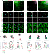
Figure 1
Figure 1
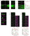
Early endosome fission occurs at ER contact sites. (A) Merged images of a live Cos-7 cell expressing mCh-Rab5 (EE in cyan), GFP-Rab4 (EE in red), and BFP-Sec61β (ER in green) that is pulse-labeled with EGF conjugated to Alexa Fluor® 647 (cargo in blue). (B) Magnified time-lapse images of the region boxed in A shows an example of early endosome fission. Merged images show the relative location of Rab4, Rab5, ER and EGF, as indicated over time. The exiting Rab4+ bud is marked by a yellow arrowhead. See also Movie S1. (C) Trace outline of the endosome shown in B and the corresponding line-scan analysis of the relative fluorescence intensity (FI) of Rab4, Rab5, ER and EGF for time points: t=0s (Pre-ER), t=10s (Pre-Fission), and t=15s (Post-fission). Images in B and line-scan in C reveal that a dynamic ER tubule (marked by a blue arrow) is recruited to the divide between Rab4 compartments just before fission (at t=10s). (D) Merged images taken of a live Cos-7 cell transfected as in A that is instead pulse-labeled with Tf conjugated to Alexa Fluor® 647 (cargo in blue). (E) Zoom from D shows the relative localization of Rab4, Rab5, ER and Tf as indicated over time. An ER tubule (marked by blue arrow) is again recruited to the divide between Rab4 compartments just before fission (t=35s). An exiting Rab4+/Tf+ bud is marked by a yellow arrowhead. See Movie S2. (F) Line-scan analysis of relative FI depicts the position of a dynamic ER tubule to the position and timing of endosome fission for time points: t=0s (Pre-ER), t=35s (Pre-Fission), and t=45s (Post-Fission). Scale bars = 5 μm in A and D; 1 μm in B and E. s = seconds.
Figure 2
Figure 2
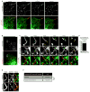
ER tubules define functional constrictions along dynasore-stalled tubular endosomes. (A) Images of live Cos-7 cells expressing mCh-Rab5 and GFP-Sec61β show that dynasore treatment elongates early endosomes while ER is unaffected. (B) Merged images of live Cos-7 cells expressing GFP-Rab5, BFP-Sec61β, and mCh-FAM21 following dynasore treatment. (C) Magnified image of endosome boxed in B. Images reveal that FAM21 concentrates at positions along the tubular endosome where Rab5-labeling is reduced and ER tubules intersect these FAM21 domains. (D) Line-scan analysis of relative FI of the endosome shown in C confirms that FAM21and ER co-localize with Rab5-labeled endosome constrictions (marked by corresponding blue and yellow arrows). (E) A model outlines the FRAP technique used to test for a cargo diffusion barrier (D.B.) along tubular endosomes. (F) A live Cos-7 cell expressing BFP-Rab5, mCh-Sec61β, and GFP-TfR (membrane-bound cargo) were treated with dynasore to elongate endosomes. Endosomes were photobleached in the region indicated (dotted yellow circle). Images were taken at times indicated during the recovery (see kymograph). See also Movie S3. (G) A graph of relative FI during time points shown reveals a D.B. limits the recoveryof TfR at the position of an ER-marked constriction (at yellow arrow). The D.B./constriction maintains contact with the ER over time (marked by yellow arrows). (H-I) Another example as in F-G. Scale bars = 5 μm in A and B; 1 μm in C, F, and H.
Figure 3
Figure 3
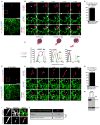
Dynasore-stalled tubular early endosomes undergo fission at ER contact sites. (A) The elongated tubular endosome phenotype that results from dynasore treatment can be reversed with the addition of FBS. Time-lapsed images of live Cos-7 cells expressing mCh-Rab5 and GFP-Sec61β following dynasore treatment (t=0min) and after FBS addition at times indicated (right panels). (B)Cells treated as in A. (C) Magnified time-lapse images of region boxed in B shows two fission events that occur on a single tubular endosome with each event marked by an ER tubule crossing (blue arrows). Yellow arrowheads mark fission products after each division. See also Movie S4. (D) The percent of tubular EE fission events marked by ER tubule crossings (out of 31 events from 9 cells). (E) Method for determining the amount of endosome image surface covered by ER tubule crossing for all 31 fission events. An example is shown of endosome and ER tracing from an image immediately preceding fission. Top row shows indicated fluorescence markers and bottom row shows thresholded images. (F) Table shows the percentage (22.36%) of endosomal pixels co-localized with pixels from ER tubules crossing the endosome. The right panel shows that the measured frequency of ER-marked fission (80.6%) is significantly higher than that due to chance (22.36%). ***, P<0.001, Fisher’s exact test. Scale bars = 5 μm in A and B; 1 μm in C and E. min = minutes.
Figure 4
Figure 4
Late endosome division occurs at ER contact sites. (A) A Cos-7 cell expressing mCh-Rab7 (late endosome) and GFP-Sec61β was pulse-labeled with EGF conjugated to Alexa Fluor® 647 (cargo in blue). (B) Magnified image of the region boxed in A shows an example of lateendosome fission. Merged images show the relative location of Rab7, EGF and the ER, as indicated over time. See also Movie S5. (C) Traced outline and the corresponding line-scan analysis of relative FI through the equator of the dividing endosome shown in B. Relative FI of Rab7, ER, and EGF were performed for time points: t=0s (Pre-ER), t=35s (Pre-fission), and t=40s (Post-fission). Note that a dynamic ER tubule is recruited to the position and timing of endosome constriction and fission (compare position of the ER tubule marked by an arrow at t=0s to t=20s in B and C). (D) Percent of late endosome division events that co-localize with ER tubules (n=29 from 24 cells). (E) Image of a Dnm2-depleted cell expressing mCh-Rab7 (red) and GFP-Sec61β (green) and pulse-labeled with EGF conjugated to Alexa Fluor® 647 (blue). (F) Magnified image of box in E shows an elongated endosome that moves until it becomes associated with an ER tubule (white arrow) at the site of endosome division (blue arrow). The location of the Rab7 endosome bud (yellow arrowhead) is shown. See also Movie S6. (G) The percent of late endosome fission events that co-localize with ER tubules in the absence of dynamin-2 (out of 12 events from 8 cells). (H) Immunoblot analysis shows efficient depletion of Dnm2 in cells transfected with Dnm2 siRNA (right lane) relative to control (left lane). (I) The endosome image surface covered by ER tubule crossing was measured for all 12 fission events. In example shown, the top row shows indicated fluorescence markers and bottom row shows thresholded images. (J) Table summarizes predicted frequency of ER-marked LE fission (21.24% based on coverage) versus the actual frequency of ER-marked fission (100%). ***, P<0.001, Fisher’s exact test. Scale bars = 5 μm in A and E; 1 μm in B and F.
Figure 5
Figure 5
The ER is recruited to FAM21-marked sorting domains prior to fission. (A) Merged image of a live Cos-7 cell expressing GFP-Rab7, BFP-Sec61β, and mCh-FAM21 that was also pulse-labeled with EGF conjugated to Alexa Fluor® 647 (cargo in blue). (B) Magnified time-lapse images of an endosome in A shows the tip of an ER tubule tracking with a FAM21 punctum at the bud of a late endosome (yellow arrowhead). See also Movie S7. (C) The number of FAM21 puncta on late endosome buds that maintain contact with ER over a 2 min time course (165 puncta from 31 cells). (D) Merged image of a live Cos-7 treated as in A. (E) Magnified images of boxed region in D shows a late endosome undergoing fission. A bud labeled by FAM21 extends from the late endosome and undergoes constriction and division. An ER tubule (follow white arrow) is recruited to the FAM21 bud just before fission (at the blue arrow). The location of the exiting Rab7 endosome bud is marked by a yellow arrowhead. See also Movie S8. (F) Line-scan analysis of endosome shown in E relates the timing and position of ER tubule recruitment relative to Rab7, FAM21, and EGF. (G) The percent of FAM21-marked late endosome division events that co-localize with ER tubules during cargo segregation, (n=36 from 31 cells). Scale bars = 5 μm in A and D; 1 μm in B and E.
Figure 6
Figure 6
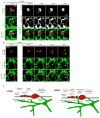
Model of endosome sorting and fission factors. (A) A live Cos-7 cell expressing GFP-Rab7, mCh-Sec61β, and BFP-FAM21.Endosome fission occurs after a dynamic ER tubule contacts the endosome at a constricted sorting domain. (B) A Cos-7 cell expressing mCh-Rab7 and GFP-Sec61β was pulse-labeled with EGF conjugated to Alexa Fluor® 647.The ER tubule contacts the endosome, fission occurs at the point of contact, the budding domain leaves, maintaining ER contact. (C) The many factors implicated in endosome sorting and fission are indicated in the model pre- and post-ER recruitment. Following sorting, the ER establishes contact with the endosome at sites that are spatially and temporally linked to endosome fission.
Figure 7
Figure 7
ER structure and dynamics affect endosome fission. (A) Live Cos-7cells expressing BFP-KDEL and mCh-Rab7 with (top panel) or without (bottom panel) Rtn4a-GFP. Note that Rtn4a-GFP tubules are less branched (top first panel) and exclude KDEL from the periphery (top second panel). (B) Zoom from A shows overlay of t=0sec (red) with t=60sec (green) to show ER movement over time “dynamics”. (C) Graph of average Pearson’s correlation coefficient shows greater co-localization and therefore fewer dynamic ER movements for Rtn4a expressing cells (control n=22 cells, Rtn4a n=20 cells, ***, p<0.001, two-tailed test). (D) Graph showing no significant difference in endosome diameter between control and Rtn4a expressing cells (control n=216 endosomes, Rtn4a n=215 endosomes). (E) Graph showing no significant difference in endosome bud length between control and Rtn4a expressing cells (control n=257 buds in 22 cells, Rtn4a n=257 buds in 20 cells). (F) Graph showing a significant (**, p<0.01, two-tailed test) decrease in the percent of endosome buds from E that undergo productive fission in Rtn4a expressing cells. Scale bars = 5μm in A, and 1μm B and G. Error bars represent SEM.

Comment in

Similar articles

Cited by

References

    1. Alpy F, Rousseau A, Schwab Y, Legueux F, Stoll I, Wendling C, Spiegelhalter C, Kessler P, Mathelin C, Rio MC, et al. STARD3 or STARD3NL and VAP form a novel molecular tether between late endosomes and the ER. J Cell Sci. 2013;126:5500–5512. - PubMed
    1. Arighi CN, Hartnell LM, Aguilar RC, Haft CR, Bonifacino JS. Role of the mammalian retromer in sorting of the cation-independent mannose 6-phosphate receptor. J Cell Biol. 2004;165:123–133. - PMC - PubMed
    1. Barbero P, Bittova L, Pfeffer SR. Visualization of Rab9-mediated vesicle transport from endosomes to the trans-Golgi in living cells. J Cell Biol. 2002;156:511–518. - PMC - PubMed
    1. Cullen PJ. Endosomal sorting and signalling: an emerging role for sorting nexins. Nat Rev Mol Cell Biol. 2008;9:574–582. - PubMed
    1. Derivery E, Sousa C, Gautier JJ, Lombard B, Loew D, Gautreau A. The Arp2/3 activator WASH controls the fission of endosomes through a large multiprotein complex. Dev Cell. 2009;17:712–723. - PubMed

Publication types