. 2014 Nov 20;10(11):e1004797.
doi: 10.1371/journal.pgen.1004797. eCollection 2014 Nov.

Three RNA binding proteins form a complex to promote differentiation of germline stem cell lineage in Drosophila

Affiliations

Three RNA binding proteins form a complex to promote differentiation of germline stem cell lineage in Drosophila

Di Chen et al. PLoS Genet. .

Abstract

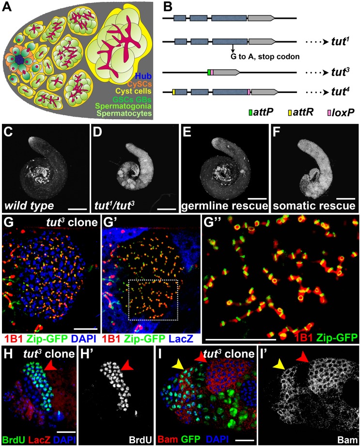
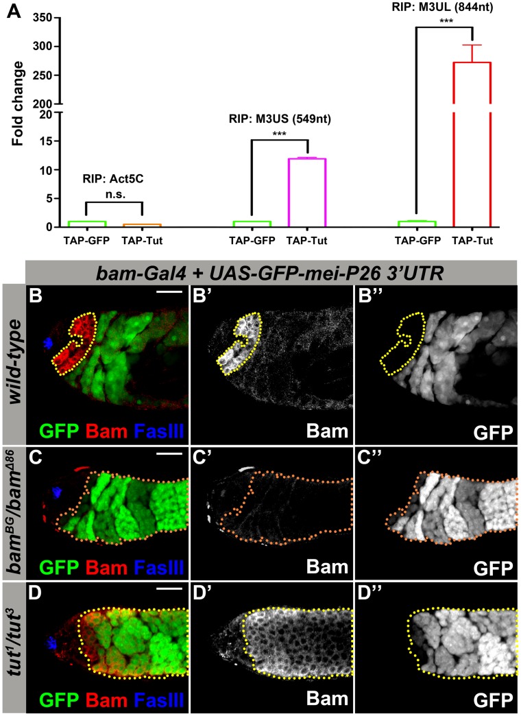
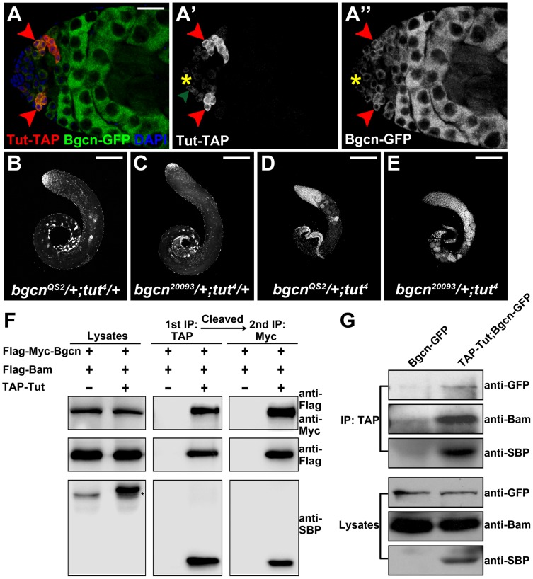
In regenerative tissues, one of the strategies to protect stem cells from genetic aberrations, potentially caused by frequent cell division, is to transiently expand the stem cell daughters before further differentiation. However, failure to exit the transit amplification may lead to overgrowth, and the molecular mechanism governing this regulation remains vague. In a Drosophila mutagenesis screen for factors involved in the regulation of germline stem cell (GSC) lineage, we isolated a mutation in the gene CG32364, which encodes a putative RNA-binding protein (RBP) and is designated as tumorous testis (tut). In tut mutant, spermatogonia fail to differentiate and over-amplify, a phenotype similar to that in mei-P26 mutant. Mei-P26 is a TRIM-NHL tumor suppressor homolog required for the differentiation of GSC lineage. We found that Tut binds preferentially a long isoform of mei-P26 3'UTR, and is essential for the translational repression of mei-P26 reporter. Bam and Bgcn are both RBPs that have also been shown to repress mei-P26 expression. Our genetic analyses indicate that tut, bam, or bgcn is required to repress mei-P26 and to promote the differentiation of GSCs. Biochemically, we demonstrate that Tut, Bam, and Bgcn can form a physical complex in which Bam holds Tut on its N-terminus and Bgcn on its C-terminus. Our in vivo and in vitro evidence illustrate that Tut acts with Bam, Bgcn to accurately coordinate proliferation and differentiation in Drosophila germline stem cell lineage.


Conflict of interest statement

The authors have declared that no competing interests exist.

Figures

Figure 1
Figure 1. tut restricts spermatogonial proliferation cell-autonomously.
(A) Schematic illustration of the cellular architecture at the apical part of Drosophila testis. CySC: cyst stem cell; GSC: germline stem cell; GB: gonialblast. Note the morphological differences of fusome (red) in different cells: dot in GSC and GB, or branched network in spermatogonia and spermatocytes. (B) Molecular information of tut alleles. tut3, a null allele, was generated by replacing tut genomic region with attP and loxP elements. tut4 is a weak allele, with tut coding region flanked by attR (before start codon) and loxP (after stop codon). (C–F) Low magnification images showing the testes of w1118 (C), tut1/3 (D), bam-Gal4/Y; UAS-GFP-tut/+; tut1/3 (E), and tj-Gal4/UAS-GFP-tut; tut1/3 (F) stained with DNA dye DAPI. (G–I) Immunofluorescence images of the apical part of the testes containing tut mutant clones. tut3 clones were marked by the absence of LacZ (G′, H) or GFP (I). (G) A clone of over-proliferating germ cells with branched fusome (1B1) running through ring canal (Zip-GFP). G″ shows the high magnification view of the boxed region in G′. (H–H′) A testis stained for clone marker LacZ and S phase marker BrdU. Red arrowhead points to the tut3 mutant clone with all cells entering S phase in synchrony. (I–I′) A testis containing tut3 mutant clones was stained for Bam, GFP, and DNA (DAPI). Bam was expressed both in wild-type spermatogonia (yellow arrowhead) and tut3 mutant cells (red arrowhead). Scale bars: 200 µm (C–F) and 25 µm (G–I). See also Figure S1.
Figure 2
Figure 2. Tut is required to repress mei-P26 expression via mei-P26 3′UTR.
(A) Physical interaction of Tut protein and mei-P26 3′UTR detected by RNA immunoprecipitation (RIP). TAP-Tut or TAP-GFP and mei-P26 3′UTR were co-expressed in S2 cells. IgG beads were used to enrich TAP-Tut or TAP-GFP followed by TEV digestion to release Tut or GFP and bound RNA. RNA was extracted and reverse transcribed. The quantity of Actin5C mRNA, short (M3US) or long (M3UL) isoform of mei-P26 3′UTR was determined by real-time PCR. The Y axis represents the ratio of RIP/Input, which was normalized to 1 for TAP-GFP. Error bar indicates SD. ***, p<0.0001 in t test. n.s., not significant. (B–B″) Genotype: bam-Gal4/Y; UAS-GFP-mei-P26 3′UTR (2k)/+. A genomic region of 2 kb in length downstream of mei-P26 stop codon was selected to cover both short and long isoforms (see Figure S2A). Yellow dots outline Bam-expressing spermatogonia. GFP was repressed in most Bam-expressing spermatogonia. (C–C″) Genotype: bam-Gal4/Y; UAS-GFP-mei-P26 3′UTR (2k)/+; bamBG/bamΔ86. Orange dots outline GFP-positive spermatogonia. (D–D″) Genotype: bam-Gal4/Y; UAS-GFP-mei-P26 3′UTR (2k)/+; tut1/tut3. Yellow dotted outline indicates Bam-expressing spermatogonia. GFP was de-repressed in tut mutant even in the presence of Bam. Scale bars: 25 µm. See also Figure S2-S3.
Figure 3
Figure 3. tut interacts with bam both genetically and physically.
(A–B) Low magnification view of tut3 (A) and tutP-tut-GFP/+; tut3 (B) testes stained with DAPI. (C&C′) tutP-tut-GFP/+; tut3 testis stained for GFP, germline marker Vasa, and hub cell marker FasIII. Yellow asterisk indicates hub and hereafter. Green arrowhead points to a GSC expressing weak Tut-GFP signal. (D–D″) Immunofluorescence images of tutP-tut-GFP/+; tut3 testis. Tut-GFP and Bam were both expressed in spermatogonia. (E–I) tut4/+ (E), bamΔ86 /+ (F), tut4 (G), tut4, bamΔ86/+ (H), and tut4, bamBG/+ (I) testes stained with DAPI. Red arrowheads point to over-proliferational cysts. (J&J′) Immunofluorescence images of tut4, bamBG/+ testis. Note the branched fusome. (K–L) Bam and Tut coimmunoprecipitated from S2 cells expressing tagged proteins. Flag-Bam and Myc-Tut were over-expressed in S2 cells and the cell lysates were used for anti-Myc (K) or anti-Flag (L) immunoprecipitation. Western analysis with corresponding antibodies was performed to detect the presence of Flag-Bam and Myc-Tut. Asterisk indicates a nonspecific band. Scale bars: 25 µm (A, B, E–I); 200 µm (C,D,J). See also Figure S4.
Figure 4
Figure 4. Tut, Bam, and Bgcn form a protein complex.
(A–A″) Immunofluorescence images of tutP-tut-TAP/+; bgcnP-bgcn-GFP/+ testis. Red arrowheads point to spermatogonial cells expressing both Tut-TAP and Bgcn-GFP. Green arrowhead points to a germline stem cell expressing weak Tut-TAP. (B-E) Genetic interactions between tut and bgcn. bgcnQS2/+; tut4/+ (A), bgcn20093/+; tut4/+ (B), bgcnQS2/+; tut4 (C), and bgcn20093/+; tut4 (D) testes were stained with DAPI. (F) S2 cells were transfected with different combinations of DNA constructs as indicated. Lysates from transfected S2 cells were used in a two-step immunoprecipitation method employing IgG and anti-Myc beads successively. Western analyses with anti-SBP (streptavidin binding protein), anti-Flag, and anti-Myc were performed to detect the presence of TAP-Tut, Flag-Bam, and Flag-Myc-Bgcn, respectively. TAP tag contains two TEV cleavage sites joining Protein G and SBP. After TEV digestion, the size of TAP-Tut changed from 48.35 kD to 32.86 kD. * indicates a nonspecific band. (G) Testes extracts of bamP-bgcnGFP/+ or tutP-tutTAP/Y; tutP-tutTAP/+;bamP-bgcnGFP/+ were immunoprecipitated with IgG beads. Tut-TAP was detected by anti-SBP on Western blot. Scale bars: 25 µm (A); 200 µm (B–E). See also Figure S5.
Figure 5
Figure 5. Tut and Bgcn are recruited by Bam to form a complex.
(A–B) S2 cells were transfected with the combinations of DNA constructs as indicated. Lysates from transfected S2 cells were immunoprecipitated with IgG (A) or anti-Myc (B) beads. Western blots were used to analyze the presence of TAP-, Flag-, or Myc-tagged proteins. Interaction between Tut and Bgcn was not detected in the absence of Bam. Asterisk in (A) indicates non-specific bands. (C–F) S2 cells were transfected with different combinations of DNA constructs as indicated. Lysates from transfected S2 cells were immunoprecipitated with anti-HA beads. Western blots were used to analyze the presence of Flag- or HA-tagged proteins. See also Figure S6.
Figure 6
Figure 6. Tut is required for Bam to drive germline stem cell differentiation.
(A–B&B′) Genotype: UASp-bam-GFP/+; nos-Gal4/+. Forced expression of Bam in GSCs eliminated all germ cells. (C–D&D′) Genotype: UASp-bam-GFP/+; tut1 nos-Gal4/tut3. Germ cells were present in tut mutant background. Green dots outline the hub-adjacent germ cells expressing Bam-GFP. (E–E″) Immunofluorescence images of UASp-bam-GFP/+; tut1 nos-Gal4/tut3 testis. Green dots outline hub-adjacent germ cells expressing Bam-GFP and yellow arrowhead points to a dot-shape spectrosome. (F–F″) Immunofluorescence images of UASp-bam-GFP/+; tut1 nos-Gal4/tut3 testis. Green dots outline the hub-adjacent germ cells expressing Stat92E and Bam-GFP. (G&G′) 1 day old UASp-bam-GFP/+;tut1 nos-Gal4/tut3 testis labeled with BrdU for 1 hour. Green dotted-line highlights three hub-adjacent germ cells positive for Bam-GFP and two of them incorporated BrdU. (H) 10 day old UASp-bam-GFP/+;tut1 nos-Gal4/tut3 testis labeled with BrdU for 1 hour. Green dotted-line indicates two hub-adjacent germ cells positive for Bam-GFP and BrdU. Scale bars: 50 µm (A,C); 25 µm (B, D–H). See also Figure S7.

Similar articles

Cited by

References

    1. Alonso L, Fuchs E (2003) Stem cells in the skin: waste not, Wnt not. Genes & Development 17: 1189–1200. - PubMed
    1. Clarke MF, Fuller M (2006) Stem cells and cancer: Two faces of eve. Cell 124: 1111–1115. - PubMed
    1. Kondo M, Wagers AJ, Manz MG, Prohaska SS, Scherer DC, et al. (2003) Biology of hematopoietic stem cells and progenitors: Implications for clinical application. Annual Review of Immunology 21: 759–806. - PubMed
    1. Schuijers J, Clevers H (2012) Adult mammalian stem cells: the role of Wnt, Lgr5 and R-spondins. Embo Journal 31: 2685–2696. - PMC - PubMed
    1. Jamieson CHM, Ailles LE, Dylla SJ, Muijtjens M, Jones C, et al. (2004) Granulocyte-macrophage progenitors as candidate leukemic stem cells in blast-crisis CML. New England Journal of Medicine 351: 657–667. - PubMed

Publication types

Grants and funding

This study was supported by National Key Basic Research Program of China (2013CB945000) and National Science Foundation China (31271539). The funders had no role in study design, data collection and analysis, decision to publish, or preparation of the manuscript.