. 2014 Nov 20;10(11):e1004512.
doi: 10.1371/journal.ppat.1004512. eCollection 2014 Nov.

Crystal structure of cytomegalovirus IE1 protein reveals targeting of TRIM family member PML via coiled-coil interactions

Affiliations

Crystal structure of cytomegalovirus IE1 protein reveals targeting of TRIM family member PML via coiled-coil interactions

Myriam Scherer et al. PLoS Pathog. .

Abstract

PML nuclear bodies (PML-NBs) are enigmatic structures of the cell nucleus that act as key mediators of intrinsic immunity against viral pathogens. PML itself is a member of the E3-ligase TRIM family of proteins that regulates a variety of innate immune signaling pathways. Consequently, viruses have evolved effector proteins to modify PML-NBs; however, little is known concerning structure-function relationships of viral antagonists. The herpesvirus human cytomegalovirus (HCMV) expresses the abundant immediate-early protein IE1 that colocalizes with PML-NBs and induces their dispersal, which correlates with the antagonization of NB-mediated intrinsic immunity. Here, we delineate the molecular basis for this antagonization by presenting the first crystal structure for the evolutionary conserved primate cytomegalovirus IE1 proteins. We show that IE1 consists of a globular core (IE1CORE) flanked by intrinsically disordered regions. The 2.3 Å crystal structure of IE1CORE displays an all α-helical, femur-shaped fold, which lacks overall fold similarity with known protein structures, but shares secondary structure features recently observed in the coiled-coil domain of TRIM proteins. Yeast two-hybrid and coimmunoprecipitation experiments demonstrate that IE1CORE binds efficiently to the TRIM family member PML, and is able to induce PML deSUMOylation. Intriguingly, this results in the release of NB-associated proteins into the nucleoplasm, but not of PML itself. Importantly, we show that PML deSUMOylation by IE1CORE is sufficient to antagonize PML-NB-instituted intrinsic immunity. Moreover, co-immunoprecipitation experiments demonstrate that IE1CORE binds via the coiled-coil domain to PML and also interacts with TRIM5α We propose that IE1CORE sequesters PML and possibly other TRIM family members via structural mimicry using an extended binding surface formed by the coiled-coil region. This mode of interaction might render the antagonizing activity less susceptible to mutational escape.


Conflict of interest statement

The authors have declared that no competing intests exist.

Figures

Figure 1
Figure 1. The IE1 proteins of the primate cytomegaloviruses human (h), chimpanzee (c) and rhesus (rh) cytomegalovirus contain a conserved, stably folded globular core domain.
(A) In silico analysis of the hIE1, cIE1 and rhIE1 sequences using IUPred . Disorder score for IE1 suggesting the presence of a central folded globular domain (scores <0.5; gray box). Scores ≥0.5 (gray line) indicate disorder. (B) Prokaryotic expression and purification of hIE1, cIE1 and rhIE1 proteins used for crystallization attempts. The panel shows a Coomassie blue stained SDS-PAGE of the purified proteins as indicated. (C) Experimental confirmation of the folded globular IE1 domain (IE1CORE) by limited proteolysis. Full-length (1–491), C-terminally truncated (1–377) or N/C-terminally truncated (20–382) hIE1 protein was incubated with subtilisin and analyzed at different time points (e.g. at 1, 10, 30, 60, 120, 180, 240 and 300 min) by SDS-PAGE and Coomassie blue staining. (D) Circular dichroism analysis of the hIE1(14–382), cIE1(15–383) and rhIE1(36–395) core domains indicating a conserved, highly α-helical fold.
Figure 2
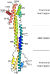
Figure 2. Structure of IE1CORE.
Ribbon representation of the rhIE1 monomer (residues 41–393) illustrating the elongated, femur-like fold of IE1. The eleven α-helices (H1 to H11) are colored from blue to red. The location of the two head regions and the central stalk are indicated by black lines.
Figure 3
Figure 3. Structural comparison of the rhIE1 with TRIM25.
(A) Overlay of dimeric TRIM25 (in blue and red) with monomeric rhIE1 (gold). (B) Dimeric TRIM25. (C) Topological arrangement of helices H1 to H3 of rhIE1. (D) Structure of the TRIM25 monomer.
Figure 4
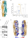
Figure 4. Oligomerization of IE1.
(A) Dimeric assembly of rhIE1 in the crystal. The two monomers (in cyan and gold) are related by a local 2-fold rotation axis indicated by a black ellipse. (B) Self-association of recombinant IE1 and IE1CORE in gel filtration experiments. Overlay of the elution profiles of full length hIE1(1–491) (blue) and hIE1CORE(14–382) (gold). Full length hIE1 and hIE1CORE eluted as symmetric main peaks with apparent molecular weights of ∼390 kDa and ∼194 kDa, respectively. It currently remains unclear whether the elution behavior of hIE1CORE merely reflects the elongated molecular shape of the dimer as observed in the crystal structure of rhIE1CORE or whether hIE1CORE assembles in solution into oligomers that comprise more than two molecules. Molecular weight estimates are based on commercially available calibration proteins, as indicated by the grey curve. (C) Self-interaction of the IE1CORE in human cells. HEK293T cells were co-transfected with expression plasmids encoding Myc-tagged hIE1(1–382) and FLAG-tagged hIE1 variants comprising residues 1–382 or 1–377. After cell lysis, immunoprecipitation was performed with an anti-FLAG antibody. Proteins within the cell lysate (input) and co-precipitated PML (IP) were analyzed by Western blotting as indicated. (D) Evolutionary sequence conservation of IE1 mapped onto the surface of rhIE1. The chains of dimeric rhIE1 are shown in space-filled and ribbon representation, respectively. Residues are colored according to their degree of conservation ranging from highly conserved (dark blue) to highly variable (red). The two views differ by a rotation of 180° around a vertical axis.
Figure 5
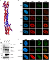
Figure 5. Structural and functional similarity between hIE1 and rhIE1.
(A) Homology model of hIE1 (blue) superimposed onto the rhIE1 template crystal structure (red). The insertions (green) and deletions (yellow) of hIE1 compared to rhIE1 are mainly located in loop regions and do therefore not disrupt the secondary structure elements. Structure validation using ProSA indicates that the model exhibits a good global and local quality and the respective structural parameters are close to that of the template crystal structure (Figure S7) suggesting that hIE1 and rhIE1 adopt a highly similar fold. (B) Dispersal of PML-NBs after RhCMV infection of primary human fibroblasts (HFFs). HFF cells were infected with RhCMV at an MOI of 0.1 and harvested at indicated times for immunofluorescence analysis of rhIE1 and endogenous PML. Cell nuclei were counterstained with DAPI. (C) DeSUMOylation of PML after RhCMV infection of HFF cells. Cell lysates harvested either from mock infected cells or from cells infected with RhCMV at an MOI of 3 for indicated times (24, 48 h) were separated by SDS-PAGE and analyzed by Western blotting for expression of PML (upper panel), rhIE1 (middle panel) and β-actin as internal loading control (lower panel). (D) Dispersal of PML-NBs after transient expression of rhIE1 in HFFs. HFFs were transfected with a eukaryotic expression vector encoding rhIE1 fused to an N-terminal FLAG-tag and, after 48 h, subjected to indirect immunofluorescence analysis of rhIE1 using an anti-FLAG antibody and of endogenous PML; cell nuclei were stained with DAPI. Upper panel: mock transfected cells. Middle panel: colocalization of rhIE1 and PML, as observed in only few transfected cells. Lower panel: dispersed pattern of rhIE1 and PML, as observed in the majority of transfected cells.
Figure 6
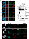
Figure 6. Effect of IE1CORE on the integrity of PML-NBs and the SUMOylation of PML.
(A) Dispersal of PML requires the IE1IDRc. HFF cells were transiently transfected with expression plasmids encoding FLAG-tagged full-length hIE1(1–491) or truncated hIE1 variants followed by immunodetection of hIE1 proteins using an anti-FLAG antibody and of endogenous PML. (B) DeSUMOylation of PML by IE1CORE. HEK293T cells were cotransfected with expression plasmids encoding FLAG-PML (isoform VI), FLAG-hIE1 variants as indicated and Myc-SUMO3. PML (upper panel), SUMOylated proteins (second panel), IE1 (third panel) and β-actin (lower panel) were detected by Western blot analysis. (C) Release of Sp100, hDaxx and ATRX from PML foci by IE1CORE. The hIE1CORE(1–382) was stably expressed in HFFs followed by immunodetection of hIE1, PML, Sp100, hDaxx or ATRX by triple staining as indicated.
Figure 7
Figure 7. Interaction of IE1CORE with PML and TRIM5α.
(A) Enhanced binding of IE1CORE to PML in coimmunoprecipitation analysis. HEK293T cells were cotransfected with expression plasmids encoding FLAG-hIE1 variants (as indicated) and Myc-PML (isoform VI). Upper two panels: Western blot detection of PML and hIE1 after immunoprecipitation with an anti-FLAG antibody. Lower two panels: detection of PML and hIE1 in cell lysates before precipitation (input). (B) Enhanced binding of IE1CORE to PML in yeast two-hybrid assays. The graph shows the quantification of β-galactosidase reporter activity after coexpression of PML and full-length hIE1 or the hIE1CORE(14–382) in yeast cells. (C) The PML coiled-coil is required for IE1 binding in yeast two-hybrid assays. Full-length hIE1 and hIE1CORE(14–382) were tested for interaction with various C-terminal PML deletion mutants (shown in the upper scheme) by XGal filter lift assays. (D) The PML coiled-coil is sufficient for binding to hIE1CORE in coimmunoprecipitation analysis. HEK293T cells were transfected with various FLAG-tagged PML deletion mutants, as shown in the upper scheme, together with Myc-hIE1CORE(1–382). Upper panel: Western blot detection of hIE1CORE after immunoprecipitation with an anti-FLAG antibody. Lower two panels: Western blot detection of PML variants and hIE1CORE in cell lysates before precipitation (input). (E) Interaction of hIE1CORE with TRIM5α. HEK293T cells were cotransfected with expression plasmids encoding Myc-tagged hIE1CORE(1–382) and either FLAG-tagged PML (isoform VI) or FLAG-tagged rhesus macaque TRIM5α (rhTRIM5α). Upper panel: Western blot detection of hIE1CORE after coimmunoprecipitation using an anti-FLAG antibody. Lower two panels: Western blot detection of PML, rhTRIM5α and hIE1CORE in cell lysates before precipitation (input). R, RING domain; B, B-boxes; CC, coiled-coil domain; NLS, nuclear localization signal; S, SUMO.
Figure 8
Figure 8. Analysis of IE1CORE during human cytomegalovirus replication.
(A) Western blot analysis showing the expression kinetics of full-length hIE1 or hIE1CORE(1–382) after infection of HFFs with wild-type HCMV (AD169) or recombinant HCMV expressing truncated hIE1 (AD169/hIE1 1–382) (MOI 0.2). (B) PML-NB dispersal after infection of HFFs with either AD169 or AD169/hIE1 1–382. Upper panel: immunofluorescence detection of hIE1 and PML at 2 and 8 hpi with the respective viruses (MOI 0.5). Lower graph: quantification of the number of infected cells containing either intact or dispersed PML foci during the first 24 h after infection. (C) DeSUMOylation of PML and Sp100 after infection of HFFs with AD169/hIE1 1–382 (MOI 3). Western blot detection of PML and Sp100 (upper panels) as well as hIE1 1–382 and β-actin (lower panels) during the first 24 hpi. (D) Growth curve analysis of wild-type AD169, AD169/hIE1 1–382 and IE1-deleted HCMV (AD169ΔhIE1). HFF cells were infected with wild-type AD169 at a low MOI (MOI 0.01) and equivalent genome copies of AD169/hIE1 1–382 and AD169ΔhIE1. Cell supernatants were harvested at indicated times and analyzed for genome copy numbers by quantitative real-time PCR. (E) Complementation of AD169ΔhIE1 by hIE1 1–382. Left panel: HFFs or HFFs expressing either hIE1 or hIE1 1–382 were infected with AD169ΔhIE1 at a low MOI (MOI 0.01). The cells were fixed at 48 hpi followed by immunofluorescence staining of UL44 and quantification of cells exhibiting viral gene expression using the ImageJ software. Middle and right panel: Western blot analysis of UL44, UL69, β-actin and IE1 expression in HFF, HFF/hIE1 or HFF/hIE1 1–382 cells after either mock infection or infection of with AD169ΔhIE1 as indicated.

Similar articles

Cited by

References

    1. Ishov AM, Sotnikov AG, Negorev D, Vladimirova OV, Neff N, et al. (1999) PML is critical for ND10 formation and recruits the PML-interacting protein daxx to this nuclear structure when modified by SUMO-1. J Cell Biol 147: 221–234. - PMC - PubMed
    1. Jensen K, Shiels C, Freemont PS (2001) PML protein isoforms and the RBCC/TRIM motif. Oncogene 20: 7223–7233. - PubMed
    1. Rajsbaum R, Garcia-Sastre A, Versteeg GA (2014) TRIMmunity: the roles of the TRIM E3-ubiquitin ligase family in innate antiviral immunity. J Mol Biol 426: 1265–1284. - PMC - PubMed
    1. Versteeg GA, Rajsbaum R, Sanchez-Aparicio MT, Maestre AM, Valdiviezo J, et al. (2013) The E3-ligase TRIM family of proteins regulates signaling pathways triggered by innate immune pattern-recognition receptors. Immunity 38: 384–398. - PMC - PubMed
    1. Chu Y, Yang X (2011) SUMO E3 ligase activity of TRIM proteins. Oncogene 30: 1108–1116. - PMC - PubMed

Publication types

MeSH terms

Grants and funding

This work was supported by the Deutsche Forschungsgemeinschaft, SFB796 (projects A2, A3 and B3) and the Interdisziplinäres Zentrum für Klinische Forschung Erlangen (project A62). The funders had no role in study design, data collection and analysis, decision to publish, or preparation of the manuscript.