. 2015 Feb;89(3):1673-87.
doi: 10.1128/JVI.02520-14. Epub 2014 Nov 19.

Syntaxin 5-dependent retrograde transport to the trans-Golgi network is required for adeno-associated virus transduction

Affiliations

Syntaxin 5-dependent retrograde transport to the trans-Golgi network is required for adeno-associated virus transduction

Mathieu E Nonnenmacher et al. J Virol. 2015 Feb.

Abstract

Intracellular transport of recombinant adeno-associated virus (AAV) is still incompletely understood. In particular, the trafficking steps preceding the release of incoming AAV particles from the endosomal system into the cytoplasm, allowing subsequent nuclear import and the initiation of gene expression, remain to be elucidated fully. Others and we previously showed that a significant proportion of viral particles are transported to the Golgi apparatus and that Golgi apparatus disruption caused by the drug brefeldin A efficiently blocks AAV serotype 2 (AAV2) transduction. However, because brefeldin A is known to exert pleiotropic effects on the entire endosomal system, the functional relevance of transport to the Golgi apparatus for AAV transduction remains to be established definitively. Here, we show that AAV2 trafficking toward the trans-Golgi network (TGN) and the Golgi apparatus correlates with transduction efficiency and relies on a nonclassical retrograde transport pathway that is independent of the retromer complex, late endosomes, and recycling endosomes. AAV2 transduction is unaffected by the knockdown of syntaxins 6 and 16, which are two major effectors in the retrograde transport of both exogenous and endogenous cargo. On the other hand, inhibition of syntaxin 5 function by small interfering RNA silencing or treatment with cyclized Retro-2 strongly decreases AAV2 transduction and transport to the Golgi apparatus. This inhibition of transduction is observed with several AAV serotypes and a number of primary and immortalized cells. Together, our data strongly suggest that syntaxin 5-mediated retrograde transport to the Golgi apparatus is a broadly conserved feature of AAV trafficking that appears to be independent of the identity of the receptors used for viral attachment.

Importance: Gene therapy constitutes a promising approach for the treatment of life-threatening conditions refractory to any other form of remedy. Adeno-associated virus (AAV) vectors are currently being evaluated for the treatment of diseases such as Duchenne muscular dystrophy, hemophilia, heart failure, Parkinson's disease, and others. Despite their promise as gene delivery vehicles, a better understanding of the biology of AAV-based vectors is necessary to improve further their efficacy. AAV vectors must reach the nucleus in order to deliver their genome, and their intracellular transport is not fully understood. Here, we dissect an important step of the intracellular journey of AAV by showing that retrograde transport of capsids to the trans-Golgi network is necessary for gene delivery. We show that the AAV trafficking route differs from that of known Golgi apparatus-targeted cargos, and we raise the possibility that this nonclassical pathway is shared by most AAV variants, regardless of their attachment receptors.


Figures

FIG 1
FIG 1
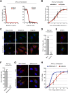
Inhibition of AAV2 transduction by Golgi apparatus-targeting drugs. (A) HeLa or HEK293T cells pretreated with various concentrations of brefeldin A or golgicide A were infected with AAV2-Luc at an MOI of 10,000. Luciferase activity was measured at 24 h postinfection and normalized to that for the dimethyl sulfoxide (DMSO)-treated controls (ctrl). RLU, relative light units. (B) Kinetics of drug addition. HeLa cells were allowed to bind AAV2-Luc on ice for 30 min, washed, and transferred to 37°C. Drugs were added at different times postwarming, and luciferase activity was measured at 24 h postwarming. Luciferase reads were normalized to the maximum plateau value rather than the value for the dimethyl sulfoxide-treated controls in order to compensate for the long-term cytotoxicity of brefeldin A and golgicide, which both reduce cell viability by 60 to 80% after 24 h of incubation. p.i., postinfection. (C) Subcellular localization of AAV2 capsids (green) and the Golgi apparatus marker giantin (red) at 3 h postinfection in cells treated by Golgi apparatus-targeting drugs. (D) AAV2-Luc transduction of poorly permissive NIH 3T3 cells compared to that of permissive HeLa cells. Identical numbers of cells were infected to allow direct comparison. (E) Subcellular localization of AAV2 capsids (red) and giantin (green) in poorly permissive NIH 3T3 cells at 3 h postinfection. (F) AAV2-Luc transduction of HeLa cells treated with bafilomycin A1. Values represent luciferase activity normalized to that for the dimethyl sulfoxide-treated controls. (G) Subcellular localization of AAV2 capsids (red) and giantin (green) in HeLa cells treated with bafilomycin A1. (H) Comparison of the addition kinetics of bafilomycin A1 and golgicide. Drugs were added at various times following AAV2-Luc infection. Luciferase values were normalized to the maximum plateau values, as described in the legend to panel B.
FIG 2
FIG 2
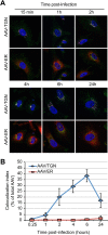
Lack of evidence for AAV ER transport. (A) HeLa cells transfected with the ER marker plasmid pER-dTmT (red) were infected with AAV2-Luc at an MOI of 10,000 and fixed at various times postinfection. Cells were immunostained with an anti-golgin-97 antibody (purple) and an anti-AAV2 capsid antibody (green). Cells were imaged by confocal microscopy. (B) Index of colocalization of AAV2 capsids with TGN or ER, expressed as the percentage of green pixels (AAV2 capsids) colocalized with TGN (purple) or ER (red) pixels. Numbers represent the mean ± SD from triplicate experiments.
FIG 3
FIG 3
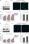
AAV transduction is independent of retromer function. (A) Validation of siRNA-mediated knockdown of Vps35 by immunoblotting. HeLa cell lysates were harvested 48 h after siRNA transfection. Scram, scrambled; siVps35-1 to siVps35-3, three different siRNAs against Vps35. (B) Steady-state localization of Ci-MPR in HeLa cells transfected with Vps35 siRNA. Cells were fixed at 48 h posttransfection, stained with an anti-Ci-MPR antibody, and analyzed by confocal microscopy. (C) AAV2-Luc transduction of HeLa cells treated with Vps35 siRNAs. Cells were infected at various MOIs (10,000, 1,000, or 100 vg/cell) 48 h after siRNA transfection. Values represent absolute luciferase activity normalized to the protein content. Data represent the mean ± SD from triplicate experiments. (D) Relative transduction values normalized to the values for cells transfected with scrambled siRNA. *, P < 0.05.
FIG 4
FIG 4
AAV transduction is independent of late endosome function. (A) Validation of Rab9 knockdown by multiple siRNAs. HeLa cells were transfected with three different siRNAs against Rab9 (siRab9-1 to siRab9-3). Protein extracts were obtained at 48 h posttransfection and analyzed by immunoblotting with a monoclonal Rab9 antibody. (B) Steady-state localization of Ci-MPR in HeLa cells treated with Rab9 siRNA1 and analyzed by confocal microscopy 48 h after siRNA transfection. (C) AAV2-Luc transduction of HeLa cells treated with Rab9 siRNAs. Cells were infected at various MOIs (10,000, 1,000, or 100 vg/cell) 48 h after siRNA transfection. (D) AAV-Luc transduction (MOI = 10,000) of HEK293T cells transfected with a dominant negative Rab9 plasmid (Rab9-DN). Luciferase values are normalized to the values for cells transfected with a control EGFP plasmid. (E) Validation of Rab7 knockdown by three siRNAs against Rab7 (siRab7-1 to siRab7-3) at 48 h posttransfection. (F) Steady-state localization of Ci-MPR in HeLa cells treated with Rab7 siRNA1 and analyzed by confocal microscopy 48 h after siRNA transfection. (G) AAV2-Luc transduction of HeLa cells treated with Rab7 siRNAs. Cells were infected at various MOIs (10,000, 1,000, or 100 vg/cell) 48 h after siRNA transfection. (H) AAV2-Luc transduction (MOI = 10,000) of HEK293T cells transfected with a dominant negative Rab7 plasmid. Luciferase values are normalized to the values for cells transfected with a control EGFP plasmid. *, P < 0.05.
FIG 5
FIG 5
AAV transduction is independent of recycling endosomes. (A) Validation of siRNA-mediated knockdown of Rab11 by multiple siRNAs (siRab11-1 to siRab11-3). Protein extracts were prepared 48 h after transfection of HeLa cells and analyzed by immunoblotting with an anti-Rab11 antibody. (B) Perturbation of TGN46 retrograde transport in HeLa cells transfected with Rab11 siRNA. Cells were fixed at 48 h posttransfection, stained with an anti-TGN46 antibody, and analyzed by confocal microscopy. (C) AAV2-Luc transduction of HeLa cells treated with Rab11 siRNAs. Cells were infected at various MOIs (10,000, 1,000, or 100 vg/cell) 48 h after siRNA transfection. Luciferase reads are normalized to the amount of control siRNA for each MOI. (D) AAV2-Luc transduction of HEK293T cells transfected with a dominant negative Rab11 plasmid. Values represent the mean and SD from triplicate experiments. *, P < 0.05.
FIG 6
FIG 6
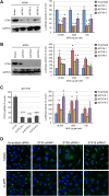
AAV transduction requires STX5, but not STX6 or STX16. (A) STX5 expression and AAV2-Luc transduction in HeLa cells treated with STX5 siRNAs (siSTX5-1 to siSTX5-3). Protein expression and viral transduction were performed 48 h after siRNA transfection. (B) STX6 expression and AAV2-Luc transduction in HeLa cells treated with STX6 siRNAs (siSTX6-1 to siSTX6-3). (C) STX16 expression and AAV2-Luc transduction in HeLa cells treated with STX16 siRNAs (siSTX16-1 to siSTX16-3). Luciferase activity values on the y axes of panels A to C are normalized to those for cells treated with scrambled siRNA and represent the mean ± SD from triplicate experiments. *, P < 0.05. (D) Steady-state localization of TGN46 and Ci-MPR in HeLa cells transfected with siRNA against Golgi apparatus-resident STXs. Cells were fixed at 48 h posttransfection and analyzed by confocal microscopy.
FIG 7
FIG 7
Inhibitors of STX5 decrease AAV2 transduction and retrograde transport. (A) AAV2-Luc transduction of HeLa cells treated with various concentrations of Retro-2 or Retro-2.1. Cells were pretreated for 30 min at 37°C before addition of AAV2-Luc at an MOI of 10,000 vg/cell. Luciferase activity values are normalized to those for the dimethyl sulfoxide-treated control. (B) Retro-2 and Retro-2.1 are not cytotoxic. HeLa cells were treated with the maximal tested concentrations of Retro-2 (100 μM) and Retro-2.1 (20 μM) for 24 h, and cell viability was measured using the luciferase-based CellTiter-Glo method. (C) AAV2-Luc transduction of HeLa cells at various MOIs in the presence of 10 μM Retro-2.1. Absolute luciferase output and inhibition ratios are indicated. (D) Endocytosis of AAV2-Luc is not reduced in HeLa cells treated with 100 μM Retro-2 or 20 μM Retro-2.1. Intracellular viral DNA was quantified by real-time PCR, and values are normalized to those for the dimethyl sulfoxide-treated control. NS, not significant. (E) Transduction of HeLa cells by Ad5-EGFP in the presence of Retro-2 or Retro-2.1. Values indicate the average ± SD of the green fluorescence intensity per microscope field, expressed in arbitrary units (AU). (F) Kinetics of Retro-2.1 addition. HeLa cells were incubated for 1 h on ice with AAV2-Luc, washed, and transferred to 37°C to trigger infection. Drug was added at various time points, and luciferase activity was measured after 24 h. Dotted lines, the kinetics of golgicide and bafilomycin A1 addition presented in Fig. 1H. Luciferase activity values were normalized to the maximum (plateau) values, as described in the legend of Fig. 1. The values in all panels represent mean and SD from triplicate experiments.
FIG 8
FIG 8
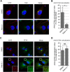
Retro-2.1 blocks endosome-to-TGN transport of AAV2. (A) HeLa cells were infected with AAV2-Luc at an MOI of 10,000 vg/cell in the presence of 20 μM Retro-2.1 or dimethyl sulfoxide. Cells were fixed at 3 h after infection and stained for AAV2 capsids (red) and golgin-97 (green) before imaging by confocal microscopy. (B) Quantification of the colocalization index between AAV2 capsids and golgin-97. Values represent the percentage of red pixels overlapping with green pixels. *, P < 0.05. (C) HeLa cells were treated as described in the legend to panel A and stained for AAV2 capsids (red) and STX5 (green). (Insets) Higher magnifications of boxed areas. (D) Quantification of the colocalization index between AAV2 capsids and STX5. Values represent the percentage of red pixels overlapping with green pixels. NS, not significant.
FIG 9
FIG 9
STX5-dependent transport is conserved among AAV serotypes and cell types. (A) Transduction of HeLa cells by different AAV serotypes containing a luciferase expression cassette, performed 48 h after STX5 siRNA transfection. Values indicate the luciferase activity normalized to the activity for scrambled siRNA-treated cells. (B to F) AAV serotypes 1, 2, 3, 4, 5, 6, 8, and 9 containing a luciferase expression cassette (MOI = 10,000 vg/cell) were used to infect HeLa cells (B), Huh7 cells (C), HUVECs (D), HCAECs (E), or RACMs (F) pretreated with 100 μM Retro-2. Luciferase values are normalized to those for dimethyl sulfoxide-treated cells. All values represent the mean ± SD from triplicate experiments. *, P < 0.05 by two-tailed Student's t test. B/D, below the detection limit.
FIG 10
FIG 10
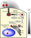
Hypothetical model of syntaxin 5-dependent transport of AAV to the TGN/Golgi apparatus. Following receptor attachment and endocytosis, early endocytic vesicles are rapidly acidified, which can be inhibited by the proton ATPase inhibitor bafilomycin A1. This endosomal acidification is dependent on the depletion of endosomal calcium. The acidic environment of the endosomes and, presumably, the proteolytic cleavage of AAV capsid proteins trigger the extrusion of the unique, PLA2-containing region of the largest capsid protein, VP1. However, as a result of the minute calcium concentrations in the endosomes, the PLA2 is enzymatically inactive. AAV is then transported to the TGN/Golgi apparatus, and this transport can be inhibited with the GBF1 inhibitor golgicide. This endosome-to-TGN/Golgi apparatus transport step is also dependent on the tSNARE STX5, whose function can be inhibited by Retro-2 and its derivatives or by siRNA-mediated knockdown of STX5. The calcium concentration in the TGN/Golgi apparatus, which is near the optimal level for VP1-PLA2 activity, then allows escape into the cytoplasm, followed by nuclear import and uncoating of the AAV capsid.

Similar articles

Cited by

References

    1. Grimm D, Kay MA. 2003. From virus evolution to vector revolution: use of naturally occurring serotypes of adeno-associated virus (AAV) as novel vectors for human gene therapy. Curr Gene Ther 3:281–304. doi:10.2174/1566523034578285. - DOI - PubMed
    1. Nonnenmacher M, Weber T. 2012. Intracellular transport of recombinant adeno-associated virus vectors. Gene Ther 19:649–658. doi:10.1038/gt.2012.6. - DOI - PMC - PubMed
    1. Bartlett JS, Wilcher R, Samulski RJ. 2000. Infectious entry pathway of adeno-associated virus and adeno-associated virus vectors. J Virol 74:2777–2785. doi:10.1128/JVI.74.6.2777-2785.2000. - DOI - PMC - PubMed
    1. Duan D, Li Q, Kao AW, Yue Y, Pessin JE, Engelhardt JF. 1999. Dynamin is required for recombinant adeno-associated virus type 2 infection. J Virol 73:10371–10376. - PMC - PubMed
    1. Nonnenmacher M, Weber T. 2011. Adeno-associated virus 2 infection requires endocytosis through the CLIC/GEEC pathway. Cell Host Microbe 10:563–576. doi:10.1016/j.chom.2011.10.014. - DOI - PMC - PubMed

Publication types

Substances

LinkOut - more resources