. 2015 Aug;26(8):1905-24.
doi: 10.1681/ASN.2014050493. Epub 2014 Nov 14.

Glomerular Autoimmune Multicomponents of Human Lupus Nephritis In Vivo (2): Planted Antigens

Affiliations

Glomerular Autoimmune Multicomponents of Human Lupus Nephritis In Vivo (2): Planted Antigens

Maurizio Bruschi et al. J Am Soc Nephrol. 2015 Aug.

Abstract

Glomerular planted antigens (histones, DNA, and C1q) are potential targets of autoimmunity in lupus nephritis (LN). However, the characterization of these antigens in human glomeruli in vivo remains inconsistent. We eluted glomerular autoantibodies recognizing planted antigens from laser-microdissected renal biopsy samples of 20 patients with LN. Prevalent antibody isotypes were defined, levels were determined, and glomerular colocalization was investigated. Renal and circulating antibodies were matched, and serum levels were compared in 104 patients with LN, 84 patients with SLE without LN, and 50 patients with rheumatoid arthritis (RA). Autoantibodies against podocyte antigens (anti-α-enolase/antiannexin AI) were also investigated. IgG2 autoantibodies against DNA, histones (H2A, H3, and H4), and C1q were detected in 50%, 55%, and 70% of biopsy samples, respectively. Anti-DNA IgG3 was the unique non-IgG2 anti-DNA deposit, and anti-C1q IgG4 was mainly detected in subepithelial membranous deposits. Anti-H3, anti-DNA, and anti-C1q IgG2 autoantibodies were also prevalent in LN serum, which also contained IgG3 against the antigen panel and anti-C1q IgG4. Serum and glomerular levels of autoantibodies were not strictly associated. High serum levels of all autoantibodies detected, including anti-α-enolase and antiannexin AI, identified LN versus SLE and RA. Anti-H3 and anti-α-enolase IgG2 levels had the most remarkable increase in LN serum and represented a discriminating feature of LN in principal component analysis. The highest levels of these two autoantibodies were also associated with proteinuria>3.5 g/24 hours and creatinine>1.2 mg/dl. Our findings suggest that timely autoantibody characterization might allow outcome prediction and targeted therapies for patients with nephritis.

Keywords: SLE; clinical immunology; immunology and pathology; lupus nephritis.


Figures

Figure 1.
Figure 1.
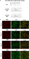
Glomerular autoantibodies and implanted antigens: isotypes in single biopsies. Renal biopsies obtained from 20 patients with LN (Supplemental Table 1) at the time of appearance of urinary symptoms were processed with laser capture, and microdissected glomeruli were used for elution of autoantibodies. (A, C, and E) Definition of most representative isotypes on pooled samples was done with dot blot analysis and followed by single-biopsy analysis. The same glomerular eluates used for anti–α-enolase and antiannexin AI antibodies were processed for definition of isotypes of antibodies versus implanted antigens (DNA, H2A, H3, H4, and C1q). In all cases, after the definition of prevalent isotype, competition experiments using the same glomerular eluates and increasing amounts of the specific antigen from 5 to 15 ng to saturate antibodies were done (not shown). In B, D, and F, the colocalization of each implanted antigen with the most relevant antibodies in single renal biopsy samples (IgG2 and IgG3 for DNA; IgG2 for H2A, H3, and H4; and IgG2 and IgG4 for C1q) is reported. In B and D, double IF staining was evaluated for each antigen (red) and IgG2 (green), whereas in F, the antigen is in green and IgG is in red. Merged images are reported in yellow. The last box in Figure 1 D is the magnification of white frame. For anti-C1q IgG4, confocal images include different renal biopsy specimens with LN classes III–IV and V. Colocalization was absent in the case of class IV LN, whereas it was well evident in subepithelia deposits in biopsies of patients with class V LN. Magnification of the same figures is in Supplemental Figure 3. Scale bar, 20 µm in A–C; 10 µm. Original magnification, ×630. In G, the hierarchical cluster analysis heat map for single antibody in each renal biopsy is shown: antibody intensities (colors from black [high] to gray [medium] to white [low]) are reported in lines and refer to single patient biopsies that are reported at the bottom of the figure. Results are given for 20 patients with LN of our study cohort and four normal kidneys; a semiquantitative evaluation of the same data is reported in Tables 1 and 2. A preliminary heat map analysis of autoantibody expressed in biopsies of 20 patients enrolled in the study has been already presented in the first part of this study; this figure includes anti-DNA IgG3 and anti-C1q IgG4, which have been only now characterized. In H, the serum levels of antibodies are reported considering two different population of patients with detectable (high) and undetectable (low) glomerular levels. The numbers of positive/negative considering the overall numbers of 20 biopsies are shown. Glomerular levels of each antibody were calculated from the dot blot and the standard curve that included different concentrations of each antibody (Supplemental Figure 4). For serum levels, specific ELISAs were used as described in Concise Methods.
Figure 1.
Figure 1.
Glomerular autoantibodies and implanted antigens: isotypes in single biopsies. Renal biopsies obtained from 20 patients with LN (Supplemental Table 1) at the time of appearance of urinary symptoms were processed with laser capture, and microdissected glomeruli were used for elution of autoantibodies. (A, C, and E) Definition of most representative isotypes on pooled samples was done with dot blot analysis and followed by single-biopsy analysis. The same glomerular eluates used for anti–α-enolase and antiannexin AI antibodies were processed for definition of isotypes of antibodies versus implanted antigens (DNA, H2A, H3, H4, and C1q). In all cases, after the definition of prevalent isotype, competition experiments using the same glomerular eluates and increasing amounts of the specific antigen from 5 to 15 ng to saturate antibodies were done (not shown). In B, D, and F, the colocalization of each implanted antigen with the most relevant antibodies in single renal biopsy samples (IgG2 and IgG3 for DNA; IgG2 for H2A, H3, and H4; and IgG2 and IgG4 for C1q) is reported. In B and D, double IF staining was evaluated for each antigen (red) and IgG2 (green), whereas in F, the antigen is in green and IgG is in red. Merged images are reported in yellow. The last box in Figure 1 D is the magnification of white frame. For anti-C1q IgG4, confocal images include different renal biopsy specimens with LN classes III–IV and V. Colocalization was absent in the case of class IV LN, whereas it was well evident in subepithelia deposits in biopsies of patients with class V LN. Magnification of the same figures is in Supplemental Figure 3. Scale bar, 20 µm in A–C; 10 µm. Original magnification, ×630. In G, the hierarchical cluster analysis heat map for single antibody in each renal biopsy is shown: antibody intensities (colors from black [high] to gray [medium] to white [low]) are reported in lines and refer to single patient biopsies that are reported at the bottom of the figure. Results are given for 20 patients with LN of our study cohort and four normal kidneys; a semiquantitative evaluation of the same data is reported in Tables 1 and 2. A preliminary heat map analysis of autoantibody expressed in biopsies of 20 patients enrolled in the study has been already presented in the first part of this study; this figure includes anti-DNA IgG3 and anti-C1q IgG4, which have been only now characterized. In H, the serum levels of antibodies are reported considering two different population of patients with detectable (high) and undetectable (low) glomerular levels. The numbers of positive/negative considering the overall numbers of 20 biopsies are shown. Glomerular levels of each antibody were calculated from the dot blot and the standard curve that included different concentrations of each antibody (Supplemental Figure 4). For serum levels, specific ELISAs were used as described in Concise Methods.
Figure 2.
Figure 2.
Circulating autoantibodies and implanted antigens in serum of patients with LN. (A) For each antibody detected in serum (including all implanted antigens), the definition of isotype was done with dot blot analysis. (B) Determination of antibody levels in serum was done with self-made ELISAs in 184 patients with SLE, 104 of whom had LN. Results are expressed as medians and interquartile ranges. (C–G) Comparison of serum levels for each antibody (including ROC curves) in SLE, LN, and AR. The same data are reported in Table 3. ROC curves for antibodies with high circulating levels were calculated to determine specificity and sensitivity versus SLE, RA, and normal subjects. *P<0.05; **P<0.01; ***P<0.001.
Figure 2.
Figure 2.
Circulating autoantibodies and implanted antigens in serum of patients with LN. (A) For each antibody detected in serum (including all implanted antigens), the definition of isotype was done with dot blot analysis. (B) Determination of antibody levels in serum was done with self-made ELISAs in 184 patients with SLE, 104 of whom had LN. Results are expressed as medians and interquartile ranges. (C–G) Comparison of serum levels for each antibody (including ROC curves) in SLE, LN, and AR. The same data are reported in Table 3. ROC curves for antibodies with high circulating levels were calculated to determine specificity and sensitivity versus SLE, RA, and normal subjects. *P<0.05; **P<0.01; ***P<0.001.
Figure 3.
Figure 3.
Clinical characteristics within different patients with LN on the basis of serum antibody levels. For this comparison, data already shown in Figures 1 and 2 and Tables 1 and 2 were analyzed with hierarchical cluster analysis and PCA. (A) The former test combines and compares simultaneously serum levels of each specific antibody (and includes different isotypes); the resulting heat map, where colors from red (maximum) to green (minimum) indicate the relative abundances, gives an estimate of how different parameters separate patients on the basis of the presence of nephritis. It seems that serum anti–α-enolase and antiannexin AI IgG2 separate LN and AR better than other antibodies, whereas anti-H3 IgG2/IgG3 and anti-DNA do it for LN versus SLE. (B) PCA gives an overall picture of statistical power of every single clinical and laboratory parameter. When all patients were analyzed with respect to autoantibody levels (here including antipodocyte antibodies), the four population of LN, SLE, AR, and normal controls were clearly separated (different colors). When proteinuria was included in the analysis, the same result was obtained according to which of the four population was comparably separated. (C) Considering LN alone, anti–α-enolase IgG2 and anti-H3 IgG2/IgG3 contributed significantly in characterizing these patients, whereas proteinuria and other clinical tests (VES, CPR, renal function, etc.) were of no value. (D) Serum antibodies presented in some case a notable fold increment that reached 10 times the 95° of normality. Anti-H3IgG2 and anti–α-enolase IgG2 had the highest performances.
Figure 4.
Figure 4.
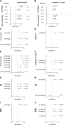
Clinical associations of each antibody with relevant clinical characteristics. Odd ratios and confidence intervals were calculated for the association of each antibody with relevant clinical characteristics, such as proteinuria, renal function, and lupus flare (A-J). Anti–α-enolase IgG2 and anti-H3 IgG3 had the best performance in relation to renal parameters (proteinuria and renal function), and anti-C1q and anti-DNA better correlated with lupus flares.

Similar articles

Cited by

References

    1. Cameron JS: Lupus nephritis. J Am Soc Nephrol 10: 413–424, 1999 - PubMed
    1. Madaio MP: The relevance of antigen binding to the pathogenicity of lupus autoantibodies. Kidney Int 82: 125–127, 2012 - PubMed
    1. Waldman M, Madaio MP: Pathogenic autoantibodies in lupus nephritis. Lupus 14: 19–24, 2005 - PubMed
    1. Bagavant H, Fu SM: Pathogenesis of kidney disease in systemic lupus erythematosus. Curr Opin Rheumatol 21: 489–494, 2009 - PMC - PubMed
    1. Hanrotel-Saliou C, Segalen I, Le Meur Y, Youinou P, Renaudineau Y: Glomerular antibodies in lupus nephritis. Clin Rev Allergy Immunol 40: 151–158, 2011 - PubMed

Publication types