. 2014 Nov 13;10(11):e1004756.
doi: 10.1371/journal.pgen.1004756. eCollection 2014 Nov.

The talin head domain reinforces integrin-mediated adhesion by promoting adhesion complex stability and clustering

Affiliations

The talin head domain reinforces integrin-mediated adhesion by promoting adhesion complex stability and clustering

Stephanie J Ellis et al. PLoS Genet. .

Abstract

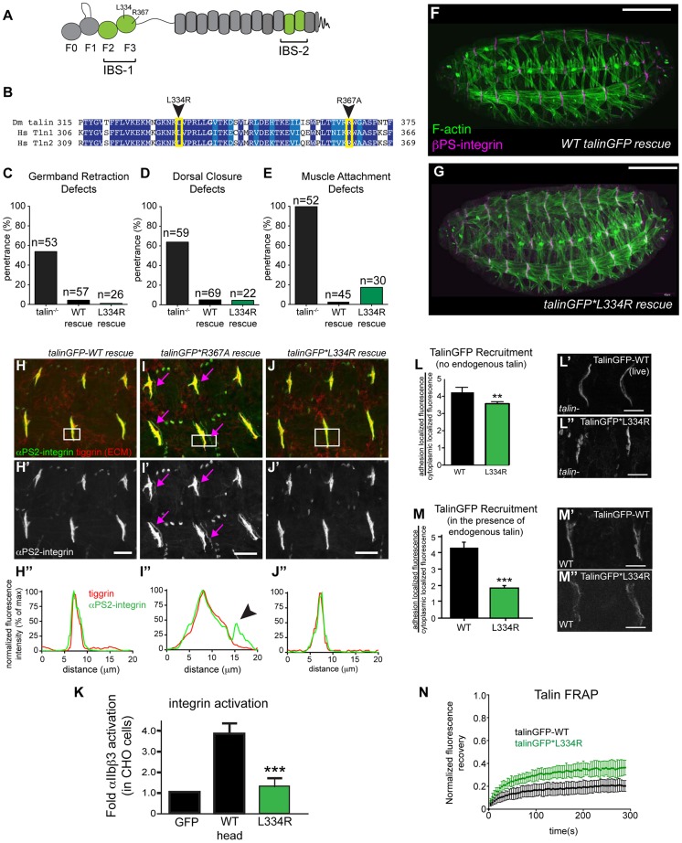
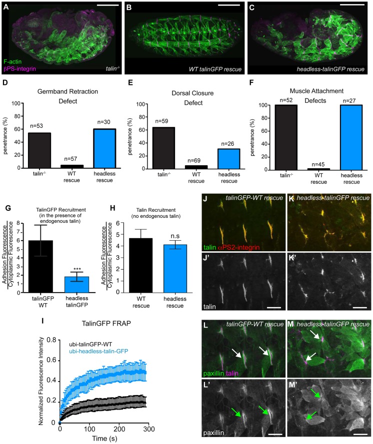
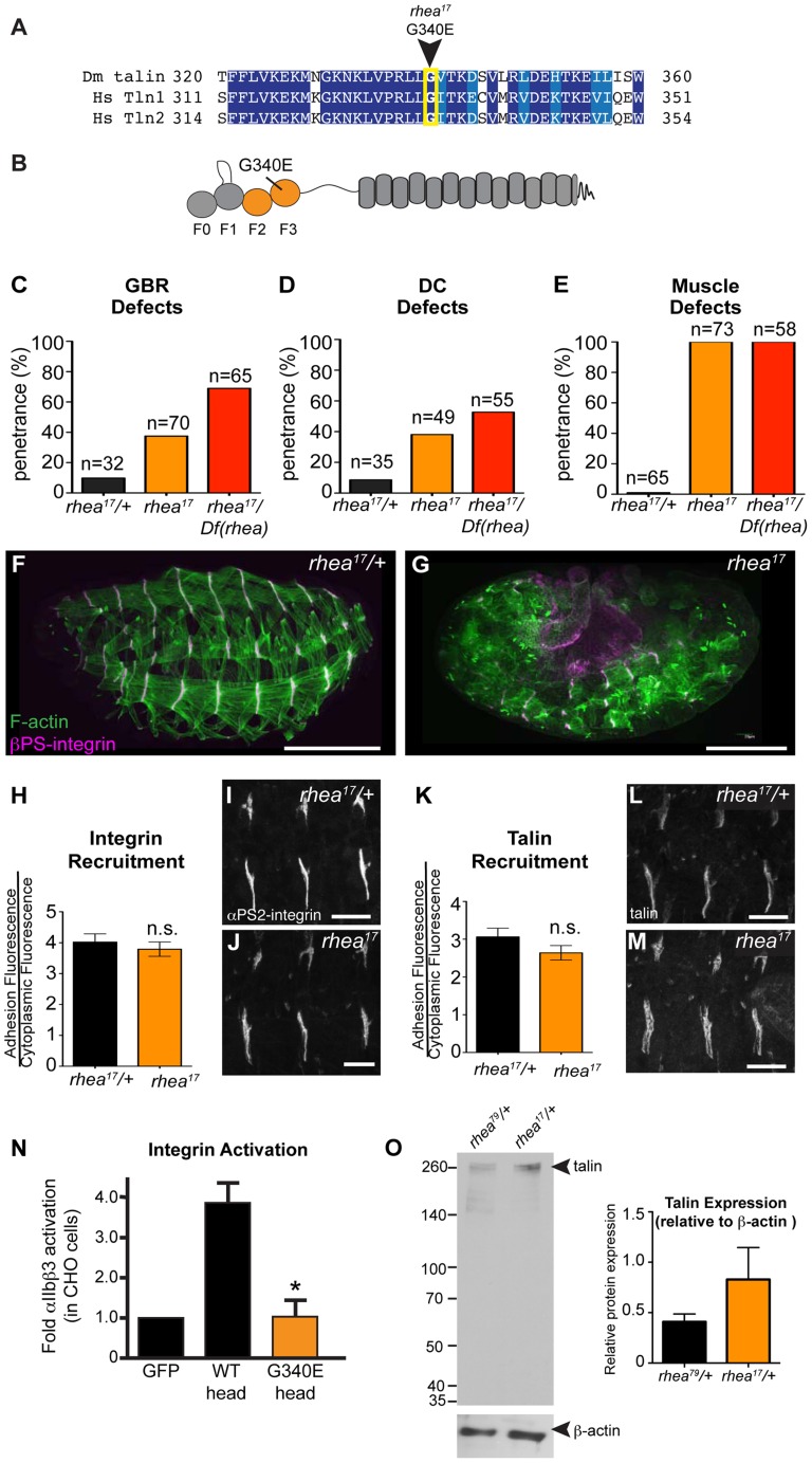
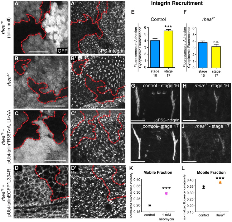
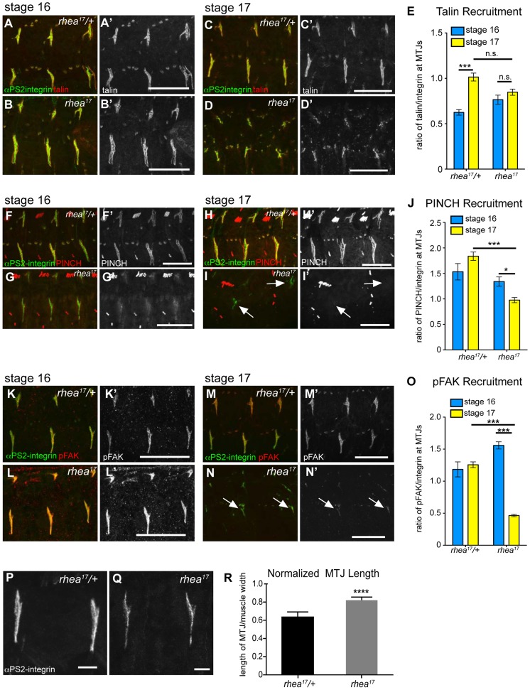
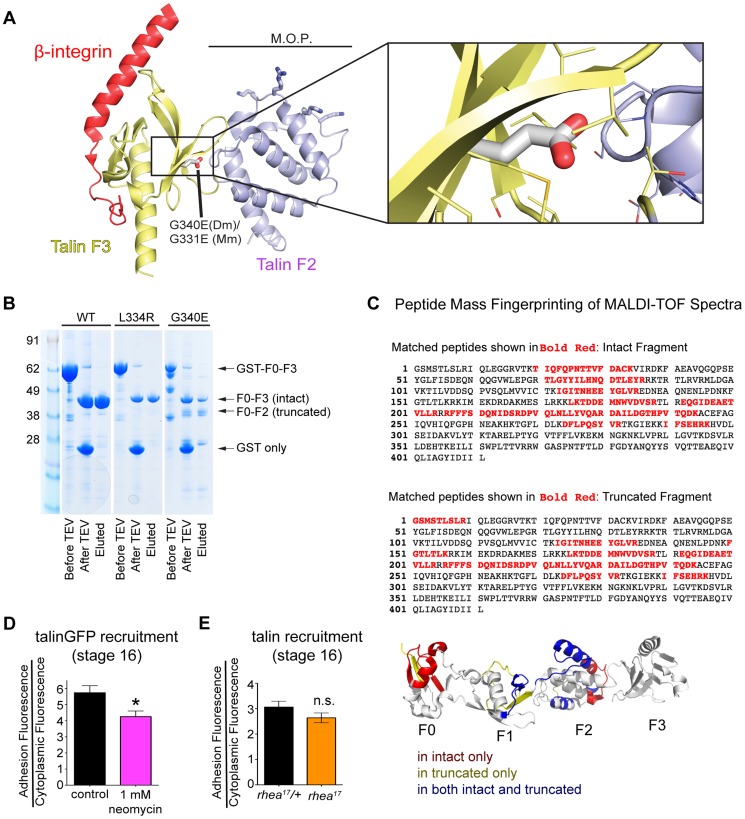
Talin serves an essential function during integrin-mediated adhesion in linking integrins to actin via the intracellular adhesion complex. In addition, the N-terminal head domain of talin regulates the affinity of integrins for their ECM-ligands, a process known as inside-out activation. We previously showed that in Drosophila, mutating the integrin binding site in the talin head domain resulted in weakened adhesion to the ECM. Intriguingly, subsequent studies showed that canonical inside-out activation of integrin might not take place in flies. Consistent with this, a mutation in talin that specifically blocks its ability to activate mammalian integrins does not significantly impinge on talin function during fly development. Here, we describe results suggesting that the talin head domain reinforces and stabilizes the integrin adhesion complex by promoting integrin clustering distinct from its ability to support inside-out activation. Specifically, we show that an allele of talin containing a mutation that disrupts intramolecular interactions within the talin head attenuates the assembly and reinforcement of the integrin adhesion complex. Importantly, we provide evidence that this mutation blocks integrin clustering in vivo. We propose that the talin head domain is essential for regulating integrin avidity in Drosophila and that this is crucial for integrin-mediated adhesion during animal development.


Conflict of interest statement

The authors have declared that no competing interests exist.

Figures

Figure 1
Figure 1. Integrin-binding to the talin head, but not integrin activation, is required for muscle attachment.
(a) Schematic of key domains in talin for this study. The talin head is contains an N-terminal atypical FERM domain and a C-terminal rod domain comprised of 13 helical bundles . (b) Alignment of residues 325–375 of fly talin F3 domain with human talin isoforms. Dark blue indicates identical residues between homologues, lighter blue indicates similar residues. The mutations utilized to study integrin activation are indicated with an arrowhead. (c–e) Integrin-dependent phenotypes germband retraction (c), dorsal closure (d) and muscle attachment (e) were assayed in talin-null embryos, WT talinGFP-rescued embryos, and talinGFP*L334R-rescued embryos. Apart from mild muscle detachment in about 20% of embryos, the talinGFP*L334R transgene was able to rescue all phenotypes such that the embryos hatched to the larval stages. (f–g) Maternal zygotic talin null embryos rescued with either full-length WT talinGFP transgene (f) or talinGFP*L334R mutant transgene (g) and stained for F-actin (green) and βPS-integrin (magenta). (h–j) MTJs of talin null embryos rescued with either talinGFP-WT (h), talinGFP*R367A (i), talinGFP*L334R (j). Embryos were stained for anti- αPS2-integrin (green in h–j; grey in h′–j′) and tiggrin, a Drosophila ECM molecule (red in h–j). (h″–j″) Average intensity profiles for integrin and tiggrin across the widths of the boxed areas in h–j. Tiggrin and integrin completely overlapped at MTJs in WT talin rescue embryos (h″), but were separated from one another in talin-null embryos rescued with talinGFP*R367A (i–i″). Overlap between tiggrin and integrin was maintained in talin-null embryos rescued with talinGFP*L334R (j–j″). The pink arrowheads mark the sites of separated integrin and ECM signal. (k) Activation of human integrins by fly talin head constructs was measured in CHO cells. The L334R mutation was sufficient to abrogate integrin activation. (l–m) Recruitment of ubi-promoter driven full-length WT talinGFP and talinGFP*L334R to sites of adhesion was assayed in talin null (l) and in wild-type embryos (m). Compared to WT TalinGFP, TalinGFP*L334R was well recruited in a background devoid of any endogenous talin (l–l″; **p<0.01), but competed less well in the presence of endogenous talin and was only weakly recruited to sites of adhesion compared to WT, which was robustly recruited (m–m″; ***p<0.001). (n) FRAP experiments on WT talinGFP and talinGFP*L334R reveal that talinGFP*L334R is much less stable at sites of adhesion than WT talinGFP. Scale bars: f–g  = 100 µm; h–j;l–m = 20 µm.
Figure 2
Figure 2. The talin head is essential for integrin function in Drosophila.
(a–c) Maternal-zygotic talin null embryos (shown in a) rescued with either full-length WT talinGFP transgene (b) or headless talinGFP transgene (c) and stained for F-actin (green) and integrin (magenta). (d–f) Integrin-dependent phenotypes germband retraction (d), dorsal closure (e) and muscle attachment (f) were assayed in talin-null embryos, WT-talin-rescued embryos, and headless-talin-rescued embryos. The talin head was required for all three processess assayed. Scale bar = 100 µm. (g–h) Recruitment of ubi-promoter driven, GFP-tagged full-length WT talin and headless talin to sites of adhesion was assayed in wild-type embryos (g) and in a talin null background (h). In a WT background, headless-talinGFP competed less well with endogenous talin and was only weakly recruited to sites of adhesion compared to WT (***p<0.001); in the absence of endogenous talin, headless-talin was well recruited to sites of adhesion. (i) FRAP experiments on talinGFP-WT and headless-talinGFP reveal that headless talin is much less stable at sites of adhesion than talinGFP-WT. (j–m) Confocal z-stacks of stage 17 maternal/zygotic-mutant embryos rescued with either full length WT talin (j,l) or headless-talin (k,m). (j–k) adhesions stained for talin (green in j–k; grey in j′–k′) and integrin (red in j–k). In the absence of the talin head, talin was still well recruited. (l–m) Muscles stained for talin (magenta in l–m) and paxillin (green in l–m; grey in l′–m′). Paxillin was not well recruited to adhesions. Scale bars: a–e = 100 µm; j–m = 20 µm.
Figure 3
Figure 3. rhea17 encodes a hypomorphic talin protein which disrupts talin head function.
(a–b) The rhea17 allele is characterized by a missense mutation in a conserved glycine residue in the F3 lobe of the talin head FERM domain, G340E. (c–g) Whole mount stage 17 embryos stained for F-actin (green) and integrin (magenta) reveal that rhea17 mutant embryos (g) harbour severe morphogenetic phenotypes in GBR (c) and DC (d), as well as muscle detachment defects (e) compared to WT heterozygous embryos (f). Phenotypic analysis of rhea17 over the rhea79 deficiency increased the penetrance of all phenotypes. (h–j) αPS2-integrin recruitment was measured in WT (h,i) and rhea17 (h,j) stage 16 embryonic muscles stained for integrin. Integrin was recruited at WT levels in rhea17 embryos. (k–m) Talin recruitment was measured in WT (k,i) and rhea17 (k,m) stage 16 embryonic muscles stained for talin. Talin was well recruited in rhea17 embryos. (n) Activation of human integrins by fly talin head constructs was measured in CHO cells. The G340E mutation was sufficient to abrogate integrin activation compared to WT. (o) Quantitative Western blot analyses of relative levels of talin normalized to beta-actin levels in flies heterozygous for either the rhea79 talin null mutation (left lane) or the rhea17 mutant allele (right lane). Scale bars: f–g = 100 µm; i–j; l-m = 20 µm.
Figure 4
Figure 4. rhea17 disrupts integrin clustering.
(a–a′) Clones of cells lacking talin, marked by the absence of GFP (a), failed to cluster integrins into adhesions (a′). (b,b′) Clones of cells expressing the rhea17 mutant allele of talin (marked by absence of GFP in b) also failed to cluster integrins (b′). (c–d) Expression of a full length talin point mutant that specifically disrupts IBS-1 binding (c, talinGFP*R367A, LI>AA, see Ellis et al, 2011) or that specifically disrupts integrin activation (talinGFP*L334R) restored integrin adhesions (c′, d′) within the clones of cells (arrow) lacking endogenous talin and the GFP marker (c,d). The red outline demarcates the position of the clones. Note that in d, cell outlines are also marked with GFP due to localization of the talinGFP*L334R protein to basolateral membranes. (e–j) Recruitment of integrins to MTJs was measured in stage 16 and stage 17 for both control (e,g,i) and rhea17 mutant embryos (f,h,j). In contrast to control embryos (***p<0.001), rhea17 mutant embryos did not exhibit an increase in integrin recruitment to MTJs during this developmental transition. (k–l) FRAP analysis revealed the mobile fraction of integrin-YFP was higher than respective controls in embryos treated with neomycin (k; ***p<0.001) or in rhea17 zygotic mutant embryos (l; ***p<0.001). Since these two FRAP experiments employed different genetic backgrounds and protocols in preparation for FRAP (ie. embryos in k were subjected to a drug delivery protocol), they necessitated two separate controls. In k, the control was established from vehicle-treated wild type embryos expressing integrin-YFP. In l, the controls were taken from heterozygous talin mutant embryos. Scale bars: a–d = 10 µm; g–i; h–j = 20 µm.
Figure 5
Figure 5. rhea17 disrupts adhesion complex reinforcement and adhesion consolidation.
WT and rhea17 embryonic muscles stained for talin (red in a–d; grey in a′–d′) and integrin (green in a–d) at stage 16 (a–b) and stage 17 (c–d). (e) The recruitment of talin to adhesions (normalized to integrin levels; see materials and methods) was comparable between WT and rhea17 in stage 16 embryos. However, although talin was maintained at sites of adhesion, its recruitment was not reinforced in rhea17 embryos in stage 17 embryos (e). (f–j). WT and rhea17 embryos stained for integrin (green in f–i) and PINCH (red in f–i, grey in f′–i′) at stage 16 (f–g) and stage 17 (h–i). PINCH recruitment was not reinforced in stage 17 rhea17 embryos as determined by measuring the ratio of anti-PINCH fluorescence intensity relative to integrin intensity at MTJs. (j; see Materials and Methods). (k–o) WT and rhea17 embryos stained for integrin (green in k–n) and pFAK (red in k–n; grey in k′–n′). pFAK recruitment was not reinforced in stage 17 rhea17 embryos as determined by measuring the ratio of anti-pFAK fluorescence intensity relative to integrin intensity at MTJs (o; see Materials and Methods). (p–r) MTJ length was measured in control heterozygous (p) and rhea17 mutant (q) embryos (see materials and methods). MTJs were significantly longer in rhea17 mutants compared to control embryos (****p<0.0001). Scale bars: a–n = 50 µm; p–q = 10 µm.
Figure 6
Figure 6. G340 maintains an intermolecular interaction between F2 and F3 that couples their activity.
(a) The conserved role of G340 (G331 in mammalian talin2, shown here) is to stabilize the domain orientation of F2 and F3 (a), which work together to induce integrin activation and stabilize that talin head at the plasma membrane. Modelling based on known structures (see [15]) of mouse talin2 and the integrin cytoplasmic tail suggests that the G340E mutation would disrupt the tight apposition of F2 and F3, thus allowing them to behave as independent modules. (b) In vitro expression of WT (left lane), L334R (middle lane), and G340E (right lane) constructs reveals proteolytic sensitivity of G340E compared to WT and L334R. (c) MALDI-TOF mass spectrometry and peptide mass fingerprinting were used to identify that the sequence of the truncated fragment of the talin head observed in (b) corresponded to the F0-F2 domains indicating F3 was often cleaved in the G340E mutant. (d–e) The recruitment of talin was measured in neomycin treated embryos (d) and in rhea17 embryos (e). Talin recruitment was significantly reduced in neomycin-treated compared to controls (*p<0.05). In contrast there was no such reduction in the rhea17 embryos suggesting the G340E mutant talin protein interacts with the membrane as well as the WT protein.

Similar articles

Cited by

References

    1. Bokel C, Brown NH (2002) Integrins in development: moving on, responding to, and sticking to the extracellular matrix. Dev Cell 3: 311–321. - PubMed
    1. Wolfenson H, Lavelin I, Geiger B (2013) Dynamic regulation of the structure and functions of integrin adhesions. Dev Cell 24: 447–458. - PMC - PubMed
    1. Haling JR, Monkley SJ, Critchley DR, Petrich BG (2011) Talin-dependent integrin activation is required for fibrin clot retraction by platelets. Blood 117: 1719–1722. - PMC - PubMed
    1. Hato T, Pampori N, Shattil SJ (1998) Complementary roles for receptor clustering and conformational change in the adhesive and signaling functions of integrin alphaIIb beta3. J Cell Biol 141: 1685–1695. - PMC - PubMed
    1. Araki E, Momota Y, Togo T, Tanioka M, Hozumi K, et al. (2009) Clustering of syndecan-4 and integrin beta1 by laminin alpha 3 chain-derived peptide promotes keratinocyte migration. Mol Biol Cell 20: 3012–3024. - PMC - PubMed

Publication types

LinkOut - more resources