

Fc gamma receptor-TLR cross-talk elicits pro-inflammatory cytokine production by human M2 macrophages
- PMID: 25392121
- PMCID: PMC4243215
- DOI: 10.1038/ncomms6444
Fc gamma receptor-TLR cross-talk elicits pro-inflammatory cytokine production by human M2 macrophages
Abstract
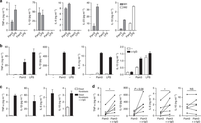
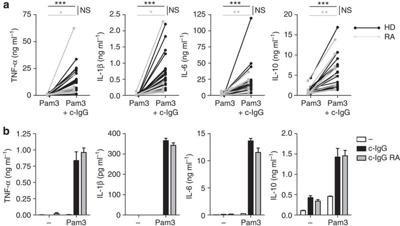
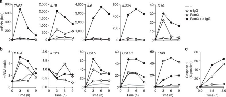
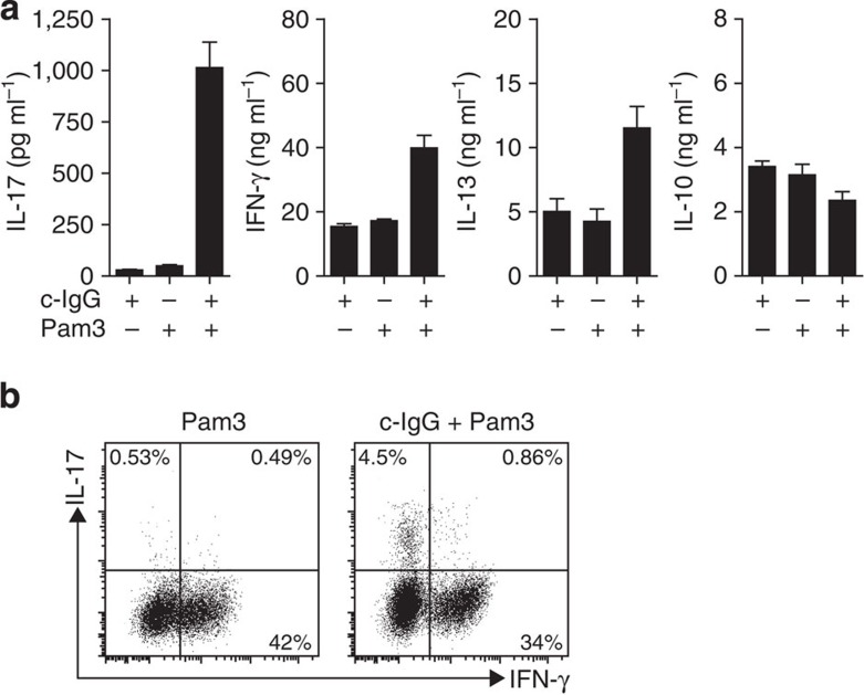
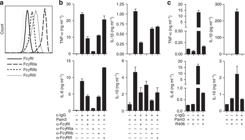
M2 macrophages suppress inflammation in numerous disorders, including tumour formation, infection and obesity. However, the exact role of M2 macrophages in the context of several other diseases is still largely undefined. We here show that human M2 macrophages promote inflammation instead of suppressing inflammation on simultaneous exposure to complexed IgG (c-IgG) and TLR ligands, as occurs in the context of diseases such as rheumatoid arthritis (RA). c-IgG-TLR ligand co-stimulation of M2 macrophages selectively amplifies production of pro-inflammatory cytokines TNF-α, IL-1β and IL-6 and promotes Th17 responses, which all play a critical role in RA pathology. Induction of pro-inflammatory cytokines on c-IgG co-stimulation mainly depends on Fc gamma receptor IIa (FcγRIIa), which selectively amplifies cytokine gene transcription and induces caspase-1 activation. These data indicate that FcγR-TLR cross-talk may be targeted for treatment to attenuate inflammation in RA, by restoring the anti-inflammatory function of M2 macrophages.
Figures
Similar articles
-
FcγR-TLR Cross-Talk Enhances TNF Production by Human Monocyte-Derived DCs via IRF5-Dependent Gene Transcription and Glycolytic Reprogramming.Front Immunol. 2019 Apr 8;10:739. doi: 10.3389/fimmu.2019.00739. eCollection 2019. Front Immunol. 2019. PMID: 31024565 Free PMC article.
-
FcγRIIa cross-talk with TLRs, IL-1R, and IFNγR selectively modulates cytokine production in human myeloid cells.Immunobiology. 2015 Feb;220(2):193-9. doi: 10.1016/j.imbio.2014.07.016. Epub 2014 Jul 25. Immunobiology. 2015. PMID: 25108563
-
Expression of Toll-like receptor 2 on CD16+ blood monocytes and synovial tissue macrophages in rheumatoid arthritis.Arthritis Rheum. 2004 May;50(5):1457-67. doi: 10.1002/art.20219. Arthritis Rheum. 2004. PMID: 15146415
-
Metabolic inflammation: role of cytokines in the crosstalk between adipose tissue and liver.Can J Physiol Pharmacol. 2013 Nov;91(11):867-72. doi: 10.1139/cjpp-2013-0050. Epub 2013 May 8. Can J Physiol Pharmacol. 2013. PMID: 24117253 Review.
-
Modulation of inflammatory cytokines by omega-3 fatty acids.Subcell Biochem. 2008;49:133-43. doi: 10.1007/978-1-4020-8831-5_5. Subcell Biochem. 2008. PMID: 18751910 Review.
Cited by
-
Macrophage Immunometabolism and Inflammaging: Roles of Mitochondrial Dysfunction, Cellular Senescence, CD38, and NAD.Immunometabolism. 2020;2(3):e200026. doi: 10.20900/immunometab20200026. Epub 2020 Jul 1. Immunometabolism. 2020. PMID: 32774895 Free PMC article.
-
Neutrophil Microvesicles from Healthy Control and Rheumatoid Arthritis Patients Prevent the Inflammatory Activation of Macrophages.EBioMedicine. 2018 Mar;29:60-69. doi: 10.1016/j.ebiom.2018.02.003. Epub 2018 Feb 7. EBioMedicine. 2018. PMID: 29449195 Free PMC article.
-
Dietary Iron and Heme Iron Consumption, Genetic Susceptibility, and Risk of Crohn's Disease and Ulcerative Colitis.Inflamm Bowel Dis. 2017 Jul;23(7):1088-1095. doi: 10.1097/MIB.0000000000001161. Inflamm Bowel Dis. 2017. PMID: 28604414 Free PMC article. Review.
-
Potential different immune phenotypes of macrophages in oral lichen planus by integrating immunofluorescence double staining and single-cell RNA sequencing.J Dent Sci. 2024 Oct;19(4):2210-2217. doi: 10.1016/j.jds.2024.03.002. Epub 2024 Mar 11. J Dent Sci. 2024. PMID: 39347036 Free PMC article.
-
Macrophage-Based Combination Therapies as a New Strategy for Cancer Immunotherapy.Kidney Dis (Basel). 2021 Sep 28;8(1):26-43. doi: 10.1159/000518664. eCollection 2022 Jan. Kidney Dis (Basel). 2021. PMID: 35224005 Free PMC article. Review.
References
-
- Biswas S. K. & Mantovani A. Macrophage plasticity and interaction with lymphocyte subsets: cancer as a paradigm. Nat. Immunol. 11, 889–896 (2010). - PubMed
Publication types
MeSH terms
Substances
LinkOut - more resources
Full Text Sources
Other Literature Sources
