Ellagic acid inhibits melanoma growth in vitro
- PMID: 25386288
- PMCID: PMC4211504
- DOI: 10.4081/dr.2011.e36
Ellagic acid inhibits melanoma growth in vitro
Abstract
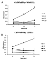
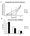
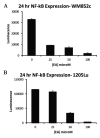
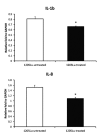
Ellagic is a polyphenolic compound with anti-fibrotic and antioxidant properties, and exhibits antitumor properties against various cancer cells in vitro. There are few studies, however, which examine the effects of ellagic acid on melanoma. In the present study, we observe effects of ellagic acid on melanoma cells in vitro. Three metastatic melanoma cell lines (1205Lu, WM852c and A375) were examined to determine the effects of ellagic acid on melanoma cell viability, cell-cycle, apoptosis, NF-κβ activity, and IL-1β & IL-8 secretion. Cell viability assays demonstrated that ellagic acid possesses an inhibitory effect on cell proliferation at concentrations between 25 and 100 µM. In addition, ellagic acid promoted G1 cell cycle arrest, increased levels of apoptosis and decreased synthesis of IL-1β and IL-8 in melanoma cells. Ellagic acid also decreased NF-κβ activity, suggesting at least one potential mechanism by which ellagic acid may exert its effects in melanoma cells. Our findings support further investigation into prospective roles for ellagic acid as a therapeutic, adjuvant, or preventive agent for melanoma.
Keywords: IL-1β; IL-8.; NF-κB; ellagic acid; melanoma.
Figures


Similar articles
-
Ellagic acid inhibits cell proliferation, migration, and invasion in melanoma via EGFR pathway.Am J Transl Res. 2020 May 15;12(5):2295-2304. eCollection 2020. Am J Transl Res. 2020. PMID: 32509220 Free PMC article.
-
Inhibitory effect and mechanism of mesenchymal stem cells on melanoma cells.Clin Transl Oncol. 2017 Nov;19(11):1358-1374. doi: 10.1007/s12094-017-1677-3. Epub 2017 Jul 21. Clin Transl Oncol. 2017. PMID: 28733866
-
Ellagic acid induced p53/p21 expression, G1 arrest and apoptosis in human bladder cancer T24 cells.Anticancer Res. 2005 Mar-Apr;25(2A):971-9. Anticancer Res. 2005. PMID: 15868936
-
Ellagic acid inhibits the proliferation of human pancreatic carcinoma PANC-1 cells in vitro and in vivo.Oncotarget. 2017 Feb 14;8(7):12301-12310. doi: 10.18632/oncotarget.14811. Oncotarget. 2017. PMID: 28135203 Free PMC article.
-
A Pharmacological Update of Ellagic Acid.Planta Med. 2018 Oct;84(15):1068-1093. doi: 10.1055/a-0633-9492. Epub 2018 May 30. Planta Med. 2018. PMID: 29847844 Review.
Cited by
-
Experimental Evidence of the Antitumor, Antimetastatic and Antiangiogenic Activity of Ellagic Acid.Nutrients. 2018 Nov 14;10(11):1756. doi: 10.3390/nu10111756. Nutrients. 2018. PMID: 30441769 Free PMC article. Review.
-
Possible Mechanisms of Oxidative Stress-Induced Skin Cellular Senescence, Inflammation, and Cancer and the Therapeutic Potential of Plant Polyphenols.Int J Mol Sci. 2023 Feb 13;24(4):3755. doi: 10.3390/ijms24043755. Int J Mol Sci. 2023. PMID: 36835162 Free PMC article. Review.
-
A Comprehensive Analysis of Diversity, Structure, Biosynthesis and Extraction of Biologically Active Tannins from Various Plant-Based Materials Using Deep Eutectic Solvents.Molecules. 2024 Jun 2;29(11):2615. doi: 10.3390/molecules29112615. Molecules. 2024. PMID: 38893491 Free PMC article. Review.
-
Ellagic acid alleviates adjuvant induced arthritis by modulation of pro- and anti-inflammatory cytokines.Cent Eur J Immunol. 2016;41(4):339-349. doi: 10.5114/ceji.2016.65132. Epub 2017 Jan 24. Cent Eur J Immunol. 2016. PMID: 28450796 Free PMC article.
-
Spice-Derived Phenolic Compounds: Potential for Skin Cancer Prevention and Therapy.Molecules. 2023 Aug 25;28(17):6251. doi: 10.3390/molecules28176251. Molecules. 2023. PMID: 37687080 Free PMC article. Review.
References
-
- Thresiamma KC, Kuttan R. Inhibition of liver fibrosis by ellagic acid. Indian J Physiol Pharmacol. 1996;40:363–6. - PubMed
-
- Devipriya N, Sudheer AR, Srinivasan M, Menon VP. Effect of Ellagic acid, a plant polyphenol, on fibrotic markers (MMPs and TIMPs) during alcohol-induced hepatotoxicity. Toxicol Mech Methods. 2007;17:349–56. - PubMed
-
- Losso JN, Bansode RR, Trappey A, 2nd, et al. In vitro anti-proliferative activities of ellagic acid. J Nutr Biochem. 2004;15:672–8. - PubMed
-
- Narayanan BA, Geoffroy O, Willingham MC, et al. p53/p21(WAF1/CIP1) expression and its possible role in G1 arrest and apoptosis in ellagic acid treated cancer cells. Cancer Lett. 1999;136:215–21. - PubMed
-
- Mukhtar H, Das M, Khan WA, et al. Exceptional activity of tannic acid among naturally occurring plant phenols in protecting against 7,12 dimethylbenz(a)-anthracene-, benzo(a)pyrene-, 3-methyl-cholanthrene-, and N-methyl-N-nitro-sourea-induced skin tumorigenesis in mice. Cancer Res. 1988;48:2361–5. - PubMed
LinkOut - more resources
Full Text Sources
