. 2014 Nov 6;10(11):e1004502.
doi: 10.1371/journal.ppat.1004502. eCollection 2014 Nov.

Coronavirus cell entry occurs through the endo-/lysosomal pathway in a proteolysis-dependent manner

Affiliations

Coronavirus cell entry occurs through the endo-/lysosomal pathway in a proteolysis-dependent manner

Christine Burkard et al. PLoS Pathog. .

Erratum in

Abstract

Enveloped viruses need to fuse with a host cell membrane in order to deliver their genome into the host cell. While some viruses fuse with the plasma membrane, many viruses are endocytosed prior to fusion. Specific cues in the endosomal microenvironment induce conformational changes in the viral fusion proteins leading to viral and host membrane fusion. In the present study we investigated the entry of coronaviruses (CoVs). Using siRNA gene silencing, we found that proteins known to be important for late endosomal maturation and endosome-lysosome fusion profoundly promote infection of cells with mouse hepatitis coronavirus (MHV). Using recombinant MHVs expressing reporter genes as well as a novel, replication-independent fusion assay we confirmed the importance of clathrin-mediated endocytosis and demonstrated that trafficking of MHV to lysosomes is required for fusion and productive entry to occur. Nevertheless, MHV was shown to be less sensitive to perturbation of endosomal pH than vesicular stomatitis virus and influenza A virus, which fuse in early and late endosomes, respectively. Our results indicate that entry of MHV depends on proteolytic processing of its fusion protein S by lysosomal proteases. Fusion of MHV was severely inhibited by a pan-lysosomal protease inhibitor, while trafficking of MHV to lysosomes and processing by lysosomal proteases was no longer required when a furin cleavage site was introduced in the S protein immediately upstream of the fusion peptide. Also entry of feline CoV was shown to depend on trafficking to lysosomes and processing by lysosomal proteases. In contrast, MERS-CoV, which contains a minimal furin cleavage site just upstream of the fusion peptide, was negatively affected by inhibition of furin, but not of lysosomal proteases. We conclude that a proteolytic cleavage site in the CoV S protein directly upstream of the fusion peptide is an essential determinant of the intracellular site of fusion.


Conflict of interest statement

The authors have declared that no competing interests exist.

Figures

Figure 1
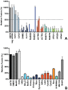
Figure 1. RNAi-mediated downregulation of endocytosis-associated proteins affects MHV infection.
A) Confirmation of endocytosis-associated hits from druggable genome-wide siRNA screen. Gene silencing was performed using individual transfection of three different siRNAs per gene in HeLa-mCC1a cells. Cells were infected with MHV-EGFPM at MOI = 0.5 for 8 h and analyzed by FACS for cell viability and virus replication. The effect of downregulation of expression on MHV infection was studied for the actin cytoskeleton-associated proteins ACTR2 and ACTR3 (orange), late endosomal proteins RAB7A and RAB7B (turquoise), HOPS complex sububit VPS39 (light green), ER/Golgi secretion-associated protein SNX1, Integrin/Actin-associated protein VCL, and Serine/Threonine-protein kinase PAK1 (grey). Error bars represent SEM, n = 4. B) Confirmation of siRNA-mediated reduction in mRNA levels. mRNA levels at 72 h post transfection were measured by qRT-PCR in comparison to non-transfected cells. Error bars represent SEM, n = 3*3. C) The effect of the RNAi-mediated downregulation of an extended set of endocytosis-associated proteins on MHV infection. Infection of MHV-EGFPM was analyzed after downregulation of proteins associated with caveolae-mediated endocytosis (light blue), clathrin-mediated endocytosis (dark blue), early endosomes (cerulean), actin cytoskeleton (dark orange), microtubule cytoskeleton (orange), late endosomes (turquoise), and late endosome-to-lysosome trafficking (light green) as described above. Error bars represent SEM, n = 3. A, C) Dotted lines show the lower 95% confidence interval of the negative siRNA controls.
Figure 2
Figure 2. Endocytosis affecting agents indicate clathrin-mediated endocytosis and endosome maturation to be important in MHV infection.
HeLa-mCC1a cells, inoculated with MHV-EGFPM at MOI = 0.5, were treated with the different inhibitors from 30 min prior to 8 h post inoculation (0–8 h) or from 2–8 h post inoculation (2–8 h; hatched bars): ammonium chloride (NH4Cl), Bafilomycin A1 (BafA1), Chloroquine (Chloq), Chlorpromazine (Chlopro), Monensin (Mon), Dynasore, Dyngo-4A, EIPA, Latrunculin A (LatA), Jasplakinolide (Jasp), Cytochalasin B (CytoB), Cytochalasin D (DytoD), Nocodazole (Noc), MG132, Brefeldin A (BrefA), as well as solvents dimethyl sulfoxide (DMSO) and methanol (MeOH). Infection was determined by FACS and displayed relative to the infection level observed in mock-treated cells (UNTR). Error bars represent SEM, n = 3.
Figure 3
Figure 3. Clathrin-mediated endocytosis and late endosome-to-lysosome trafficking is required for MHV fusion.
A) Fusion assay upon siRNA-mediated gene silencing. Three different siRNAs per gene were transfected individually into HeLa-mCC1a-ΔM15. 72 h post transfection, cells were pre-loaded with FDG by hypotonic shock. MHV-αN was allowed to bind to the cells on ice at MOI = 20 for 90 min. 100 min post warming to 37°C, cells were collected and analyzed by FACS. Fusion was determined relative to the number of FIC-positive cells observed upon mock treatment of infected cells (UNTR). Error bars represent SEM, n = 3. B) Fusion of MHV upon treatment of cells with different inhibitors was studied as in A. Cells were pretreated with ammonium chloride (NH4Cl), Bafilomycin A1 (BafA1), Chloroquine (Chloq), Chlorpromazine (Chlopro), Monensin (Mon), Dynasore, Dyngo-4A, EIPA, Latrunculin A, (LatA), Jasplakinolide (Jasp), Cytochalasin B (CytoB), Cytochalasin D (DytoD), Nocodazole (Noc), U18666A, MG132, Brefelding A (BrefA), as well as with the solvents dimethyl sulfoxide (DMSO) and methanol (MeOH), protein synthesis inhibitor cyclohexamide (CHX), and MHV fusion inhibitor HR2 peptide (HR2) for 30 min at 37°C. The inhibitors were kept present during binding of MHV-αN to cells and during warming to 37°C cells for 100 min. Fusion was determined relative to the number of FIC-positive cells after mock treatment (UNTR). Error bars represent SEM, n = 3.
Figure 4
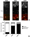
Figure 4. Live-cell microscopy demonstrates co-localization and co-tracking of MHV with endosomal vesicles and fusion of MHV in these vesicles.
HeLa-mCC1a cells transfected with plasmids encoding RAB5-mRFP, RAB7-mRFP, or dsRed-LAMP1 were inoculated with DyLight 488-labeled MHV. Live cell imaging was performed to track internalized particles. A) Examples of MHV particles co-localizing with RAB5-, RAB7-, and LAMP1-positive endosomal vesicles. Size bars indicate 0.2 µM B) Virus particles that could be tracked were classified as ‘fusing’ (Fusing) ‘associating/dissociating’ (Assoc/Dissoc), or ‘non-fusing’ (Non-fusing) as described in the Materials and Methods section.
Figure 5
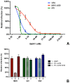
Figure 5. MHV infection depends on endosomal maturation.
A) HeLa-mCC1a cells were pretreated with increasing concentrations of Bafilomycin A1 (BafA1) for 30 min and subsequently infected with luciferase expressing MHV, VSV, or IAV in the presence of BafA1. Infection levels were determined by assaying the luciferase activity in cell lysates relative to lysates of infected cells that had been mock treated. Error bars represent SEM, n = 3*3. B) Haploid cells (HAP1), haploid cells lacking VPS33A (H1-ΔV33) or VPS33A-lacking haploid cells retransfected with FLAG-tagged VLP33A (H1-ΔV33-fV33) were infected with luciferase expressing MHV, VSV, or IAV. Cells were lysed at 7 h (MHV and VSV) or 16 h post infection. Infection is displayed relative to virus-driven luciferase expression levels in HAP1 cells. Error bars represent SEM, n = 3*3.
Figure 6
Figure 6. Inhibition of lysosomal proteases prevents MHV fusion.
The MHV fusion assay was performed on HeLa-mCC1a-ΔM15 cells as described in the legend to Figure 3, in the presence of the protease inhibitors CPI, AEBSF, Aprotinin, Leupeptin, Pepstatin A, Camostat, and Phosphoramidon. As controls, cells were treated with solvent DMSO, MHV fusion inhibitor HR2 peptide (HR2), and lysosomotropic agent ammonium chloride (NH4Cl). Fusion was determined relative to the number of FIC-positive cells after mock treatment (UNTR). Error bars represent SEM, n = 3.
Figure 7
Figure 7. Introduction of a furin cleavage site just upstream of the fusion peptide renders MHV independent of lysosomal proteases.
A) Schematic representation of the MHV spike protein. The MHV S proteins are partially processed by furin at the S1/S2 boundary (S1/S2) as indicated by the arrow. The furin cleavage site sequence at this position (RRAHR) is shown. The signal sequence (SS) at the amino-terminal end of the S1 subunit and the approximate positions of the fusion peptide (FP), heptad repeat regions 1 and 2 (HR1 and HR2) and the transmembrane domain (TM) in the S2 subunit are indicated. MHV-S2′FCS virus contains an optimal furin cleavage site (RRRRR) immediately upstream of the FP (S2′, indicated by the arrow. B) Effect of pan-lysosomal protease inhibitor (CPI) on MHV and MHV-S2′FCS infection. HeLa-mCC1a cells were pretreated with CPI for 30 min and inoculated at MOI = 0.2 with luciferase expression cassette containing MHV-EFLM or MHV-S2′FCS in the presence of CPI, after which incubations were continued in the presence of CPI until 7 hpi. Infection levels were determined by measuring the luciferase activity in cell lysates relative to mock-treated cells. Error bars represent SEM, n = 3*3.
Figure 8
Figure 8. Furin inhibitor reduces infection with MHV-S2′FCS and renders the virus sensitive to endosomal maturation.
Haploid HAP1 cells (HAP1), haploid cells lacking VPS33A (H1-ΔV33) or VPS33A-lacking haploid cells retransfected with FLAG-tagged VLP33A (H1-ΔV33-fV33) were infected (MOI = 0.2) with MHV-EFLM (MHV-wt) or MHV-S2′FCS for 7 h. Where indicated, cells were treated with furin inhibitor (FI). Infection levels were determined by measuring the luciferase activity in cell lysates relative to mock-treated cells. Error bars represent SEM, n = 3*3.
Figure 9
Figure 9. MHV-S2′FCS fuses in early endosomes.
siRNA-mediated gene silencing was performed as described in the legend to Figure 1. At 72 h post transfection, HeLa-mCC1a were inoculated with MHV-EFLM or MHV-S2′FCS at MOI = 0.2 and incubated until 7 hpi. Infection levels were determined by measuring the luciferase activity in cell lysates relative to mock-treated cells. Dotted line shows the lower 95% confidence interval of the negative siRNA controls. Error bars represent SEM, n = 3*3.
Figure 10
Figure 10. Entry of FIPV.
A) Clustal W alignment of spike proteins from several coronaviruses. Displayed is the fusion peptide (boxed) and the area upstream thereof. The area immediately upstream of the fusion peptide that contains the optimal FCS site (RRRRR) in MHV-S2′FCS is also boxed. B) siRNA-mediated gene silencing was performed as described in the legend to Figure 1. At 72 h post transfection, HeLa-fAPN cells were inoculated at MOI = 0.2 with luciferase expressing FIPV-RLuc. At 7 hpi infection was determined by measuring the luciferase activity in cell lysates and displayed relative to mock treated infection (inf). Error bars represent SEM, n = 3*3. Dotted line shows the lower 95% confidence interval of the negative siRNA controls. C) HeLa-fAPN cells inoculated with FIPV-Rluc at MOI = 0.1 were treated with pan-lysosomal protease inhibitor (CPI) or furin inhibitor (FI) from 30 min prior to 7 h post inoculation (0–7 h) or from 2–7 h post inoculation (2–7 h; hatched bars). Infection levels were determined by measuring the luciferase activity in cell lysates relative to mock-treated cells. Error bars represent SEM, n = 3*3.
Figure 11
Figure 11. MERS-CoV requires cleavage by furin but not by lysosomal proteases for infection.
Huh-7 cells inoculated with MERS-CoV were treated with furin inhibitor (FI) or pan-lysosomal protease inhibitor (CPI) starting from 30 min prior to inoculation. Numbers of infected cells was determined by immunocytochemical staining. Error bars represent SEM, n = 3.
Figure 12
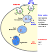
Figure 12. Model of early and late coronavirus fusion.
MHV and MHV-S2′FCS are taken up by DAB2-dependent clathrin-mediated endocytosis to end up in RAB5-containing early endosomes. The FCS of MHV-S2′FCS is cleaved by furin or furin-like enzymes to allow fusion of the virus in early endosomes. Trafficking of MHV from late endosomes to lysosomes (RAB7/LAMP1-positive compartments) is required for processing of MHV by lysosomal proteases and viral fusion to occur. We propose that the sequence immediately upstream of the FP is a key determinant of the intracellular site of fusion. MERS-CoV and FIPV enter cells via fusion in early endosomes or lysosomes, respectively. MERS-CoV, which contains a minimal FCS, is inhibited by furin inhibitor (FI) but not by the pan-lysosomal protease inhibitor (CPI). The opposite holds true for FIPV. Based on this model, we predict that IBV strain Beaudette and HCoV-NL63, which contain FCSs (Fig. 10), to fuse in early endosomes in a furin-dependent manner. Other CoVs that do not contain a FCS at this position are predicted to fuse in lysosomes.

Similar articles

Cited by

References

    1. Fuller AO, Spear PG (1987) Anti-glycoprotein D antibodies that permit adsorption but block infection by herpes simplex virus 1 prevent virion-cell fusion at the cell surface. Proceedings of the National Academy of Sciences of the United States of America 84: 5454–5458. - PMC - PubMed
    1. Sodeik B, Ebersold MW, Helenius A (1997) Microtubule-mediated transport of incoming herpes simplex virus 1 capsids to the nucleus. The Journal of cell biology 136: 1007–1021. - PMC - PubMed
    1. Okada Y (1969) Factors in fusion of cells by HVJ. Current topics in microbiology and immunology 48: 102–128. - PubMed
    1. Permanyer M, Ballana E, Este JA (2010) Endocytosis of HIV: anything goes. Trends in microbiology 18: 543–551. - PubMed
    1. Stein BS, Gowda SD, Lifson JD, Penhallow RC, Bensch KG, et al. (1987) pH-independent HIV entry into CD4-positive T cells via virus envelope fusion to the plasma membrane. Cell 49: 659–668. - PubMed

Publication types

Substances

Grants and funding

This work was supported by the EU 7th Framework Programme (Virus Entry, project 235649, PJMR) and by a Utrecht University High potential grant to CAMdH. The funders had no role in study design, data collection and analysis, decision to publish, or preparation of the manuscript.