

Altered resting-state amygdala functional connectivity after 36 hours of total sleep deprivation
- PMID: 25372882
- PMCID: PMC4221616
- DOI: 10.1371/journal.pone.0112222
Altered resting-state amygdala functional connectivity after 36 hours of total sleep deprivation
Abstract
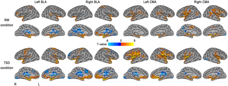
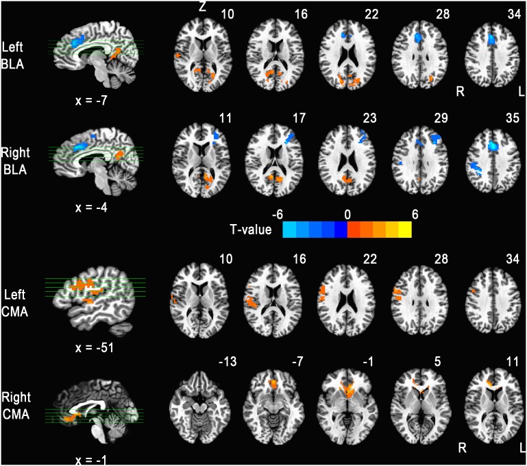
Objectives: Recent neuroimaging studies have identified a potentially critical role of the amygdala in disrupted emotion neurocircuitry in individuals after total sleep deprivation (TSD). However, connectivity between the amygdala and cerebral cortex due to TSD remains to be elucidated. In this study, we used resting-state functional MRI (fMRI) to investigate the functional connectivity changes of the basolateral amygdala (BLA) and centromedial amygdala (CMA) in the brain after 36 h of TSD.
Materials and methods: Fourteen healthy adult men aged 25.9 ± 2.3 years (range, 18-28 years) were enrolled in a within-subject crossover study. Using the BLA and CMA as separate seed regions, we examined resting-state functional connectivity with fMRI during rested wakefulness (RW) and after 36 h of TSD.
Results: TSD resulted in a significant decrease in the functional connectivity between the BLA and several executive control regions (left dorsolateral prefrontal cortex [DLPFC], right dorsal anterior cingulate cortex [ACC], right inferior frontal gyrus [IFG]). Increased functional connectivity was found between the BLA and areas including the left posterior cingulate cortex/precuneus (PCC/PrCu) and right parahippocampal gyrus. With regard to CMA, increased functional connectivity was observed with the rostral anterior cingulate cortex (rACC) and right precentral gyrus.
Conclusion: These findings demonstrate that disturbance in amygdala related circuits may contribute to TSD psychophysiology and suggest that functional connectivity studies of the amygdala during the resting state may be used to discern aberrant patterns of coupling within these circuits after TSD.
Conflict of interest statement
Figures
Similar articles
-
Altered insula-prefrontal functional connectivity correlates to decreased vigilant attention after total sleep deprivation.Sleep Med. 2021 Aug;84:187-194. doi: 10.1016/j.sleep.2021.05.037. Epub 2021 Jun 5. Sleep Med. 2021. PMID: 34166985
-
Decreased thalamocortical functional connectivity after 36 hours of total sleep deprivation: evidence from resting state FMRI.PLoS One. 2013 Oct 25;8(10):e78830. doi: 10.1371/journal.pone.0078830. eCollection 2013. PLoS One. 2013. PMID: 24205327 Free PMC article.
-
Altered superficial amygdala-cortical functional link in resting state after 36 hours of total sleep deprivation.J Neurosci Res. 2015 Dec;93(12):1795-803. doi: 10.1002/jnr.23601. Epub 2015 Sep 8. J Neurosci Res. 2015. PMID: 26346195
-
Altered functional connectivity between the nucleus basalis of Meynert and anterior cingulate cortex is associated with declined attentional performance after total sleep deprivation.Behav Brain Res. 2021 Jul 9;409:113321. doi: 10.1016/j.bbr.2021.113321. Epub 2021 Apr 25. Behav Brain Res. 2021. PMID: 33910027
-
Neuroimaging Predictors and Mechanisms of Treatment Response in Social Anxiety Disorder: an Overview of the Amygdala.Curr Psychiatry Rep. 2018 Aug 28;20(10):89. doi: 10.1007/s11920-018-0948-1. Curr Psychiatry Rep. 2018. PMID: 30155657 Free PMC article. Review.
Cited by
-
Regional cerebral hypoperfusion after acute sleep deprivation: A STROBE-compliant study of arterial spin labeling fMRI.Medicine (Baltimore). 2019 Jan;98(2):e14008. doi: 10.1097/MD.0000000000014008. Medicine (Baltimore). 2019. PMID: 30633191 Free PMC article.
-
A challenge for psychocardiology: Addressing the causes and consequences of patients' perceptions of enduring somatic threat.Am Psychol. 2018 Dec;73(9):1160-1171. doi: 10.1037/amp0000418. Am Psychol. 2018. PMID: 30525797 Free PMC article.
-
Intrinsic brain connectivity after partial sleep deprivation in young and older adults: results from the Stockholm Sleepy Brain study.Sci Rep. 2017 Aug 25;7(1):9422. doi: 10.1038/s41598-017-09744-7. Sci Rep. 2017. PMID: 28842597 Free PMC article.
-
Effects of Chronic Sleep Restriction on the Brain Functional Network, as Revealed by Graph Theory.Front Neurosci. 2019 Oct 11;13:1087. doi: 10.3389/fnins.2019.01087. eCollection 2019. Front Neurosci. 2019. PMID: 31680823 Free PMC article.
-
Age effect on gray matter volume changes after sleep restriction.PLoS One. 2020 Feb 6;15(2):e0228473. doi: 10.1371/journal.pone.0228473. eCollection 2020. PLoS One. 2020. PMID: 32027695 Free PMC article. Clinical Trial.
References
-
- Durmer JS, Dinges DF (2005) Neurocognitive consequences of sleep deprivation. Seminars in neurology. Vol. 25: pp. 117–129. - PubMed
-
- Mu Q, Nahas Z, Johnson KA, Yamanaka K, Mishory A, et al... (2005) Decreased Cortical Response to Verbal Working Memory Following Sleep Deprivation: 55–67. - PubMed
-
- Scott JPR, McNaughton LR, Polman RCJ (2006) Effects of sleep deprivation and exercise on cognitive, motor performance and mood. Physiol Behav 87: 396–408. - PubMed
-
- Anderson C, Platten CR (2011) Sleep deprivation lowers inhibition and enhances impulsivity to negative stimuli. Behav Brain Res 217: 463–466. - PubMed
Publication types
MeSH terms
Grants and funding
LinkOut - more resources
Full Text Sources
Other Literature Sources
Medical
