NPRL-Z-1, as a new topoisomerase II poison, induces cell apoptosis and ROS generation in human renal carcinoma cells
- PMID: 25372714
- PMCID: PMC4221609
- DOI: 10.1371/journal.pone.0112220
NPRL-Z-1, as a new topoisomerase II poison, induces cell apoptosis and ROS generation in human renal carcinoma cells
Abstract
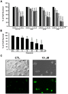
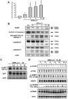
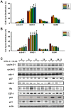
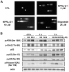
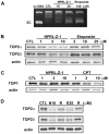
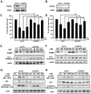
NPRL-Z-1 is a 4β-[(4"-benzamido)-amino]-4'-O-demethyl-epipodophyllotoxin derivative. Previous reports have shown that NPRL-Z-1 possesses anticancer activity. Here NPRL-Z-1 displayed cytotoxic effects against four human cancer cell lines (HCT 116, A549, ACHN, and A498) and exhibited potent activity in A498 human renal carcinoma cells, with an IC50 value of 2.38 µM via the MTT assay. We also found that NPRL-Z-1 induced cell cycle arrest in G1-phase and detected DNA double-strand breaks in A498 cells. NPRL-Z-1 induced ataxia telangiectasia-mutated (ATM) protein kinase phosphorylation at serine 1981, leading to the activation of DNA damage signaling pathways, including Chk2, histone H2AX, and p53/p21. By ICE assay, the data suggested that NPRL-Z-1 acted on and stabilized the topoisomerase II (TOP2)-DNA complex, leading to TOP2cc formation. NPRL-Z-1-induced DNA damage signaling and apoptotic death was also reversed by TOP2α or TOP2β knockdown. In addition, NPRL-Z-1 inhibited the Akt signaling pathway and induced reactive oxygen species (ROS) generation. These results demonstrated that NPRL-Z-1 appeared to be a novel TOP2 poison and ROS generator. Thus, NPRL-Z-1 may present a significant potential anticancer candidate against renal carcinoma.
Conflict of interest statement
Figures


Similar articles
-
The catalytic topoisomerase II inhibitor dexrazoxane induces DNA breaks, ATF3 and the DNA damage response in cancer cells.Br J Pharmacol. 2015 May;172(9):2246-57. doi: 10.1111/bph.13046. Epub 2015 Feb 27. Br J Pharmacol. 2015. PMID: 25521189 Free PMC article.
-
QS-ZYX-1-61 induces apoptosis through topoisomerase II in human non-small-cell lung cancer A549 cells.Cancer Sci. 2012 Jan;103(1):80-7. doi: 10.1111/j.1349-7006.2011.02103.x. Epub 2011 Oct 17. Cancer Sci. 2012. PMID: 21920000 Free PMC article.
-
Phosphorylation of p53 on Ser15 during cell cycle caused by Topo I and Topo II inhibitors in relation to ATM and Chk2 activation.Cell Cycle. 2008 Oct;7(19):3048-55. doi: 10.4161/cc.7.19.6750. Epub 2008 Oct 6. Cell Cycle. 2008. PMID: 18802408 Free PMC article.
-
Regulation of topoisomerase II stability and activity by ubiquitination and SUMOylation: clinical implications for cancer chemotherapy.Mol Biol Rep. 2021 Sep;48(9):6589-6601. doi: 10.1007/s11033-021-06665-7. Epub 2021 Sep 2. Mol Biol Rep. 2021. PMID: 34476738 Free PMC article. Review.
-
Topoisomerase IIα, rather than IIβ, is a promising target in development of anti-cancer drugs.Drug Discov Ther. 2012 Oct;6(5):230-7. Drug Discov Ther. 2012. PMID: 23229142 Review.
Cited by
-
Design, synthesis, docking, and anticancer evaluations of new thiazolo[3,2-a] pyrimidines as topoisomerase II inhibitors.J Enzyme Inhib Med Chem. 2023 Dec;38(1):2175209. doi: 10.1080/14756366.2023.2175209. J Enzyme Inhib Med Chem. 2023. PMID: 36776024 Free PMC article.
-
DIVERSet JAG Compounds Inhibit Topoisomerase II and Are Effective Against Adult and Pediatric High-Grade Gliomas.Transl Oncol. 2019 Oct;12(10):1375-1385. doi: 10.1016/j.tranon.2019.07.007. Epub 2019 Jul 30. Transl Oncol. 2019. PMID: 31374406 Free PMC article.
-
Predicting blood-brain barrier permeability of molecules with a large language model and machine learning.Sci Rep. 2024 Jul 9;14(1):15844. doi: 10.1038/s41598-024-66897-y. Sci Rep. 2024. PMID: 38982309 Free PMC article.
-
The Impact of Oxidative Stress and AKT Pathway on Cancer Cell Functions and Its Application to Natural Products.Antioxidants (Basel). 2022 Sep 19;11(9):1845. doi: 10.3390/antiox11091845. Antioxidants (Basel). 2022. PMID: 36139919 Free PMC article. Review.
-
The Topoisomerase 1 Inhibitor Austrobailignan-1 Isolated from Koelreuteria henryi Induces a G2/M-Phase Arrest and Cell Death Independently of p53 in Non-Small Cell Lung Cancer Cells.PLoS One. 2015 Jul 6;10(7):e0132052. doi: 10.1371/journal.pone.0132052. eCollection 2015. PLoS One. 2015. PMID: 26147394 Free PMC article.
References
-
- Hurley LH (2002) DNA and its associated processes as targets for cancer therapy. Nat Rev Cancer 2: 188–200. - PubMed
-
- Wang H-K, Morris-Natschke SL, Lee K-H (1997) Recent advances in the discovery and development of topoisomerase inhibitors as antitumor agents. Medicinal Research Reviews 17: 367–425. - PubMed
-
- Haglof KJ, Popa E, Hochster HS (2006) Recent developments in the clinical activity of topoisomerase-1 inhibitors. Update on Cancer Therapeutics 1: 117–145. - PubMed
Publication types
MeSH terms
Substances
Grants and funding
LinkOut - more resources
Full Text Sources
Other Literature Sources
Medical
Molecular Biology Databases
Research Materials
Miscellaneous

