Review
. 2014 Oct 17:5:514.
doi: 10.3389/fimmu.2014.00514. eCollection 2014.

From Monocytes to M1/M2 Macrophages: Phenotypical vs. Functional Differentiation

Affiliations
Review

From Monocytes to M1/M2 Macrophages: Phenotypical vs. Functional Differentiation

Paola Italiani et al. Front Immunol. .

Abstract

Studies on monocyte and macrophage biology and differentiation have revealed the pleiotropic activities of these cells. Macrophages are tissue sentinels that maintain tissue integrity by eliminating/repairing damaged cells and matrices. In this M2-like mode, they can also promote tumor growth. Conversely, M1-like macrophages are key effector cells for the elimination of pathogens, virally infected, and cancer cells. Macrophage differentiation from monocytes occurs in the tissue in concomitance with the acquisition of a functional phenotype that depends on microenvironmental signals, thereby accounting for the many and apparently opposed macrophage functions. Many questions arise. When monocytes differentiate into macrophages in a tissue (concomitantly adopting a specific functional program, M1 or M2), do they all die during the inflammatory reaction, or do some of them survive? Do those that survive become quiescent tissue macrophages, able to react as naïve cells to a new challenge? Or, do monocyte-derived tissue macrophages conserve a "memory" of their past inflammatory activation? This review will address some of these important questions under the general framework of the role of monocytes and macrophages in the initiation, development, resolution, and chronicization of inflammation.

Keywords: functional phenotypes; inflammation; monocyte-derived macrophages; monocytes; tissue-resident macrophages.


Figures

Figure 1
Figure 1
Origin of tissue-resident macrophages in the mouse. In adult tissues, macrophages derive from three sources. The first is the yolk sac in the embryo, where primitive hematopoiesis occurs giving rise to progenitors that seed tissues with F4/80high macrophages. Later during fetal development, hematopoiesis shifts from the yolk sac to the fetal liver (that seems to contribute to the LC pool in the skin, possibly through a yolk sac-derived progenitor). It is unknown whether other resident macrophages in other tissues may also derive from fetal liver hematopoiesis. The third source is the bone marrow, where definitive hematopoiesis occurs in the fetus and in the adult, giving rise to monocytes and to monocyte-derived F4/80low macrophages. Expression of murine F4/80 (the human EMR1) is an insufficient marker to discriminate between monocyte-derived macrophages and tissue-resident macrophages. It seems that Ly6C+ monocytes are the precursors of tissue macrophages, while the exact contribution of Ly6C monocytes remains unclear. HSC, hematopoietic stem cell; GMP, granulocyte-macrophage progenitor; MDP, macrophage–dendritic cell progenitor; LC, Langerhans cell; CSF-1, colony-stimulating factor 1; IL-34, interleukin 34.
Figure 2
Figure 2
Distribution of tissue-resident macrophages and monocyte-derived macrophages in tissues and organs. Monocyte contribution to resident macrophages is highly tissue-dependent and varies from no contribution for brain microglia and epidermal LC to complete monocyte origin for intestinal lamina propria macrophages. The tissues listed in the middle are those at the center of ongoing controversy (see the main text), and for which a mixed contribution is probable. Here, we define yolk sac-derived macrophages as tissue-resident macrophages, and both fetal liver-derived macrophages and bone marrow-derived macrophages as monocyte-derived macrophages (considering that bone marrow hematopoiesis derives from fetal liver hematopoiesis). LC, Langerhans cells; LP, lamina propria; RP, red pulp.
Figure 3
Figure 3
Schematic representation of monocyte and macrophage populations in homeostasis and inflammation. Under homeostatic conditions (left panel), Ly6C+ monocytes derive from the bone marrow and circulate via the blood into the tissue. A minor fraction of these cells lose Ly6C expression and become Ly6C monocytes in the blood or in the bone marrow where some of them might return in the absence of inflammation. Ly6C+ blood monocytes enter tissues and become either macrophages, for example, in the gut, lung, and dermis (monocyte-derived macrophages or monocyte-derived tissue-resident macrophages). Some tissue macrophages derive directly from yolk sac during the embryogenesis (e.g., LC, microglia, liver Kupffer cells, and alveolar macrophages), are long lived, and are mainly maintained by self-renewal (tissue-resident macrophages). Ly6C monocytes act as resident macrophages of the vasculature, patrolling, and monitoring the endothelial surface in the blood vessel lumen. In the figure, the presence of “trained” macrophages is also considered, which we define as “memory macrophages,” i.e., the tissue macrophages that retain the memory of a previous inflammation and are in a quiescent state in the tissue. During an inflammatory reaction (right panel), the number of blood Ly6C+ monocytes recruited to an inflamed tissue increases considerably. The large majority of these cells gives rise to the inflammatory monocyte-derived macrophages, while some of them do not differentiate into macrophages and remain monocyte-like cells, are able to take up antigens, and to migrate to the draining lymph nodes (tissue monocytes). These are the antigen-uptaking and -presenting cells of the tissue. During inflammation, all macrophages (tissue-resident macrophages, monocyte-derived tissue macrophages, inflammatory monocyte-derived macrophages) are activated and differentiate into M1-like inflammatory cells following interaction with pathogenic and damaged signals/insults in surrounding microenvironment. These cells produce a series of cytokines and other inflammatory factors. Tissue-resident macrophages increase their capacity of proliferation to compensate the loss of macrophages caused by the inflammatory reaction. Recent evidence demonstrates that also inflammatory monocyte-derived macrophages are able to proliferate in a late phase of the inflammatory reaction. Memory macrophages are important players in the inflammatory reaction, as they can react to inflammatory stimuli with a faster and stronger inflammatory cytokine production. The role of circulating Ly6C cells during an inflammatory reaction is not fully identified. They probably remain in the blood vessels as sentinels, and in some cases they could enter in the tissue, as it has been reported in the case of myocardial infarction, to take up a repair function. HSC, hematopoietic stem cell; cMoP, common monocyte progenitor; Ly6C, lymphocyte antigen 6 complex; LC, Langerhans cells; MΦ, macrophages.
Figure 4
Figure 4
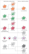
Fate of the different monocyte/macrophage populations in the tissue during the post-inflammatory phase. Tissue-resident macrophages are in general maintained locally by proliferative self-renewal, and retain an M2-like functional phenotype. The same situation is hypothesized for monocyte-derived resident macrophages, since it is not possible to fully discriminate between the two populations. A number of cells of these two populations probably die during the inflammatory reaction. Inflammatory monocyte-derived macrophages can die killed by the NO they have produced, and the surviving cells can undergo in situ phenotype conversion and become M2-like tissue-resident macrophages. In addition, a number of these cells can conserve a “memory” of their past inflammatory activation, and become trained monocytes/memory macrophages. Monocytes recruited from the blood during the post-inflammatory phase can lose the expression of Ly6C and become Ly6C cells, subsequently differentiating in M2 macrophages. They may also become memory macrophages. Memory macrophages that are present in the tissue, reminiscent of previous inflammatory events, would probably behave like naïve macrophages upon a new inflammatory challenge, except for a much quicker reaction, and will, therefore, mostly die or generate M2-like macrophages or again memory macrophages. Their life span in the tissue is presently unknown.

Similar articles

Cited by

References

    1. Matzinger P. Friendly and dangerous signals: is the tissue in control? Nat Immunol (2007) 8:11–3.10.1038/ni0107-11 - DOI - PubMed
    1. Medzhitov R. Origin and physiological roles of inflammation. Nature (2008) 454:428–35.10.1038/nature07201 - DOI - PubMed
    1. Nathan C. Points of control in inflammation. Nature (2002) 420:846–52.10.1038/nature01320 - DOI - PubMed
    1. van Furth R, Cohn ZA. The origin and kinetics of mononuclear phagocytes. J Exp Med (1968) 128:415–35.10.1084/jem.128.3.415 - DOI - PMC - PubMed
    1. Taylor PR, Gordon S. Monocytes heterogeneity and innate immunity. Immunity (2003) 19:2–4.10.1016/S1074-7613(03)00178-X - DOI - PubMed