. 2014 Oct 23;10(10):e1004569.
doi: 10.1371/journal.pgen.1004569. eCollection 2014 Oct.

The Not5 subunit of the ccr4-not complex connects transcription and translation

Affiliations

The Not5 subunit of the ccr4-not complex connects transcription and translation

Zoltan Villanyi et al. PLoS Genet. .

Abstract

Recent studies have suggested that a sub-complex of RNA polymerase II composed of Rpb4 and Rpb7 couples the nuclear and cytoplasmic stages of gene expression by associating with newly made mRNAs in the nucleus, and contributing to their translation and degradation in the cytoplasm. Here we show by yeast two hybrid and co-immunoprecipitation experiments, followed by ribosome fractionation and fluorescent microscopy, that a subunit of the Ccr4-Not complex, Not5, is essential in the nucleus for the cytoplasmic functions of Rpb4. Not5 interacts with Rpb4; it is required for the presence of Rpb4 in polysomes, for interaction of Rpb4 with the translation initiation factor eIF3 and for association of Rpb4 with mRNAs. We find that Rpb7 presence in the cytoplasm and polysomes is much less significant than that of Rpb4, and that it does not depend upon Not5. Hence Not5-dependence unlinks the cytoplasmic functions of Rpb4 and Rpb7. We additionally determine with RNA immunoprecipitation and native gel analysis that Not5 is needed in the cytoplasm for the co-translational assembly of RNA polymerase II. This stems from the importance of Not5 for the association of the R2TP Hsp90 co-chaperone with polysomes translating RPB1 mRNA to protect newly synthesized Rpb1 from aggregation. Hence taken together our results show that Not5 interconnects translation and transcription.


Conflict of interest statement

The authors have declared that no competing interests exist.

Figures

Figure 1
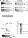
Figure 1. Rpb4 interacts with Not5 and the cytoplasmic functions of Rpb4 require Not5.
A. Serial dilutions of exponentially growing reporter cells expressing LexA-Rpb4 as a bait, and the indicated proteins fused to B42 as preys, were spotted either on medium selective for the plasmids (left panel +L) or selective for the plasmids and indicative of an interaction between bait and prey (right panel -L). B. Serial dilutions of exponentially growing cells from the indicated strains were spotted on plates and left to grow for several days at 30°C. C and D. Fractions from 7–47% sucrose gradients of extracts from wild-type or mutant strains expressing the indicated Tap-tagged (TT) proteins were precipitated with TCA and analyzed by western blotting with PAP antibodies. The positions of 40S, 60S, 80S and polysomes are indicated under the blots. The numbers of the gradient fractions tested or the total extract (TE) are indicated at the top. The polysome profiles for these experiments are available in Fig. S15 along with a typical distribution of a ribosomal protein (Rps3) in the wt and not5Δ gradients. Rpb4-TT (E) or Not1-TT (F) were immunoprecipitated from extracts of wild-type or mutant cells expressing HA-tagged Prt1. Wild-type cells expressing untagged Rpb4 or Not1 were used as a control. Similar negative controls were obtained with not5Δ cells not expressing any Tap-tagged protein (Fig. S16). The immunoblots were developed using anti-CBP or HA antibodies. G and H. Wild-type and not5Δ cells expressing Rpb4-TT (G) or the indicated (H) Not5 derivatives, were grown exponentially and stained with anti-CBP antibodies (upper panels) or DAPI (middle panels). The pictures were merged (lower panels) and the indicated section from wild-type (a) or not5Δ (b) was enlarged for better visualization. The localization of the Not5 derivatives is presented in Fig. S2.
Figure 2
Figure 2. Presence of Rpb7 in polysomes or in the cytoplasm is not affected by Not5.
A. Wild-type or mutant cells expressing Tap-tagged Rpb7 as indicated were analyzed on sucrose gradients as in Fig. 1C. The polysome profiles and protein loading for these experiments are available in Fig. S15. B. Wild-type and not5Δ cells expressing Rpb7-TT were grown exponentially and stained with anti-CBP antibodies or DAPI as for Fig. 1G. The merged pictures are displayed.
Figure 3
Figure 3. Polymerase subunits are present in polysome fractions and interact with Rpl25.
A and B. Cells expressing Tap-tagged polymerase subunits were analyzed on sucrose gradients as in Fig. 1C. The polysome profiles and protein loading for these experiments are available in Fig. S15. Extracts were treated either with CHX to preserve polysomes or with EDTA to disrupt them or C. with RNase to disrupt them. D. Rpb2-TT was immunoprecipitated from extracts of cells expressing HA-tagged Rpl25. Cells expressing untagged Rpb2 were used as a control. The immunoprecipitates were incubated with antibodies against HA or CBP to reveal Rpl25 and Rpb2, respectively. The total extract (Input) or immunoprecipitates (IP) were analyzed.
Figure 4
Figure 4. Polymerase sub-complexes lacking Rpb1 accumulate in not5Δ.
A and B. Total extracts from cells expressing the indicated Tap-tagged (TT) polymerase subunits were separated on native gels (upper panels) or SDS-PAGE (lower panels) and analyzed by western blotting with anti- CBP antibodies. C. Rpb9-TT was purified by single step affinity and the purified proteins were analyzed on native gels (upper panels) or SDS-PAGE (lower panels) and western blotting with anti-CBP antibodies (left panel) or anti-Rpb1 antibodies (right panel). D. Total extracts from cells expressing Rpb11-TT were either untreated (-) or treated with DNase or RNase as indicated and separated by Native-PAGE, and analyzed by western blotting with PAP antibodies.
Figure 5
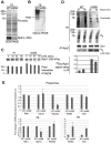
Figure 5. The levels of soluble Rpb1 are decreased in not5Δ.
A and B. Total soluble extracts were analyzed by SDS-PAGE (A) or Native-PAGE (B) and membranes were stained with Ponceau (upper panel in A) or probed with antibodies against Rpb1 (B and lower panel in A). C. Cells were grown exponentially to an OD600 of 1.0 and then CHX (100 µg ml−1) was added. 0.8 OD600 units of wild-type cells and 1.6 OD600 units of not5Δ were collected at the indicated times after protein synthesis arrest. Total proteins prepared by post-alcaline lysis were analyzed by western blotting with antibodies against Rpb1. Quantification of the blots (shown below the blots) revealed no significant difference in the reduction of Rpb1 over time in the 2 strains. D. Wild- type and not5Δ cells were metabolically labeled with 35S for 5 min then chased with cold methionine for 30 and 60 min. Total extracts (TE) were prepared and counted for 35S incorporation. The same amount of labeled total protein (20’00 cpm) from both strains (corresponding to 10 µg of protein from wt and 2.5 µg from not5Δ) was separated by SDS-PAGE. The gel was stained by Coomassie (TE, Coomassie), dried and then exposed (TE, 35S). The same amount of labeled protein from each extract (corresponding to 2 mg of protein from wt and 0.5 mg of protein from not5Δ) was also incubated with antibodies against Rpb1, and the immunoprecipitate was analyzed by western blotting (IP-Rpb1, Rpb1). The membranes were also exposed (IP-Rpb1, 35S). Quantified ratios of the 35S-Rpb1 signal and anti-Rpb1 signal from the western blotting are shown below the blots. E. Total extracts from wild-type or not5Δ were separated on sucrose gradients as in Fig. 1C, and RNA was extracted from the total extracts (TE, lower panels) or polysome fraction 14 (Fig. S15) (Polysomes, upper panels). The amount of the indicated mRNAs were evaluated by RT followed by qPCR in 1 µg of total and polysomal RNA. Values were normalized to the level of NIP1 mRNA that showed no change in abundance between the wild-type and not5Δ in total extracts or polysome fractions (Fig. S7). All mRNA levels are expressed relative to the level in the total extract of the wild-type expressed as 1. * represents statistically significant differences in mRNA abundance between wild-type and not5Δ samples at p<0.05.
Figure 6
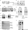
Figure 6. Assembly of Rpb1 with the R2TP Hsp90 co-chaperone is reduced and Rpb1 aggregates in not5Δ.
A. Total extracts and protein aggregates from the indicated strains were analyzed on SDS-PAGE followed by western blotting with antibodies against Rpb1. B. Rvb1-TT or Rvb2-TT were purified from wild-type and not5Δ cells. Equal amounts of the total extract (Input), and the purified proteins (Purified Rvb1 or 2) were analyzed by western blotting with anti-CBP or anti-Rpb1 antibodies. C. Rpb1 was immunoprecipitated from wild-type or not5Δ and the level of Rpb1 and Hsp90 proteins in the total extract (Input) immunoprecipitate (Ip) was evaluated by western blotting. D. Total extracts (TE) or protein aggregates (A) from wild-type cells (WT) or not5Δ cells or from not5Δ cells expressing NLS-Not5 as indicated were separated on SDS-PAGE and tested by western blotting for the levels of Hsp90 or Rpb1. E. Total extracts from wild-type or not5Δ cells grown in galactose expressing Rpb11-TT or not, and expressing or not NLS-Not5 or LexA-Rpb4, as indicated, were separated on native gels and analyzed by western blotting with anti- CBP antibodies (left panel). The same extracts were separated on sucrose gradients and the polysome profiles are shown in Fig. S1A, whereas the distribution of LexA-Rpb4 along the sucrose gradient is shown in Fig. S1B. Total extracts from not5Δ cells expressing Rpb11-TT and expressing either Myc-Not5 (Not5) or Myc-Not5-NES (NES) from episomes were separated by Native-PAGE and analyzed by western blotting with anti-CBP antibodies (upper panel) or by SDS-PAGE and analyzed by western blotting with anti-Rpb1 antibodies (lower panel). (F). Total extracts from wild-type or not5Δ cells expressing Rvb1-TT, Rvb2-TT or Hsp82-TT were analyzed as in Fig. 1C. The polysome profiles for these experiments are available in Fig. S15. G. Total extracts from WT, not5Δ or rpb4Δ cells expressing Rpb9-TT from cells were separated on native gels and analyzed by western blotting with anti-CBP antibodies.
Figure 7
Figure 7. Rpb1 accumulates in cytoplasmic speckles in RPB2+/− CNOT3+/− trans- heterozygotes.
Nurse cells (with large polyploid nuclei surrounded by a layer of follicle cells) of Drosophila melanogaster of the indicated genotypes were stained with antibodies against Rpb1, or with DAPI, and the images were merged as indicated. The indicated section was enlarged for better visualization. Scale bar 30 µM.

Similar articles

Cited by

References

    1. Hsin JP, Manley JL (2012) The RNA polymerase II CTD coordinates transcription and RNA processing. Genes & development 26: 2119–2137. - PMC - PubMed
    1. Aguilera A (2005) Cotranscriptional mRNP assembly: from the DNA to the nuclear pore. Current opinion in cell biology 17: 242–250. - PubMed
    1. de Almeida SF, Carmo-Fonseca M (2008) The CTD role in cotranscriptional RNA processing and surveillance. FEBS letters 582: 1971–1976. - PubMed
    1. Harel-Sharvit L, Eldad N, Haimovich G, Barkai O, Duek L, et al. (2010) RNA polymerase II subunits link transcription and mRNA decay to translation. Cell 143: 552–563. - PubMed
    1. Goler-Baron V, Selitrennik M, Barkai O, Haimovich G, Lotan R, et al. (2008) Transcription in the nucleus and mRNA decay in the cytoplasm are coupled processes. Genes & development 22: 2022–2027. - PMC - PubMed

MeSH terms

Grants and funding

This work was supported by grant 31003A_135794 from the Swiss National Science awarded to MAC and by a SCIEX (Scientific Exchange Program between New EU Member States and Switzerland) grant awarded to MAC for ZV. IB and ZP were supported by Hungarian State Science Fund (OTKA 77443). The funders had no role in study design, data collection and analysis, decision to publish, or preparation of the manuscript.

LinkOut - more resources