

Stable cellular senescence is associated with persistent DDR activation
- PMID: 25340529
- PMCID: PMC4207795
- DOI: 10.1371/journal.pone.0110969
Stable cellular senescence is associated with persistent DDR activation
Abstract
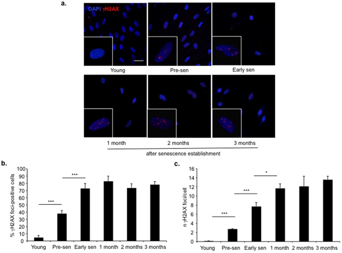
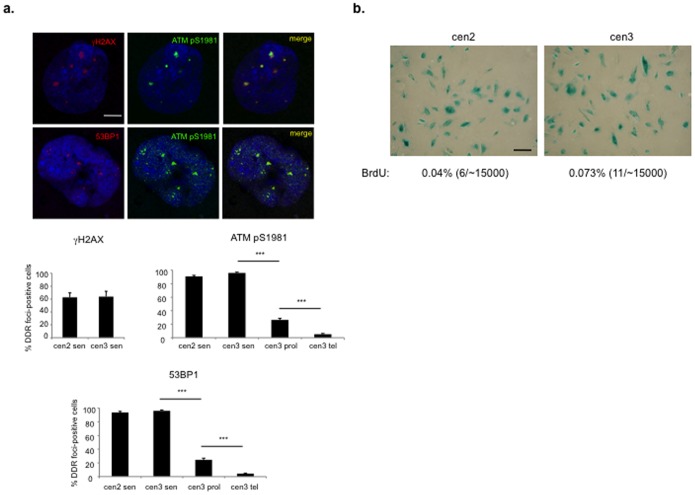
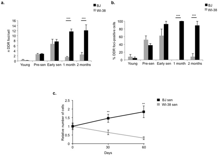
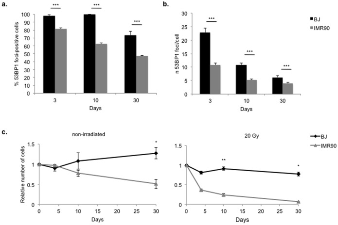
The DNA damage response (DDR) is activated upon DNA damage generation to promote DNA repair and inhibit cell cycle progression in the presence of a lesion. Cellular senescence is a permanent cell cycle arrest characterized by persistent DDR activation. However, some reports suggest that DDR activation is a feature only of early cellular senescence that is then lost with time. This challenges the hypothesis that cellular senescence is caused by persistent DDR activation. To address this issue, we studied DDR activation dynamics in senescent cells. Here we show that normal human fibroblasts retain DDR markers months after replicative senescence establishment. Consistently, human fibroblasts from healthy aged donors display markers of DDR activation even three years in culture after entry into replicative cellular senescence. However, by extending our analyses to different human cell strains, we also observed an apparent DDR loss with time following entry into cellular senescence. This though correlates with the inability of these cell strains to survive in culture upon replicative or irradiation-induced cellular senescence. We propose a model to reconcile these results. Cell strains not suffering the prolonged in vitro culture stress retain robust DDR activation that persists for years, indicating that under physiological conditions persistent DDR is causally involved in senescence establishment and maintenance. However, cell strains unable to maintain cell viability in vitro, due to their inability to cope with prolonged cell culture-associated stress, show an only-apparent reduction in DDR foci which is in fact due to selective loss of the most damaged cells.
Conflict of interest statement
Figures
Similar articles
-
DNA damage response activation in mouse embryonic fibroblasts undergoing replicative senescence and following spontaneous immortalization.Cell Cycle. 2008 Nov 15;7(22):3601-6. doi: 10.4161/cc.7.22.7152. Epub 2008 Nov 8. Cell Cycle. 2008. PMID: 19001874
-
Irreparable telomeric DNA damage and persistent DDR signalling as a shared causative mechanism of cellular senescence and ageing.Curr Opin Genet Dev. 2014 Jun;26:89-95. doi: 10.1016/j.gde.2014.06.009. Epub 2014 Aug 11. Curr Opin Genet Dev. 2014. PMID: 25104620 Free PMC article. Review.
-
Persistent DNA damage signalling triggers senescence-associated inflammatory cytokine secretion.Nat Cell Biol. 2009 Aug;11(8):973-9. doi: 10.1038/ncb1909. Epub 2009 Jul 13. Nat Cell Biol. 2009. PMID: 19597488 Free PMC article.
-
CBX4 Regulates Replicative Senescence of WI-38 Fibroblasts.Oxid Med Cell Longev. 2022 Feb 23;2022:5503575. doi: 10.1155/2022/5503575. eCollection 2022. Oxid Med Cell Longev. 2022. PMID: 35251476 Free PMC article.
-
High-LET-Radiation-Induced Persistent DNA Damage Response Signaling and Gastrointestinal Cancer Development.Curr Oncol. 2023 Jun 7;30(6):5497-5514. doi: 10.3390/curroncol30060416. Curr Oncol. 2023. PMID: 37366899 Free PMC article. Review.
Cited by
-
Uncoupling of mTORC1 from E2F activity maintains DNA damage and senescence.Nat Commun. 2024 Oct 24;15(1):9181. doi: 10.1038/s41467-024-52820-6. Nat Commun. 2024. PMID: 39448567 Free PMC article.
-
Ablation of XP-V gene causes adipose tissue senescence and metabolic abnormalities.Proc Natl Acad Sci U S A. 2015 Aug 18;112(33):E4556-64. doi: 10.1073/pnas.1506954112. Epub 2015 Aug 3. Proc Natl Acad Sci U S A. 2015. PMID: 26240351 Free PMC article.
-
The Chromatin Landscape of Cellular Senescence.Trends Genet. 2016 Nov;32(11):751-761. doi: 10.1016/j.tig.2016.09.005. Epub 2016 Sep 28. Trends Genet. 2016. PMID: 27692431 Free PMC article. Review.
-
Emerging Insight Into the Role of Circadian Clock Gene BMAL1 in Cellular Senescence.Front Endocrinol (Lausanne). 2022 Jun 6;13:915139. doi: 10.3389/fendo.2022.915139. eCollection 2022. Front Endocrinol (Lausanne). 2022. PMID: 35733785 Free PMC article. Review.
-
Persistent DNA Double-Strand Breaks After Repeated Diagnostic CT Scans in Breast Epithelial Cells and Lymphocytes.Front Oncol. 2021 Apr 23;11:634389. doi: 10.3389/fonc.2021.634389. eCollection 2021. Front Oncol. 2021. PMID: 33968734 Free PMC article.
References
-
- Campisi J, d'Adda di Fagagna F (2007) Cellular senescence: when bad things happen to good cells. Nat Rev Mol Cell Biol 8: 729–740. - PubMed
-
- d'Adda di Fagagna F (2008) Living on a break: cellular senescence as a DNA-damage response. Nat Rev Cancer 8: 512–522. - PubMed
-
- Evan GI, d'Adda di Fagagna F (2009) Cellular senescence: hot or what? Curr Opin Genet Dev 19: 25–31. - PubMed
Publication types
MeSH terms
Substances
Grants and funding
LinkOut - more resources
Full Text Sources
Other Literature Sources
Miscellaneous
