Testisimmune privilege - Assumptions versus facts
- PMID: 25309630
- PMCID: PMC4192663
Testisimmune privilege - Assumptions versus facts
Abstract
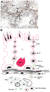
The testis has long enjoyed a reputation as an immunologically privileged site based on its ability to protect auto-antigenic germ cells and provide an optimal environment for the extended survival of transplanted allo- or xeno-grafts. Exploration of the role of anatomical, physiological, immunological and cellular components in testis immune privilege revealed that the tolerogenic environment of the testis is a result of the immunomodulatory factors expressed or secreted by testicular cells (mainly Sertoli cells, peritubular myoid cells, Leydig cells, and resident macrophages). The blood-testis barrier/Sertoli cell barrier, is also important to seclude advanced germ cells but its requirement in testis immune privilege needs further investigation. Testicular immune privilege is not permanent, as an effective immune response can be mounted against transplanted tissue, and bacterial/viral infections in the testis can be effectively eliminated. Overall, the cellular components control the fate of the immune response and can shift the response from immunodestructive to immunoprotective, resulting in immune privilege.
Keywords: immune privilege; testis; transplantation.
Figures


Similar articles
-
An overview of a Sertoli cell transplantation model to study testis morphogenesis and the role of the Sertoli cells in immune privilege.Environ Epigenet. 2017 Aug 3;3(3):dvx012. doi: 10.1093/eep/dvx012. eCollection 2017 Jul. Environ Epigenet. 2017. PMID: 29492314 Free PMC article. Review.
-
Structural, cellular and molecular aspects of immune privilege in the testis.Front Immunol. 2012 Jun 11;3:152. doi: 10.3389/fimmu.2012.00152. eCollection 2012. Front Immunol. 2012. PMID: 22701457 Free PMC article.
-
Somatic-Immune Cells Crosstalk In-The-Making of Testicular Immune Privilege.Reprod Sci. 2022 Oct;29(10):2707-2718. doi: 10.1007/s43032-021-00721-0. Epub 2021 Sep 27. Reprod Sci. 2022. PMID: 34580844 Review.
-
Sertoli cells--immunological sentinels of spermatogenesis.Semin Cell Dev Biol. 2014 Jun;30:36-44. doi: 10.1016/j.semcdb.2014.02.011. Epub 2014 Mar 3. Semin Cell Dev Biol. 2014. PMID: 24603046 Free PMC article. Review.
-
Genetically engineered immune privileged Sertoli cells: A new road to cell based gene therapy.Spermatogenesis. 2012 Jan 1;2(1):23-31. doi: 10.4161/spmg.19119. Spermatogenesis. 2012. PMID: 22553487 Free PMC article.
Cited by
-
Increased tumor vascularization is associated with the amount of immune competent PD-1 positive cells in testicular germ cell tumors.Oncol Lett. 2018 Jun;15(6):9852-9860. doi: 10.3892/ol.2018.8597. Epub 2018 Apr 27. Oncol Lett. 2018. PMID: 29928359 Free PMC article.
-
9-cis-retinoic acid signaling in Sertoli cells regulates their immunomodulatory function to control lymphocyte physiology and Treg differentiation.Reprod Biol Endocrinol. 2024 Jun 26;22(1):75. doi: 10.1186/s12958-024-01246-2. Reprod Biol Endocrinol. 2024. PMID: 38926848 Free PMC article.
-
The Spleen as an Optimal Site for Islet Transplantation and a Source of Mesenchymal Stem Cells.Int J Mol Sci. 2018 May 7;19(5):1391. doi: 10.3390/ijms19051391. Int J Mol Sci. 2018. PMID: 29735923 Free PMC article. Review.
-
The Sertoli cell: one hundred fifty years of beauty and plasticity.Andrology. 2016 Mar;4(2):189-212. doi: 10.1111/andr.12165. Epub 2016 Feb 4. Andrology. 2016. PMID: 26846984 Free PMC article. Review.
-
Testicular involvement of acute lymphoblastic leukemia in children and adolescents: Diagnosis, biology, and management.Cancer. 2021 Sep 1;127(17):3067-3081. doi: 10.1002/cncr.33609. Epub 2021 May 25. Cancer. 2021. PMID: 34031876 Free PMC article. Review.
References
-
- Akimaru K, Stuhlmiller GM, Seigler HF. Allotransplantation of insulinoma into the testis of diabetic rats. Transplantation. 1981;32:227–232. - PubMed
-
- Bajic P, Selman SH, Rees MA. Voronoff to virion: 1920s testis transplantation and AIDS. Xenotransplantation. 2012;19:337–341. - PubMed
-
- Baratelli F, Krysan K, Heuze-Vourc’h N, Zhu L, Escuadro B, Sharma S, Reckamp K, Dohadwala M, Dubinett SM. PGE2 confers survivin-dependent apoptosis resistance in human monocyte-derived dendritic cells. J Leukoc Biol. 2005;78:555–564. - PubMed
-
- Barker CF, Billingham RE. Immunologically privileged sites. Adv Immunol. 1977;25:1–54. - PubMed
-
- Bhushan S, Tchatalbachev S, Klug J, Fijak M, Pineau C, Chakraborty T, Meinhardt A. Uropathogenic Escherichia coli block MyD88-dependent and activate MyD88-independet signalling pathways in rat testicular cells. J Immunol. 2008;180:5537–5547. - PubMed
Grants and funding
LinkOut - more resources
Full Text Sources
