Review
. 2014 Jun:26:131-40.
doi: 10.1016/j.gde.2014.07.003. Epub 2014 Oct 1.

Trinucleotide expansion in disease: why is there a length threshold?

Affiliations
Review

Trinucleotide expansion in disease: why is there a length threshold?

Do-Yup Lee et al. Curr Opin Genet Dev. 2014 Jun.

Erratum in

  • Curr Opin Genet Dev. 2015 Feb;30:80

Abstract

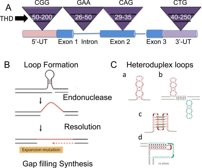
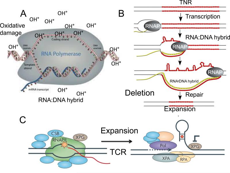
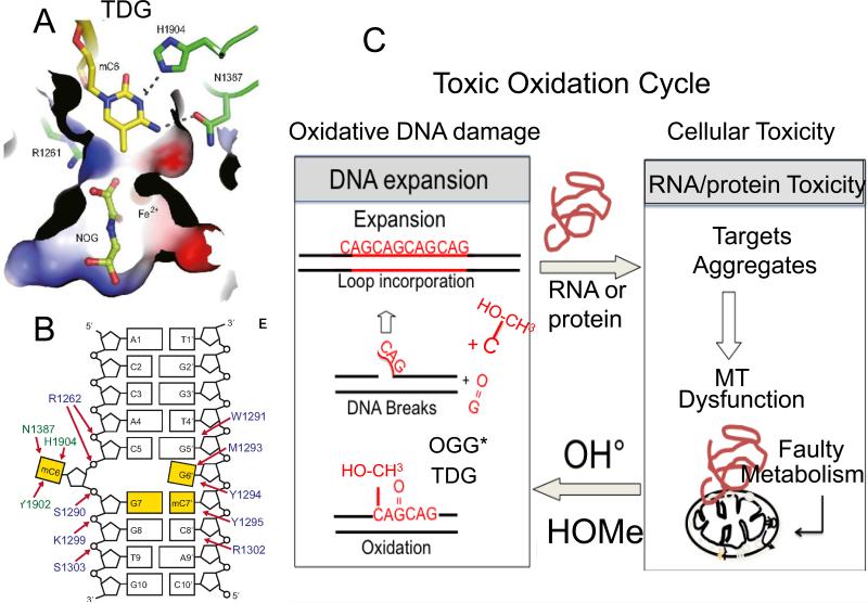
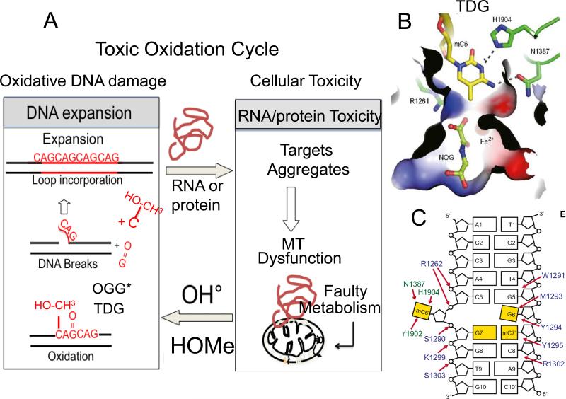
Trinucleotide repeats (TNRs) expansion disorders are severe neurodegenerative and neuromuscular disorders that arise from inheriting a long tract (30-50 copies) of a trinucleotide unit within or near an expressed gene (Figure 1a). The mutation is referred to as 'trinucleotide expansion' since the number of triplet units in a mutated gene is greater than the number found in the normal gene. Expansion becomes obvious once the number of repeating units passes a critical threshold length, but what happens at the threshold to render the repeating tract unstable? Here we discuss DNA-dependent and RNA-dependent models by which a particular DNA length permits a rapid transition to an unstable state.


Figures

Figure 1
Figure 1. Features of expansion and its threshold
(A) Generic representation of threshold limits for some representative disease alleles from distinct TNR disorders. The Inverted purple triangles represent the size ranges associated with the normal, threshold, and disease length TNR alleles. In white is the threshold length for representative TNRs: CGG in the 5’-untranslated (5’UT) region characterizes the FMR-1 gene; GAA in an intron (lines) characterizes the Friedreich's ataxia gene; CAG in a coding region (exon) characterizes the Huntington's gene; CTG in the 3’-untranslated (3’UT) region characterizes the Myotonic dystrophy 1 (DM1) gene. The threshold limit is also referred to as the premutation length, as all full mutations arise from lengths at the upper range of normal allele and the lower edge of disease allele lengths. Below that range are stable normal repeats, and above the ranges at which expansion exists. The premutation lengths as shown are approximate sizes since there is no precise range. (B) The three most basic steps of expansion. (C) Distinct types of heteroduplex DNA loops are proposed as precursors to expansion: hairpins (a); cruciform (b); quadraplex (c); H-DNA triplet helix (d). THD is threshold. (C) is taken from Mirkin SM: Nature 2007, 447: 932-40.
Figure 2
Figure 2. (A) A schematic representation of an RNA-DNA hybrid and its role in hypothetical mechanisms in TNR expansion during transcription
(A) In the FMR1 gene, the RNA-DNA hybrid is proposed to form at the promoter region including the 5’ untranslated region harboring CGG repeat. The paused RNA polymerase stabilizes the open transcription bubble, which is susceptible to oxidative DNA damage. A possible model for expansion is oxidative stress and base damage which is removed during BER [, ]. (B) TCR may also play a role by removing the RNA-DNA hybrid. At or below the threshold, the stalled polymerase may successfully recruit the TCR machinery or utilize it in some way to remove the RNA-DNA hybrid. If XPG is inhibited, a flap structure folds-back to form a structural intermediated for expansion. (B) A possible model for TNR deletion at the RNA-DNA hybrid site. The open non-transcribed strand forms self-paired DNA loops along the transcription bubble as polymerase passes [,]. These loops are most likely to result in deletion after subsequent removal, although expansion is possible if an endonuclease is able to clip the hairpin loops on the unpaired DNA strand, and the TNR duplex is restored by gap filling synthesis [,].
Figure 3
Figure 3. The structural models of TDP-43 and MBNL bound to nucleic acids are similar
(A) TDP-43 binds to both DNA and to RNA. The small angle X-ray scattering (SAXS) envelope of TDP-43 dimer is fitted with the crystal structure of RNA recognition motif (hRRM1)-DNA and mRRM2-DNA in the orientation that the DNA forms a continuous 5′–3′ strand, as it is bound from hRRM1 to mRRM2 in TDP-43. (B) SAX structure of a TDP-43 monomer bound to ssDNA. The putative RNA binding sequence of TDP-43 is derived from the sequence of two tandem RNA recognition motifs (RRM), hRRM1-DNA and mRRM2-DNA complexes. The RRMs in TDP-43 dimers participate in binding of UG-rich RNA or TG-rich DNA with RRM1 playing a dominant role and RRM2 playing a supporting role. The RRM1 binds to RNA with extensive contacts with the conserved β-sheet residues and loop residues. (C) Schematic representation of the domains structure of TDP-43 homodimer, which binds to a long UG-rich RNA via its RRM1 and RRM2 domains. (D) Ribbon diagram presentation of zinc finger domain of MBNL2, with the side chains of the ligand residues and zinc ions (Gray balls) and binds to a GU-rich sequence (E). (F) Like TDP-43, MBNL2 is also capable of forming a dimer. Each circle represents a CCCH-type zinc finger motif, and the black arrows represent the zinc finger orientation corresponding to the direction of the first helix in each of the domains. The red arrows indicate the 5′–3′ direction of the two accommodated RNA on the TZF12 of the MBNL2 dimer.
Figure 4
Figure 4. The Methylation cycle and its metabolic regulation
(A) α-ketoglutarate (α KG) is a critical co-factor for the Ten-eleven translocation dioxygenase (TET) which oxidizes 5-methylcytosine. TET regulates both DNA and histone methylation, and EGLN1 gene product, hypoxia-inducible factor prolyl hydroxylase 2 (HIF-PH2), or prolyl hydroxylase domain-containing protein 2 (PHD2), is an enzyme encoded by the EGLN1 gene. It is also known as Egl nine homolog 1.The EGLN1 gene is responsible for ubiquitin-proteasome degradation pathway by hydroxylation of proline-564 and proline-402 by PHD2. (B) The methylation cycle regulated by TET. The action of DNA demethytransferases (DNMTs) converts cytosine (C) for 5’ methyl cytosine. The TET enzymes oxidize 5’ methyC to 5’-hydroxylmethyC and 5’formylC. The 5’-hydroxylmethyC is removed by the BER enzyme, TDG, to restore C, which is subsequently methylated. 5’formylC is caboxylated by an unknown enzyme to restore C, which is subsequently methylated. Success of this cycle maintains a balance of methylation in the genome. The dotted line indicates the removal of 5-hydrolymethylcytosine and restoration to cytosine to re-set the cycle.
Figure 5
Figure 5. Two-state threshold model
(A) We propose a two-state model for expansion. Expansion arises from toxicity imparted from RNA and protein-mediated toxicity by two mechanisms. (1) The toxic oxidation cycle. RNA and protein-mediated toxicity induces mitochondrial stress and a concomitant increase in oxidative damage to DNA. The oxidative damage is removed by DNA glycolsylases. 8-oxo-guanine glycolsylase (OGG1)* is a major enzyme that removes oxidative damage, but it can also be removed by the NEILS 1* glycosylase, or the machinery of TCR*. Single strand break intermediates arise during base removal and produce flaps that fold-back to generate structural intermediates for expansion. (2) The methylation cycle. When stress overwhelms the capacity of TET dioxygenase to hydroxymethylate hemimethylated DNA in the affected region, hypermethylation will result. TET activity induces elevated hydroxymethyl cytosine (OH-CH3-cytosine), which is removed by TDG and creates the flap intermediate. Single strand break intermediates arise during base removal and produce flaps that foldback to generate structural intermediates for expansion. However, there is no direct evidence that TDG induces expansion. The star indicates that more than one enzyme can remove the oxidative DNA damage. (B) The binding pocket of the OH-CH3-cytosine in TDG. (C) The overall cartoon structure of the OH-CH3-cytosine-TDG complex.

Similar articles

Cited by

References

    1. Nelson DL, Orr HT, Warren ST. The unstable repeats--three evolving faces of neurological disease. Neuron. 2013;77:825–43. - PMC - PubMed
    1. McMurray CT. Mechanisms of trinucleotide repeat instability during human development. Nat Rev Genet. 2010;11:786–99. [A comprehensive review of recent progress in linking the features of human disease with the replication and repair-dependent mechanisms of expansion and how they are used during different stages of development.] - PMC - PubMed
    1. La Spada AR, Taylor JP. Repeat expansion disease: progress and puzzles in disease pathogenesis. Nat Rev Genet. 2010;11:247–58. [A comprehensive review of recent progress in understanding the pathophysiology of expansion disease.] - PMC - PubMed
    1. Dion V, Wilson JH. Instability and chromatin structure of expanded trinucleotide repeats. Trends Genet. 2009;25:288–97. [A comprehensive review about the chromatin aspects of triplet expansion.] - PMC - PubMed
    1. Mirkin SM. Expandable DNA repeats and human disease. Nature. 2007;447:932–40. [An excellent review about the replication and repair-dependent mechanisms of triplet expansion.] - PubMed

Publication types