Characterization of programmed death-1 homologue-1 (PD-1H) expression and function in normal and HIV infected individuals
- PMID: 25279955
- PMCID: PMC4184823
- DOI: 10.1371/journal.pone.0109103
Characterization of programmed death-1 homologue-1 (PD-1H) expression and function in normal and HIV infected individuals
Abstract
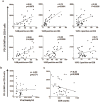
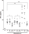
Chronic immune activation that persists despite anti-retroviral therapy (ART) is the strongest predictor of disease progression in HIV infection. Monocyte/macrophages in HIV-infected individuals are known to spontaneously secrete cytokines, although neither the mechanism nor the molecules involved are known. Here we show that overexpression of the newly described co-stimulatory molecule, PD1 homologue (PD-1H) in human monocyte/macrophages is sufficient to induce spontaneous secretion of multiple cytokines. The process requires signaling via PD-1H as cytokine secretion could be abrogated by deletion of the cytoplasmic domain. Such overexpression of PD-1H, associated with spontaneous cytokine expression is seen in monocytes from chronically HIV-infected individuals and this correlates with immune activation and CD4 depletion, but not viral load. Moreover, antigen presentation by PD-1H-overexpressing monocytes results in enhanced cytokine secretion by HIV-specific T cells. These results suggest that PD-1H might play a crucial role in modulating immune activation and immune response in HIV infection.
Conflict of interest statement
Figures


Similar articles
-
HIV-1 Tat Protein Induces Production of Proinflammatory Cytokines by Human Dendritic Cells and Monocytes/Macrophages through Engagement of TLR4-MD2-CD14 Complex and Activation of NF-κB Pathway.PLoS One. 2015 Jun 19;10(6):e0129425. doi: 10.1371/journal.pone.0129425. eCollection 2015. PLoS One. 2015. PMID: 26090662 Free PMC article.
-
Foxp3+Helios+ regulatory T cells are associated with monocyte subsets and their PD-1 expression during acute HIV-1 infection.BMC Immunol. 2019 Oct 24;20(1):38. doi: 10.1186/s12865-019-0319-7. BMC Immunol. 2019. PMID: 31651258 Free PMC article.
-
Monocyte Based Correlates of Immune Activation and Viremia in HIV-Infected Long-Term Non-Progressors.Front Immunol. 2019 Dec 6;10:2849. doi: 10.3389/fimmu.2019.02849. eCollection 2019. Front Immunol. 2019. PMID: 31867010 Free PMC article.
-
Fighting with the enemy's weapons? the role of costimulatory molecules in HIV.Curr Mol Med. 2011 Apr;11(3):172-96. doi: 10.2174/156652411795243397. Curr Mol Med. 2011. PMID: 21375494 Review.
-
Monocyte mobilization, activation markers, and unique macrophage populations in the brain: observations from SIV infected monkeys are informative with regard to pathogenic mechanisms of HIV infection in humans.J Neuroimmune Pharmacol. 2012 Jun;7(2):363-71. doi: 10.1007/s11481-011-9330-3. Epub 2011 Dec 14. J Neuroimmune Pharmacol. 2012. PMID: 22167311 Review.
Cited by
-
VISTA as a prospective immune checkpoint in gynecological malignant tumors: A review of the literature.Open Med (Wars). 2023 Dec 11;18(1):20230866. doi: 10.1515/med-2023-0866. eCollection 2023. Open Med (Wars). 2023. PMID: 38152334 Free PMC article. Review.
-
VISTA expression and patient selection for immune-based anticancer therapy.Front Immunol. 2023 Feb 20;14:1086102. doi: 10.3389/fimmu.2023.1086102. eCollection 2023. Front Immunol. 2023. PMID: 36891296 Free PMC article. Review.
-
Ablation of T cell-associated PD-1H enhances functionality and promotes adoptive immunotherapy.JCI Insight. 2022 Jan 25;7(2):e148247. doi: 10.1172/jci.insight.148247. JCI Insight. 2022. PMID: 34905507 Free PMC article.
-
Beyond CTLA-4 and PD-1, the Generation Z of Negative Checkpoint Regulators.Front Immunol. 2015 Aug 21;6:418. doi: 10.3389/fimmu.2015.00418. eCollection 2015. Front Immunol. 2015. PMID: 26347741 Free PMC article. Review.
-
A New VISTA on combination therapy for negative checkpoint regulator blockade.J Immunother Cancer. 2016 Dec 20;4:86. doi: 10.1186/s40425-016-0190-5. eCollection 2016. J Immunother Cancer. 2016. PMID: 28031817 Free PMC article. Review.
References
-
- Ariffin JK, Sweet MJ (2013) Differences in the repertoire, regulation and function of Toll-like Receptors and inflammasome-forming Nod-like Receptors between human and mouse. Curr Opin Microbiol 16: 303–310. - PubMed
-
- Cohen OJ, Kinter A, Fauci AS (1997) Host factors in the pathogenesis of HIV disease. Immunol Rev 159: 31–48. - PubMed
Publication types
MeSH terms
Substances
Grants and funding
LinkOut - more resources
Full Text Sources
Other Literature Sources
Medical
Molecular Biology Databases
Research Materials

