. 2015 Apr;26(4):896-906.
doi: 10.1681/ASN.2014020195. Epub 2014 Sep 29.

Vascular-resident CD169-positive monocytes and macrophages control neutrophil accumulation in the kidney with ischemia-reperfusion injury

Affiliations

Vascular-resident CD169-positive monocytes and macrophages control neutrophil accumulation in the kidney with ischemia-reperfusion injury

Kazunori Karasawa et al. J Am Soc Nephrol. 2015 Apr.

Abstract

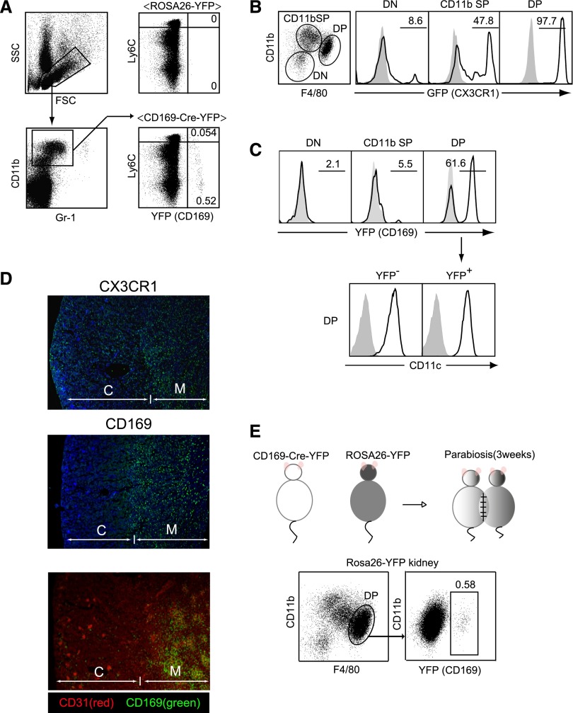
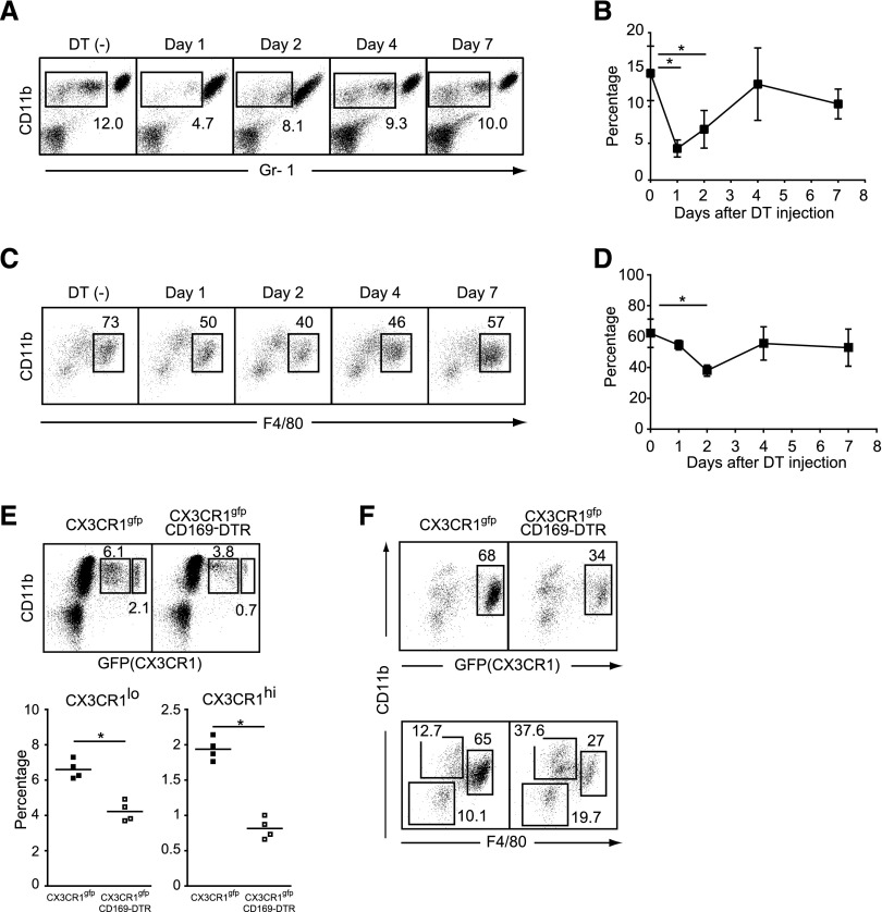
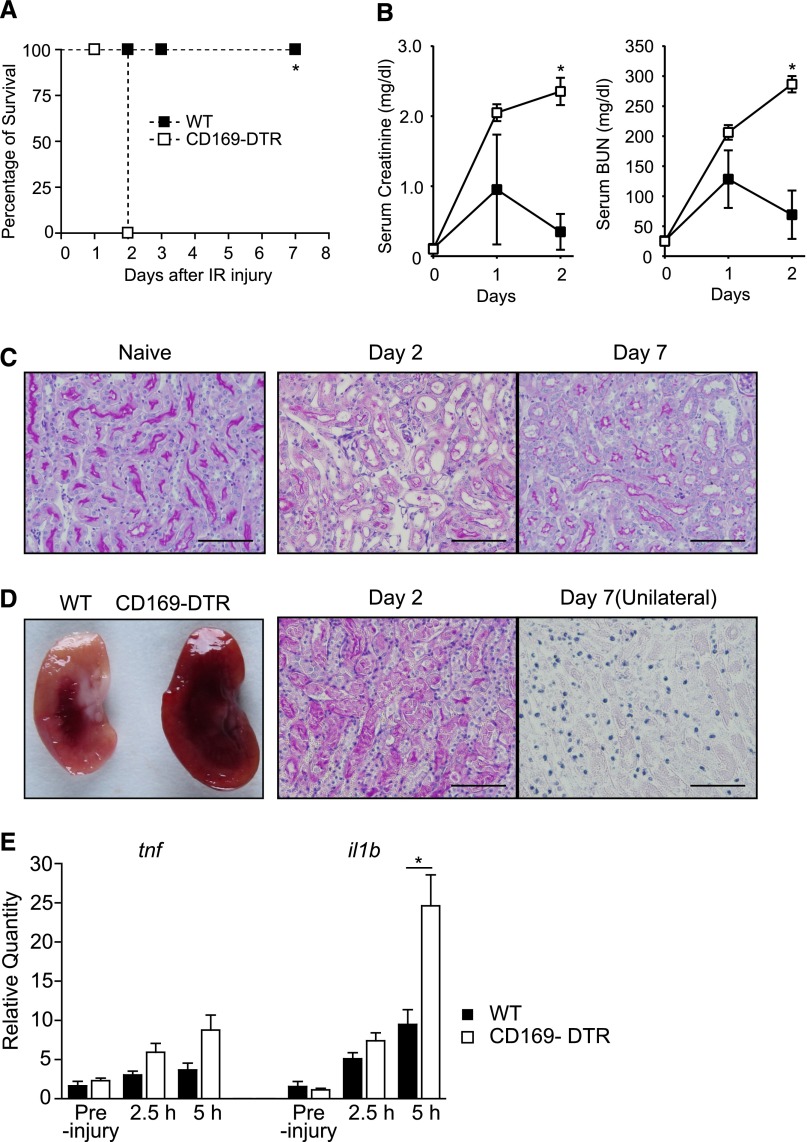
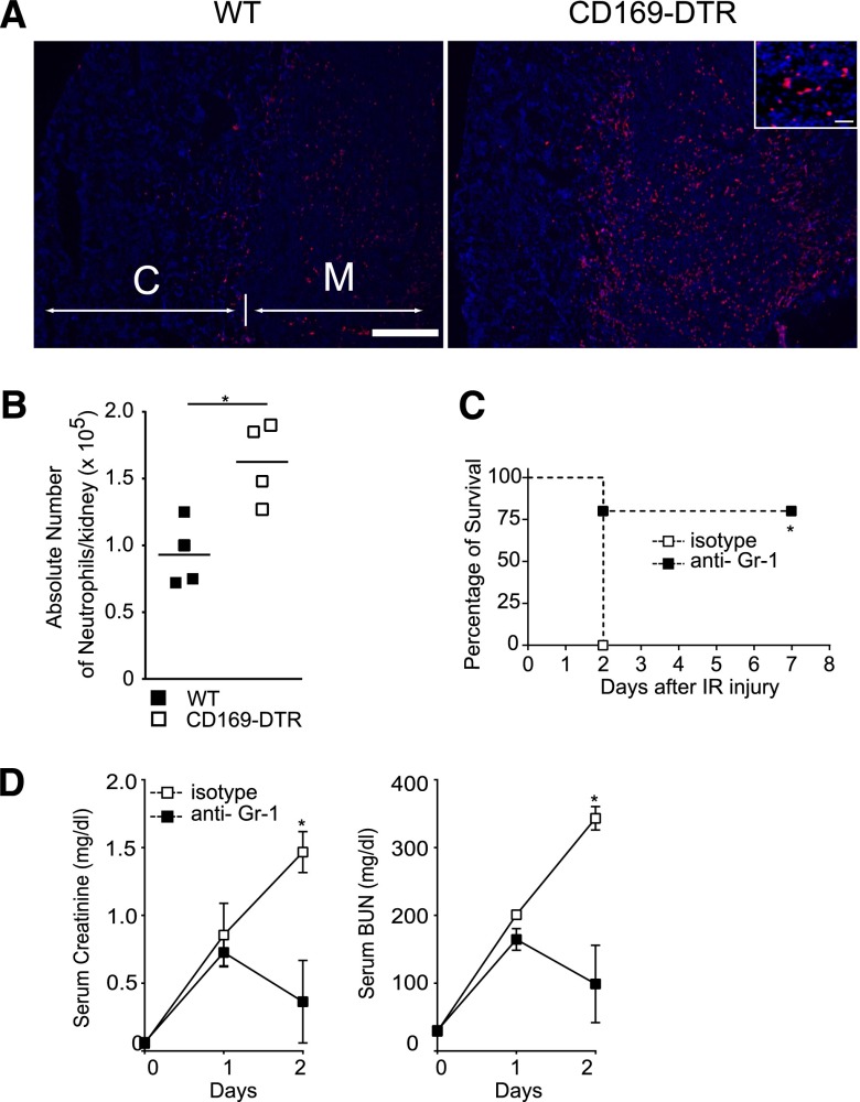
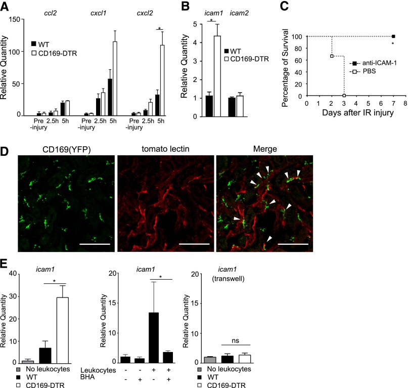
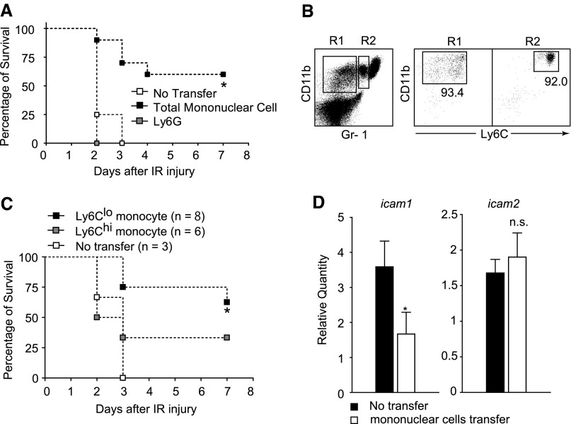
Monocytes and kidney-resident macrophages are considered to be involved in the pathogenesis of renal ischemia-reperfusion injury (IRI). Several subsets of monocytes and macrophages are localized in the injured tissue, but the pathologic roles of these cells are not fully understood. Here, we show that CD169(+) monocytes and macrophages have a critical role in preventing excessive inflammation in IRI by downregulating intercellular adhesion molecule-1 (ICAM-1) expression on vascular endothelial cells. Mice depleted of CD169(+) cells showed enhanced endothelial ICAM-1 expression and developed irreversible renal damage associated with infiltration of a large number of neutrophils. The perivascular localization of CD169(+) monocytes and macrophages indicated direct interaction with blood vessels, and coculture experiments showed that the direct interaction of CD169(+) cell-depleted peripheral blood leukocytes augments the expression levels of ICAM-1 on endothelial cells. Notably, the transfer of Ly6C(lo) monocytes into CD169(+) cell-depleted mice rescued the mice from lethal renal injury and normalized renal ICAM-1 expression levels, indicating that the Ly6C(lo) subset of CD169(+) monocytes has a major role in the regulation of inflammation. Our findings highlight the previously unknown role of CD169(+) monocytes and macrophages in the maintenance of vascular homeostasis and provide new approaches to the treatment of renal IRI.

Keywords: acute renal failure; immunology and pathology; ischemia-reperfusion; macrophages.


Figures

Figure 1.
Figure 1.
Identification of novel subsets of monocytes and kidney macrophages using CD169-Cre-YFP mice. (A) YFP-positive cells in peripheral blood. White blood cells obtained from CD169-Cre-YFP mice or ROSA26-YFP mice were stained for CD11b, Gr-1, and Ly6C. Numbers indicate the frequency (percentage) of YFP+ cells among the Gr-1int-loCD11b+ fraction. Data are representative of three independent experiments. FSC, forward scatter; SSC, side scatter. (B and C) Kidney myeloid cells in (B) CX3CR1-GFP mice and (C) CD169-Cre-YFP mice. CD11b+ and/or CD11c+ cells in kidneys were stained with Gr-1, CD11b, F4/80, and CD11c antibodies. Numbers indicate the frequency (percentage) of GFP+ or YFP+ cells in CD11b and F4/80 double-negative (DN), CD11b single-positive (SP), or CD11b and F4/80 double-positive (DP) cells. (C, lower panel) CD11c expression in DP cells in CD169-Cre-YFP mice. Shaded area represents renal cells from (B) WT or (C, upper panel) ROSA26-YFP mice or (C, lower panel) staining without CD11c. Experiments were independently repeated at least three times. (D) Localization of renal CX3CR1+ macrophages and CD169+ macrophages. (Top and middle panels) Cryosections of kidneys from (top panel) CX3CR1-GFP and (middle panel) CD169-Cre-YFP mice were stained with anti-GFP antibody (green) and 4′,6-diamidino-2-phenylindole (blue). (Bottom panel) Cryosections of kidneys from CD169-Cre-YFP mice were stained with anti-GFP antibody (green) and CD31 (clone: MEC13.3; red). Note that the glomeruli in the cortex are densely stained with the anti-CD31 Ab, a marker of vascular endothelial cells. C, cortex; M, medulla. (E) CD11b+, F4/80+ kidney macrophages are derived from blood monocytes. Parabiotic mice were generated from CD169-Cre-YFP and ROSA26-YFP mice. Three weeks later, the frequency of YFP+ cells in the DP macrophages in ROSA26-YFP mouse kidneys was analyzed. The numbers indicates the frequency (percentage) of YFP+ cells among DP macrophages.
Figure 2.
Figure 2.
Selective depletion of monocytes and kidney macrophages in CD169-DTR mice. (A) White blood cells of DT-treated CD169-DTR mice at the indicated time points were stained with CD11b and Gr-1 antibodies. Numbers indicate the frequency (percentage) of CD11b+Gr-1int-lo monocytes among white blood cells. Average percentages of CD11b+Gr-1int-lo monocytes (three to five mice per group) are plotted with SD in B. Mean percentage of CD11b+Gr-1int-lo cells was compared by one-way ANOVA. *P<0.05. (C) Kidney macrophages of DT-treated CD169-DTR mice at the indicated time points were stained for Gr-1, CD11b, and F4/80 antibodies. Numbers indicate the frequency (percentage) of CD11b+F4/80+ kidney macrophages among CD11b+ and/or CD11c+ cells in kidneys. Average percentages of CD11b+, F4/80+ kidney macrophages (four mice per group) are plotted with SD in D. Mean percentage of CD11b+F4/80+ cells was compared by one-way ANOVA. *P<0.05. (E) Depletion of monocytes in CX3CR1-GFP×CD169-DTR mice. DT was injected into CX3CR1-GFP mice (upper left panel) or CX3CR1-GFP×CD169-DTR mice (upper right panel). Twenty-four hours later, white blood cells were stained for CD11b. Numbers indicate the frequency (percentage) of CX3CR1lo or CX3CR1hi cells among white blood cells. (Lower panel) Average percentages of CX3CR1lo and CX3CR1hi cells are plotted. ▪, CX3CR1-GFP; □, CX3CR1-GFP × CD169-DTR. *P<0.05. (F) Depletion of kidney macrophages in CX3CR1-GFP × CD169-DTR mice. DT was injected into (left panel) CX3CR1-GFP or (right panel) CX3CR1-GFP×CD169-DTR mice. Two days after the DT injection, kidney macrophages were stained for CD11b and F4/80. Numbers indicate (upper panel) the frequency (percentage) of CX3CR1-positive cells among CD11b+ and/or CD11c+ cells in kidneys or (lower panel) the frequency (percentage) of CD11b and F4/80 double-negative, CD11b single-positive, or CD11b and F4/80 double-positive cells among CD11b+ and/or CD11c+ cells in kidneys. Data are representative of three independent experiments.
Figure 3.
Figure 3.
Severe IRI in CD169-DTR mice. (A and B) DT was intraperitoneally injected into WT or CD169-DTR mice. Thirty-six hours later, clamping of the unilateral renal pedicle for 1 hour followed by contralateral nephrectomy (bilateral IR) was performed in the mice. (A) Survival rate of IR mice. n=6 WT mice or n=7 CD169-DTR mice. *P< 0.001. Data are representative of three independent experiments. (B) Serum creatinine and BUN concentrations in IR mice. The average values of five WT or three CD169-DTR mice are plotted with SD. ▪, WT; □, CD169-DTR. Mean serum creatinine and BUN concentrations of WT and CD169-DTR mice at different time points were compared by two-way ANOVA. *P<0.05. (C) Microscopic observation of naïve and IR kidneys in WT mice. Bilateral IR was performed in WT mice. Paraffin sections were stained with periodic acid–Schiff (PAS). Scale bars, 50 μm. (D) Macroscopic and microscopic observations of IR kidneys. Bilateral IR was performed in WT or CD169-DTR mice as described in A and B. (Left panel) Macroscopic observation of kidneys on day 1 after IR. (Center panel) Bilateral or (Right panel) unilateral IR was performed in the DT-treated CD169-DTR mice. Paraffin sections of kidneys on (center panel) day 2 or (right panel) day 7 (unilateral) were stained with PAS. Unilateral IR was performed in CD169-DTR mice to avoid death after bilateral IR in those mice. Scale bars, 50 μm. (E) Inflammatory cytokine mRNA levels in kidneys. Bilateral IR was performed in WT or CD169-DTR mice as described in A and B. Quantity of tnfa and il1b mRNA levels in the kidneys relative to WT preinjury mice at the indicated time points after IR were determined by quantitative PCR. Mean±SD, n=5 mice/group. *P<0.05.
Figure 4.
Figure 4.
Depletion of neutrophils rescues CD169-DTR mice from lethal AKI. (A and B) Large numbers of neutrophils accumulated in kidneys of CD169-DTR mice subsequent to IR. (A) Bilateral IR was performed in DT-treated WT or CD169-DTR mice. Neutrophils in the kidneys on day 1 were detected immunohistochemically by anti-Ly6G (red) and 4′,6-diamidino-2-phenylindole (blue) staining. C, cortex; M, medulla. Scale bars, 1 mm in low power fields; 50 μm in Inset. (B) Absolute numbers of CD11b+, Gr-1+ neutrophils were quantitated by a flow cytometer. Each symbol represents data from an individual animal, and the bars indicate average values. *P<0.05. (C and D) Injection of anti–Gr-1 antibody suppresses IR-induced AKI in CD169-DTR mice. DT was injected intraperitoneally into CD169-DTR mice. Twenty-four hours later, 100 μg anti–Gr-1 antibody or rat IgG2B isotype control was injected intravenously into these mice. Bilateral IR was performed 36 hours after DT injection. (C) Survival rate of IR mice. n=5 mice/each group. *P<0.05. (D) Serum creatinine and BUN concentrations. The average values of six anti–Gr-1–injected mice or three isotype antibody-injected mice are plotted with SD. Mean serum creatinine and BUN concentrations of WT and CD169-DTR mice at different time points were compared by two-way ANOVA. *P<0.05.
Figure 5.
Figure 5.
Upregulated ICAM-1 expression in the absence of CD169+ cells exacerbates AKI. (A) Chemokine expression in IR kidneys. DT was injected intraperitoneally into WT or CD169-DTR mice. Thirty-six hours later, bilateral IR was performed. Quantities of ccl2, cxcl1, and cxcl2 mRNA levels in the kidneys relative to WT preinjury mice were determined by quantitative PCR. n=5 mice/group. Mean±SD. *P<0.05. (B) Enhanced expression of ICAM-1 in kidneys of CD169-DTR mice. Quantity of icam1 and icam2 mRNA levels in the kidneys of DT-treated WT or CD169-DTR mice relative to WT mice without DT were determined by quantitative RT-PCR. n=8 mice/each group. The average values are plotted with SD. *P<0.05. (C) Blockade of ICAM-1 suppresses the development of AKI in CD169-DTR mice. DT was injected intraperitoneally into CD169-DTR mice. Twenty-four hours later, 100 μg anti–ICAM-1 (clone KAT1) antibody or PBS was injected intravenously into these mice. Thirty-six hours after the DT injection, bilateral IR was performed. Survival rate is shown. n=3 mice/group. *P<0.05. (D) CD169+ cells localize in the perivascular area of the renal interstitium. DyLight 488-labeled tomato lectin was injected intravenously into CD169-Cre-YFP mice 5 minutes before removing the kidney. Cryosections of the kidneys were stained for YFP (green) and observed under a confocal microscope. White arrows indicate the CD169-positive macrophages, which are located along the capillary vessels. z-Stack images were processed by ImageJ software. Red signal indicates tomato lectin on endothelium. Scale bar, 100 μm. (E) Peripheral blood leukocytes from CD169-DTR mice augment ICAM-1 expression levels in cultured endothelial cells. (Left panel) Peripheral blood leukocytes from DT-treated WT or CD169-DTR mice were cocultured with UV♀2 cells for 18 hours. icam1 mRNA levels of these cells were determined by quantitative RT-PCR. (Center panel) Peripheral blood leukocytes from DT-treated CD169-DTR mice were cocultured with UV♀2 cells for 18 hours in the presence or absence of butylated hydroxyanisole (BHA). (Right panel) Peripheral blood leukocytes from DT-treated WT or DT-treated CD169-DTR mice were cocultured with UV♀2 cells for 18 hours using the transwell plate. The average values are plotted with SD. Data are representative of two or three independent experiments. *P<0.05.
Figure 6.
Figure 6.
Transfer of Ly6Clo monocytes rescues CD169-DTR mice from lethal AKI. (A) Transfer of mononuclear blood cells rescues CD169-DTR mice from lethal IRI. Gr-1 antibody (100 μg) was injected intraperitoneally into WT mice to deplete neutrophils. Residual leukocytes from these mice were injected into CD169-DTR mice (▪). Ly6Ghi neutrophils were enriched from WT mice by magnetic sorting and injected into CD169-DTR mice (gray square). Twenty-four hours later, DT was injected intraperitoneally into the mice. Bilateral IR was performed 36 hours after DT injection. Survival rate is shown. n=4 mice without transfer, n=10 mice with monocyte transfer, and n=3 mice with Ly6G+ neutrophils transfer. *P<0.05. (B and C) Ly6Clo monocytes are primarily responsible for the suppression of IR-induced AKI. (B) Ly6Clo (n=8) or Ly6Chi (n=6) monocytes prepared from WT mice by using a cell sorter were injected intravenously into CD169-DTR mice. Twenty-four hours later, DT was injected intraperitoneally into the mice. Bilateral IR was performed 36 hours after DT injection. (C) Survival rate of IR mice. *P<0.05 compared with no transfer group. (D) Transfer of mononuclear cells suppresses the augmented expression of ICAM-1 in the kidneys of CD169-DTR mice. Transfer of mononuclear cells into CD169-DTR mice was performed as described in A. Twenty-four hours later, DT was injected intraperitoneally into the mice. Thirty-six hours later, ICAM-1 mRNA levels relative to those of WT kidneys were determined by quantitative RT-PCR. n=6 mice/group. The average values are plotted with SD. *P<0.05.

Comment in

Similar articles

Cited by

References

    1. Carden DL, Granger DN: Pathophysiology of ischaemia-reperfusion injury. J Pathol 190: 255–266, 2000 - PubMed
    1. Bonventre JV, Yang L: Cellular pathophysiology of ischemic acute kidney injury. J Clin Invest 121: 4210–4221, 2011 - PMC - PubMed
    1. Eltzschig HK, Eckle T: Ischemia and reperfusion—from mechanism to translation. Nat Med 17: 1391–1401, 2011 - PMC - PubMed
    1. Ricardo SD, van Goor H, Eddy AA: Macrophage diversity in renal injury and repair. J Clin Invest 118: 3522–3530, 2008 - PMC - PubMed
    1. Day YJ, Huang L, Ye H, Linden J, Okusa MD: Renal ischemia-reperfusion injury and adenosine 2A receptor-mediated tissue protection: Role of macrophages. Am J Physiol Renal Physiol 288: F722–F731, 2005 - PubMed

Publication types

MeSH terms

Substances

LinkOut - more resources