. 2014 Nov 1;193(9):4558-67.
doi: 10.4049/jimmunol.1401372. Epub 2014 Sep 26.

miR-15a/16 regulates macrophage phagocytosis after bacterial infection

Affiliations

miR-15a/16 regulates macrophage phagocytosis after bacterial infection

Hyung-Geun Moon et al. J Immunol. .

Abstract

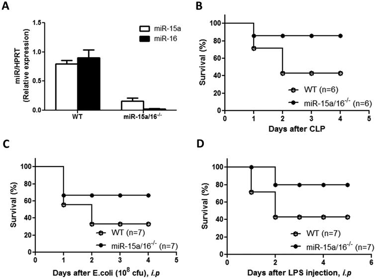
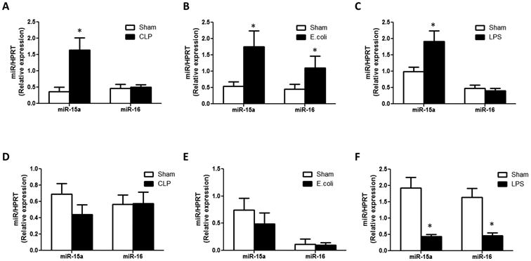
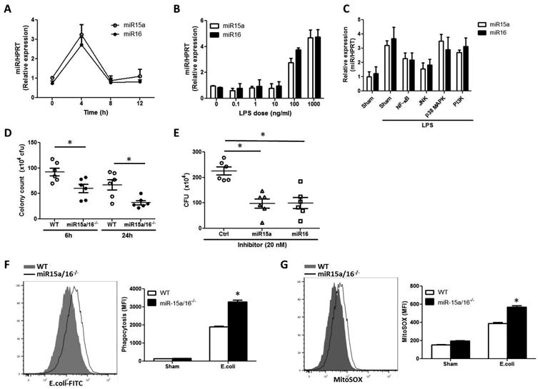
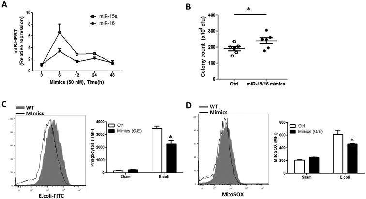
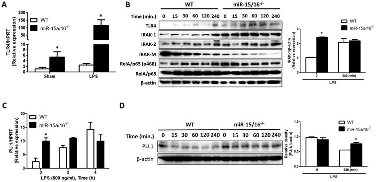
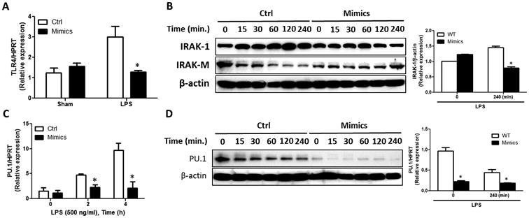
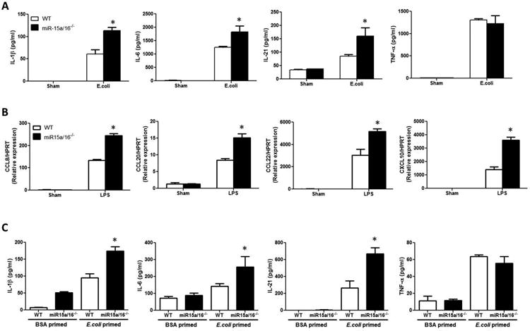
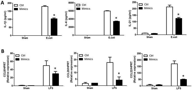
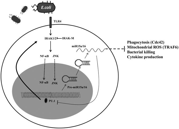
Bacterial infection and its associated sepsis are devastating clinical entities that lead to high mortality and morbidity in critically ill patients. Phagocytosis, along with other innate immune responses, exerts crucial impacts on the outcomes of these patients. MicroRNAs (miRNAs) are a novel class of regulatory noncoding RNAs that target specific mRNAs for modulation of translation and expression of a targeted protein. The roles of miRNAs in host defense against bacterial sepsis remain unclear. We found that bacterial infections and/or bacterial-derived LPS enhanced the level of miR-15a/16 in bone marrow-derived macrophages (BMDMs). Deletion of miR-15a/16 (miR-15a/16(-/-)) in myeloid cells significantly decreased the bacterial infection-associated mortality in sepsis mouse models. Moreover, miR-15a/16 deficiency (miR-15a/16(-/-)) resulted in augmented phagocytosis and generation of mitochondrial reactive oxygen species in BMDMs. Supportively, overexpression of miR-15a/16 using miRNA mimics led to decreased phagocytosis and decreased generation of mitochondrial reactive oxygen species. Mechanistically, deletion of miR-15a/16 upregulated the expression of TLR4 via targeting the principle transcriptional regulator PU.1 locating on the promoter region of TLR4, and further modulated the downstream signaling molecules of TLR4, including Rho GTPase Cdc 42 and TRAF6. In addition, deficiency of miR-15a/16 also facilitated TLR4-mediated proinflammatory cytokine/chemokine release from BMDMs at the initial phase of infections. Taken together, miR-15a/16 altered phagocytosis and bacterial clearance by targeting, at least partially, on the TLR4-associated pathways, subsequently affecting the survival of septic mice.


Figures

Figure 1
Figure 1. Deletion of miR-15a/16 conferred protective effects on sepsis using mouse models
(A) Basal level of miR-15a/16 expression in BMDMs isolated from WT and miR-15a/16-/- mice. miR-15a/16 was normalized using HPRT. (B)-(D) Survival of WT mice or mononuclear cell specific Cre-miR-15a/16-/- mice after CLP (B) E.coli (108 cfu/mouse) injection intraperitoneally (C) and LPS (25 mg/kg) injection intraperitoneally (D)
Figure 2
Figure 2. Expression of miR-15a and miR-16 in mouse tissue and cells after bacterial infection
(A)-(C): Expression of miR-15a and miR-16 in mouse lung tissue after CLP (A), E.coli injection i.p. (B) and LPS injection i.p. (C) (D)-(F): Expression of miR-15a and miR-16 in mouse spleen tissue after CLP (D), E.coli injection i.p. (E), and LPS injection i.p. (F) All figures represent two independent experiments with identical results. * p<0.05
Figure 3
Figure 3. Deletion of miR-15a/16 promoted BMDM-mediated phagocytosis
(A) Expression of miR-15a and miR-16 after LPS (500 ng/ml) in mouse BMDM (B) Dose-dependent responses of miR-15a/16 expression in mouse BMDMs after LPS treatment (C) BMDMs were pre-treated with designated pathway inhibitors, followed by LPS. Expression of miR-15a or miR-16 were determined (D) E.coli-DH5a (107 cfu) was added to the WT and miR-15a/16-/- BMDM (1×10⁶). After mixing, the cell culture supernatant (10 μl) was taken at 6h & 24h and sub-cultured in LB plates. Colony counts were determined.(E) Inhibitors of miR-15a or miR-16 were transfected into mouse BMDMs. After 24h, E.coli-DH5a (107 cfu) was added to the BMDMs. 10 μl mixture was taken at 6h and colony counts were performed on LB plate.(F) Phagocytosis of FITC-labeled E.coli particle (20 μg/ml) in WT and miR-15a/16-/- BMDMs. FITC was assayed using flow cytometer(G) Expression of Rho GTPase Cdc42 after LPS (time course) in WT and miR-15a/16-/- BMDMs (H) Mitochondrial ROS (MitoSOX) was measured 6h after E.coli-FITC stimulation (I) Expression of TRAF6 after LPS (time course) in WT and miR-15a/16-/- BMDMs All figures represent 3 independent experiments with identical results. * p<0.05
Figure 4
Figure 4. Overexpression of miR-15a/16 inhibited BMDM-mediated phagocytosis
Mimics of miR-15a and miR-16 (50 nM each) were co-transfected into the BMDMs. (A) Expression of miR-15a and miR-16 in the BMDMs after mimics transfection. (B) E.coli-DH5a (107 cfu) was added to the WT and miR-15a/16 over-expressing BMDMs (1×10⁶) and colony counts was determined after 6h (C). Phagocytosis of FITC-labeled E.coli particle (20 μg/ml) in WT and miR-15a/16 over-expressing BMDMs. FITC was assayed using flow cytometer. (D) Mitochondrial ROS (MitoSOX) was measured 6h after E.coli-FITC stimulation in WT and miR-15a/16 over-expressing BMDMs. All figures represent 2 independent experiments with identical results. * p<0.05
Figure 5
Figure 5. Deletion of miR-15a/16 up-regulated the TLR4 signaling in BMDMs
(A) TLR4 mRNA level after 4h LPS (500 ng/ml) stimulation in WT and miR-15a/16-/- BMDMs. TLR4 mRNA level was normalized by HPRT. (B) Protein level of TLR4 signaling components after LPS (500 ng/ml) in miR-15a/16-/- BMDMs detected using the Western Blot analysis. WT and miR-15a/16-/- BMDMs were treated with LPS (500 ng/ml) for the designated time points (left panel). IRAK-1 expression was normalized by β-actin (right panel). (C) PU.1 mRNA level after LPS (500 ng/ml) for 2 and 4h, respectively. PU.1 expression level was normalized using HPRT. (D) PU.1 protein level in WT and miR-15a/16-/- BMDMs after LPS stimulation (left panel), analyzed using Western Blot analysis. PU.1 expression was normalized by β-actin (right panel). All figures represent 2 independent experiments with identical results. * p<0.05
Figure 6
Figure 6. Over-expression of miR-15a/16 down-regulated the TLR4 signaling in BMDMs
(A) TLR4 mRNA level 4h after LPS (500 ng/ml) stimulation in WT and miR-15a/16 over-expressing BMDMs. TLR4 expression level was normalized by HPRT. (B) Protein expression of IRAK-1 and IRAK-M in WT and miR-15a/16 over-expressing BMDMs. Cells were treated with LPS (500 ng/ml). After the designated time points, protein levels were analyzed using the Western Blot analysis (left panel). IRAK-1 expression was normalized by β-actin (right panel). (C) PU.1 mRNA level after LPS (500 ng/ml), for 2 and 4h respectively. PU.1 expression level was normalized using HPRT. (D) PU.1 protein level in WT and miR-15a/16 over-expressing BMDMs after LPS stimulation (left panel), analyzed using Western Blot analysis. PU.1 expression was normalized by β-actin (right panel). All figures represent 2 independent experiments with identical results. * p<0.05
Figure 7
Figure 7. Cytokine and chemokine levels in WT or miR-15a/16-/- BMDMs after bacterial infections
(A) WT and miR-15a/16-/- BMDMs were treated with E.coli-FITC particles (20 μg/ml). After 6h, cytokines were measured using ELISA. (B) WT and miR-15a/16-/- BMDMs were treated with LPS (500 ng/ml). After 4h, RNA was isolated. Chemokine gene expressions were determined using real-time PCR and normalized using HPRT. (C) WT and miR-15a/16-/- BMDMs were primed with BSA-FITC or E.coli-FITC; after 4h, cells were harvested and injected into WT mice intra-peritoneally. Peritoneal fluids were collected after additional 24h and the cytokines secreted into the peritoneal fluids were measured using ELISA. (D) WT and TLR4-/- BMDMs were transfected with miR-15a/16 inhibitors, followed by stimulation with LPS (500 ng/ml, 6h). IL-1β and IL-6 were measured using ELISA All figures represent 3 independent experiments with identical results. * p<0.05 copmared to WT-LPS, **p<0.05 compared to LPS-Ctrl
Figure 8
Figure 8. Cytokine and chemokine levels in WT or miR-15a/16 over-expressing BMDMs after bacterial infections
(A) WT and miR-15a/16 over-expressing BMDMs were treated with E.coli-FITC particles (20 μg/ml). After 6h, cytokines were measured using ELISA. (B) WT and miR-15a/16 over-expressing BMDMs were treated with LPS (500 ng/ml). After 4h, RNA was isolated. Chemokine gene expressions were determined using real-time PCR and were normalized by HPRT. All figures represent 3 independent experiments with identical results. * p<0.05
Figure 9
Figure 9. Proposed mechanisms involved in the functions of miR-15a/16 in macrophages
miR-15a/16 down-regulated the expression of TLR4 via targeting PU.1 locating on the promoter region of TLR4 and modulated TLR4 down-stream signaling molecules, including the Rho GTPase Cdc 42 and TRAF6. Subsequently, miR-15a/16 altered phagocytosis and bacterial clearance, as well as cytokine releases in macrophages, via targeting at least partially, on the TLR4-associated pathways.

Similar articles

Cited by

References

    1. Deutschman CS, Tracey KJ. Sepsis: Current Dogma and New Perspectives. Immunity. 2014;40:463–475. - PubMed
    1. Shi C, Pamer EG. Monocyte recruitment during infection and inflammation. Nat Rev Immunol. 2011;11:762–774. - PMC - PubMed
    1. Andrews T, Sullivan KE. Infections in patients with inherited defects in phagocytic function. Clin Microbiol Rev. 2003;16:597–621. - PMC - PubMed
    1. Danikas DD, Karakantza M, Theodorou GL, Sakellaropoulos GC, Gogos CA. Prognostic value of phagocytic activity of neutrophils and monocytes in sepsis. Correlation to CD64 and CD14 antigen expression. Clin Exp Immunol. 2008;154:87–97. - PMC - PubMed
    1. Underhill DM, Goodridge HS. Information processing during phagocytosis. Nat Rev Immunol. 2012;12:492–802. - PMC - PubMed

Publication types

MeSH terms