The STAT5-regulated miR-193b locus restrains mammary stem and progenitor cell activity and alveolar differentiation
- PMID: 25236432
- PMCID: PMC4252501
- DOI: 10.1016/j.ydbio.2014.09.012
The STAT5-regulated miR-193b locus restrains mammary stem and progenitor cell activity and alveolar differentiation
Abstract
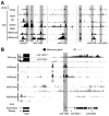
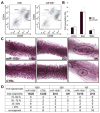
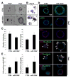
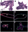
The transcription factor STAT5 mediates prolactin signaling and controls functional development of mammary tissue during pregnancy. This study has identified the miR-193b locus, also encoding miRNAs 365-1 and 6365, as a STAT5 target in mammary epithelium. While the locus was characterized by active histone marks in mammary tissue, STAT5 binding and expression during pregnancy, it was silent in most non-mammary cells. Inactivation of the miR-193b locus in mice resulted in elevated mammary stem/progenitor cell activity as judged by limiting dilution transplantation experiments of primary mammary epithelial cells. Colonies formed by mutant cells were larger and contained more Ki-67 positive cells. Differentiation of mammary epithelium lacking the miR-193b locus was accelerated during puberty and pregnancy, which coincided with the loss of Cav3 and elevated levels of Elf5. Normal colony development was partially obtained upon ectopically expressing Cav3 or upon siRNA-mediated reduction of Elf5 in miR-193b-null primary mammary epithelial cells. This study reveals a previously unknown link between the mammary-defining transcription factor STAT5 and a microRNA cluster in controlling mammary epithelial differentiation and the activity of mammary stem and progenitor cells.
Keywords: Alveoli, Differentiation; Development; Mammary; Micro RNAs; STAT5; Stem cells; miR-193b.
Published by Elsevier Inc.
Figures


Similar articles
-
The miR-17/92 cluster is targeted by STAT5 but dispensable for mammary development.Genesis. 2012 Sep;50(9):665-71. doi: 10.1002/dvg.22023. Epub 2012 Mar 31. Genesis. 2012. PMID: 22389215 Free PMC article.
-
STAT5-regulated microRNA-193b controls haematopoietic stem and progenitor cell expansion by modulating cytokine receptor signalling.Nat Commun. 2015 Nov 25;6:8928. doi: 10.1038/ncomms9928. Nat Commun. 2015. PMID: 26603207 Free PMC article.
-
Loss of EZH2 results in precocious mammary gland development and activation of STAT5-dependent genes.Nucleic Acids Res. 2015 Oct 15;43(18):8774-89. doi: 10.1093/nar/gkv776. Epub 2015 Aug 6. Nucleic Acids Res. 2015. PMID: 26250110 Free PMC article.
-
STAT5-Driven Enhancers Tightly Control Temporal Expression of Mammary-Specific Genes.J Mammary Gland Biol Neoplasia. 2019 Mar;24(1):61-71. doi: 10.1007/s10911-018-9418-y. Epub 2018 Oct 17. J Mammary Gland Biol Neoplasia. 2019. PMID: 30328555 Review.
-
Signal transducer and activator of transcription 5 as a key signaling pathway in normal mammary gland developmental biology and breast cancer.Breast Cancer Res. 2011 Oct 12;13(5):220. doi: 10.1186/bcr2921. Breast Cancer Res. 2011. PMID: 22018398 Free PMC article. Review.
Cited by
-
Mammary gland stem cells and their application in breast cancer.Oncotarget. 2017 Feb 7;8(6):10675-10691. doi: 10.18632/oncotarget.12893. Oncotarget. 2017. PMID: 27793013 Free PMC article. Review.
-
Integration of microRNA signatures of distinct mammary epithelial cell types with their gene expression and epigenetic portraits.Breast Cancer Res. 2015 Jun 18;17(1):85. doi: 10.1186/s13058-015-0585-0. Breast Cancer Res. 2015. PMID: 26080807 Free PMC article.
-
Metazoan MicroRNAs.Cell. 2018 Mar 22;173(1):20-51. doi: 10.1016/j.cell.2018.03.006. Cell. 2018. PMID: 29570994 Free PMC article. Review.
-
Facultative CTCF sites moderate mammary super-enhancer activity and regulate juxtaposed gene in non-mammary cells.Nat Commun. 2017 Jul 17;8:16069. doi: 10.1038/ncomms16069. Nat Commun. 2017. PMID: 28714474 Free PMC article.
-
Food Deprivation Affects the miRNome in the Lactating Goat Mammary Gland.PLoS One. 2015 Oct 16;10(10):e0140111. doi: 10.1371/journal.pone.0140111. eCollection 2015. PLoS One. 2015. PMID: 26473604 Free PMC article.
References
Publication types
MeSH terms
Substances
Grants and funding
LinkOut - more resources
Full Text Sources
Other Literature Sources
Medical
Molecular Biology Databases
Miscellaneous

