. 2014 Sep 11;158(6):1362-1374.
doi: 10.1016/j.cell.2014.08.012.

The unfolded protein response triggers selective mRNA release from the endoplasmic reticulum

Affiliations

The unfolded protein response triggers selective mRNA release from the endoplasmic reticulum

David W Reid et al. Cell. .

Abstract

The unfolded protein response (UPR) is a stress response program that reprograms cellular translation and gene expression in response to proteotoxic stress in the endoplasmic reticulum (ER). One of the primary means by which the UPR alleviates this stress is by reducing protein flux into the ER via a general suppression of protein synthesis and ER-specific mRNA degradation. We report here an additional UPR-induced mechanism for the reduction of protein flux into the ER, where mRNAs that encode signal sequences are released from the ER to the cytosol. By removing mRNAs from the site of translocation, this mechanism may serve as a potent means to transiently reduce ER protein folding load and restore proteostasis. These findings identify the dynamic subcellular localization of mRNAs and translation as a selective and rapid regulatory feature of the cellular response to protein folding stress.


Figures

Figure 1
Figure 1. Disruption and recovery of translation in the UPR
(A) Immunoblots against UPR-associated proteins over a time course of Tg treatment (B) Time course of translational activity during Tg treatment as measured by [35S]Met/Cys incorporation. Error bars represent SD (n=3) (C) Distribution of ribosome positions relative to the start codon at each experimental time point. (D) Translational response at each time point compared to translational responses at other time points. For each time point of Tg treatment, the log2 change in change translation was calculated for each mRNA. A correlation coefficient was then calculated between that time point and all other time points. (E) The median divergence (absolute value of change) from untreated cells in mRNA levels and in ribosome loading density (ribosomes per mRNA) was calculated for all genes and depicted over the treatment time course. See also Figure S1, Table S1.
Figure 2
Figure 2. Divergent translation trajectories of UPR-responsive mRNAs
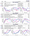
The total translation, mRNA levels, and ribosome density are plotted for mRNAs whose translation is altered in response to Tg treatment. Three categories are used to select mRNAs: down-regulated (average of translation at all Tg treatment times/0 h Tg), early response (translation at 0.5h Tg/0h Tg), and late response (translational at 4h Tg/average (0h Tg, 0.5h Tg)). For each mRNA, the fold change in translation during the given time is indicated. See also Figure S2, Table S2.
Figure 3
Figure 3. UPR activation elicits the release of mRNAs encoding ER-targeted proteins from the ER
(A) Changes in the mRNA levels, ribosome loading density, and total translation for mRNAs encoding ER-targeted proteins over time. (B) Subcellular localization of the translation of mRNAs encoding ER-targeted (red) proteins and cytosolic (blue) proteins over the treatment time course (C) Trajectories of the subcellular localization of the translation of all mRNAs encoding ER-targeted proteins over the treatment time course, where red indicates an increased ER localization, blue decreased. Each line represents the translation of an individual mRNA. (D) As in (C), depicting the subcellular localization of all mRNAs encoding ER-targeted proteins. (E) Relationships between the change in localization of translation and the change in localization of mRNAs encoding ER-targeted proteins for each time point transition. Best fit lines are depicted. (F) Relationship between the ribosome density (ribosomes per mRNA) after 0.5h Tg treatment and the change in mRNA localization at the same time point for mRNAs encoding ER-targeted proteins. Red line represents a moving average; shaded area is ± SD. (G) Selected mRNAs encoding ER-targeted proteins whose translation is retained or recruited to the ER following UPR induction. (H) Gene ontologies among mRNAs encoding ER-targeted proteins that are enriched for ER retention. See also Figure S3, Table S3.
Figure 4
Figure 4. Kinetics of UPR-induced mRNA re-localization
(A) Translational activity as measured by [35S]Met/Cys incorporation over a time course of treatment with 1mM DTT. Error bars represent SD (n=3). (B) Kinetics of eIF2α phosphorylation as assessed by immunoblot during DTT treatment. (C) Localization of an mRNA encoding a cytosolic protein (GAPDH) and an ER-targeted protein (Hsp90b1/GRP94) (n=3). (D) Fraction of ribosomes associated with the ER during DTT treatment and washout. Error bars represent SD (n=3). (E) Localization of the translation of ER-targeted proteins and cytosolic proteins as assessed by ribosome profiling during the induction and recovery from DTT treatment. Error bars represent SD (n=3). (F) Changes in gene-level translation for cytosolic and ER-associated ribosomes in UPR induction and recovery. The median value of the absolute log2 deviation in translation is calculated in each compartment, with error bars representing standard error between replicates. At the 0 time point, points represent the deviation in experimental replicates for Control samples. (G) Patterns of translational remodeling for canonical targets of UPR translational regulation. See also Figure S4, Table S2.
Figure 5
Figure 5. UPR induction effects on ribosome loading status for mRNAs encoding ER-targeted proteins
(A) The distribution of ribosomes along mRNAs encoding cytosolic (red) or ER-targeted (blue) in the cytosol and ER with or without 30min Tg treatment. (B) Ribosome loading of mRNAs encoding ER-targeted protein in the ER and cytosol with or without Tg treatment.
Figure 6
Figure 6. Divergent fates of mRNAs encoding ER-targeted proteins in the cytosol upon UPR induction
Cytosolic polyribosomes were analyzed by sucrose gradient centrifugation following (A) no treatment, (B) 10 min treatment with 150nM pactamycin, (C) 30 min treatment with 1μM Tg (D) 30 min Tg treatment with addition of 150nM pactamycin at 20 min. Gradient fractions were collected and analyzed for mRNA content by qPCR. (E) Radiolabelled, glycoslyated nascent polypeptide chains were quantified after a 10min pulse with [35S]Met/Cys or after a 30min chase with DMSO or 1μM Tg (n=3). See also Figure S5.
Figure 7
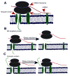
Figure 7. Model for dynamic mRNA localization to the ER
(A) Binding interactions that can confer polyribosome association with the ER are indicated by red arrowheads; red line is mRNA, green is nascent protein, black ellipses are ribosomal subunits. In addition to disruptions in mRNA, ribosome, and nascent chain interactions with the ER, an mRNA may also be released from the ER in the case where the mRNA dissociates from the ribosomes while the ribosomes itself retains ER affinity. (B) Model of the changes in binding interactions that occur for a ribosome engaged in the synthesis of an ER-targeted protein upon induction of ER stress. Here, ER:nascent chain and ER:mRNA interactions are disrupted, allowing for the release of the polyribosome. The dashed green line indicates a nascent polypeptide chain. (C) Same as (B) for a polyribosome synthesizing a cytosolic protein where the ribosome is independently associated with the ER. Having no nascent chain or mRNA interaction with the ER, the ribosome is retained on the ER due to the ongoing interaction with its ER binding partner(s).

Similar articles

Cited by

References

    1. Adesnik M, Lande M, Martin T, Sabatini DD. Retention of mRNA on the endoplasmic reticulum membranes after in vivo disassembly of polysomes by an inhibitor of initiation. J Cell Biol. 1976;71:307–313. - PMC - PubMed
    1. Blower MD. Molecular insights into intracellular RNA localization. International Review of Cell and Molecular Biology. 2013;302:1–39. - PMC - PubMed
    1. Borgese D, Blobel G, Sabatini DD. In vitro exchange of ribosomal subunits between free and membrane-bound ribosomes. J Mol Biol. 1973;74:415–438. - PubMed
    1. Braakman I, Helenius J, Helenius A. Manipulating disulfide bond formation and protein folding in the endoplasmic reticulum. EMBO J. 1992;11:1717–1722. - PMC - PubMed
    1. Campanella ME, Chu H, Low PS. Assembly and regulation of a glycolytic enzyme complex on the human erythrocyte membrane. Proc Natl Acad Sci USA. 2005;102:2402–2407. - PMC - PubMed

Publication types

Associated data