. 2014 Sep 11;10(9):e1004372.
doi: 10.1371/journal.ppat.1004372. eCollection 2014 Sep.

Incomplete deletion of IL-4Rα by LysM(Cre) reveals distinct subsets of M2 macrophages controlling inflammation and fibrosis in chronic schistosomiasis

Affiliations

Incomplete deletion of IL-4Rα by LysM(Cre) reveals distinct subsets of M2 macrophages controlling inflammation and fibrosis in chronic schistosomiasis

Kevin M Vannella et al. PLoS Pathog. .

Abstract

Mice expressing a Cre recombinase from the lysozyme M-encoding locus (Lyz2) have been widely used to dissect gene function in macrophages and neutrophils. Here, we show that while naïve resident tissue macrophages from IL-4Rαf(lox/delta)LysM(Cre) mice almost completely lose IL-4Rα function, a large fraction of macrophages elicited by sterile inflammatory stimuli, Schistosoma mansoni eggs, or S. mansoni infection, fail to excise Il4rα. These F4/80(hi)CD11b(hi) macrophages, in contrast to resident tissue macrophages, express lower levels of Lyz2 explaining why this population resists LysM(Cre)-mediated deletion. We show that in response to IL-4 and IL-13, Lyz2(lo)IL-4Rα(+) macrophages differentiate into an arginase 1-expressing alternatively-activated macrophage (AAM) population, which slows the development of lethal fibrosis in schistosomiasis. In contrast, we identified Lyz2(hi)IL-4Rα(+) macrophages as the key subset of AAMs mediating the downmodulation of granulomatous inflammation in chronic schistosomiasis. Our observations reveal a limitation on using a LysMCre mouse model to study gene function in inflammatory settings, but we utilize this limitation as a means to demonstrate that distinct populations of alternatively activated macrophages control inflammation and fibrosis in chronic schistosomiasis.


Conflict of interest statement

We have read the journal's policy and disclose the following potential conflicts: AWC was employed by a commercial company, Biomedical Research Institute. This does not alter our adherence to all PLOS Pathogens policies on sharing data and materials.

Figures

Figure 1
Figure 1. Surviving chronic S. mansoni infection depends on Il4rα allele, not LysMCre expression.
IL-4Rαflox/ΔLysMCre mice (open circles and bars), IL-4Rαflox/Δ littermate controls (solid circles and bars), IL-4Rαflox/flox mice (solid squares), and IL-4RαΔ/Δ (open triangles) were infected percutaneously with 35 Schistosoma mansoni cercariae. A. Survival kinetics through 16 weeks (n = 10–20 per group). B. Th1 response. 9 or 16 weeks post-infection, liver leukocytes were isolated, restimulated with phorbol myristate acetate/ionomycin, stained for IFN-γ, and analyzed by flow cytometry (n = 7–15, ns = not significant). C. Hepatotoxicity. Aspartate aminotransferase (AST) and alanine aminotransferase (ALT) were assayed in serum 9 or 16 weeks after infection (n = 7–8 mice, ns = not significant). Data shown are mean ±SEM and represent at least two independent experiments.
Figure 2
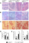
Figure 2. Inflammation but not fibrosis is exacerbated in chronically infected IL-4Rαflox/ΔLysMCre mice.
IL-4Rαflox/ΔLysMCre mice and IL-4Rαflox/Δ littermate controls were infected percutaneously with 35 S. mansoni cercariae. A–C. Representative 10× images of granuloma formation 9 weeks and 16 weeks post-infection from (A) hematoxylin and eosin-stained sections of intestinal tissue, (B) Giemsa-stained sections of liver tissue, or (C) picrosirius red-stained sections of liver tissue. D. Liver granuloma size in IL-4Rαflox/ΔLysMCre (open bars) and IL-4Rαflox/Δ littermate control (solid bars) mice. E. Liver fibrosis was assessed by hydroxyproline content, normalized to mass or worm pairs recovered by perfusion of infected mice through the portal vein. Data shown are mean ±SEM and represent two independent experiments (n = 15, ns = not significant).
Figure 3
Figure 3. Normal cytokine response in S. mansoni-infected IL-4Rαflox/ΔLysMCre mice.
IL-4Rαflox/ΔLysMCre mice and IL-4Rαflox/Δ littermate controls were infected percutaneously with 35 S. mansoni cercariae. A. Serum IL-13Rα2 levels were measured by ELISA 9 or 16 weeks post-infection. The open and solid portions of each bar correspond to unbound IL-13Rα2 and IL-13Rα2 bound to IL-13, respectively. B. Tissue cytokine levels. Expression of il12p40 and il10 was quantified by qPCR from liver tissue snips of IL-4Rαflox/ΔLysMCre mice (open bars) or IL-4Rαflox/Δ littermate controls (solid bars). C. Th2 response. Liver leukocytes were isolated from IL-4Rαflox/ΔLysMCre mice (open bars) or IL-4Rαflox/Δ littermate controls (solid bars), stimulated with phorbol myristate acetate/ionomycin, and analyzed by flow cytometry. The percentage of CD4+ leukocytes expressing intracellular IL-4 and IL-13 are shown. (n = 7-15 for each experiment, ns = not significant). Data shown are mean ±SEM and represent two independent experiments.
Figure 4
Figure 4. Normal expression of AAM-associated genes in IL-4Rαflox/ΔLysMCre liver.
IL-4Rαflox/ΔLysMCre mice (open bars) and IL-4Rαflox/Δ littermate controls (solid bars) were infected percutaneously with 35 cercariae. Expression of selected genes was measured by qPCR in liver tissue 9 weeks (A) and 16 weeks (B) post-infection and normalized to expression in naïve littermate control tissue (n = 7–15; p>0.05 except where noted, **p<0.01). Data shown are mean ±SEM and represent two independent experiments.
Figure 5
Figure 5. A population of inflammatory IL-4Rα-expressing myeloid cells resists LysMCre-mediated deletion.
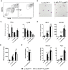
BALB/c, IL-4Rαflox/Δ, and IL-4Rαflox/ΔLysMCre mice were injected i.p. with 2 ml thioglycollate 4 d prior to harvest or were left untreated (naïve). Peritoneal cells were harvested from each group, stimulated for 30 min with 20 ng/ml IL-4 (black outline), and compared to unstimulated cells (solid gray). IL-4Rα function was assessed by IL-4-induced phosphorylation of STAT6 using flow cytometry. A, D. Gating strategy for lymphocytes and F4/80hi CD11bhi macrophages. Detection of pSTAT6 in lymphocytes (B, E) and macrophages (C, F). Each histogram peak represents an individual mouse (n = 2–6 for 2 independent experiments). G. DNA was isolated from F4/80hi CD11bhi macrophages FACS sorted from naïve and thioglycollate-treated IL-4Rαflox/flox, IL-4Rαflox/Δ, IL-4Rαflox/ΔLysMCre and IL-4RαΔ/Δ mice. Rearrangement of the Il4rα locus was measured using PCR to compare the presence of wild-type (WT) and knockout (KO) Il4rα alleles. Quantification of band intensity is shown in the right panels. Aggregate intensity of WT product plus KO product was normalized to 100 percent for each sample. H. In a separate experiment, CD11b+ F4/80+ macrophages were sorted from the same naïve or thioglycollate-elicited peritoneal cells. Lyz2 gene expression was found to be lower in thioglycollate-elicited macrophages than naïve macrophages (n = 2–5 for 2 independent experiments). Data shown are mean ±SEM and represent two independent experiments (*p<0.05).
Figure 6
Figure 6. Lyz2 lo macrophages develop features of AAMs in response to S. mansoni eggs.
IL-4Rαflox/ΔLysMCre mice (open bars) and littermate controls (solid bars) were left untreated (naïve), challenged with 5000 S. mansoni eggs i.p. 4 days before harvest (1o), 18 days before harvest (1o-rested), or challenged on both 18 days and 4 days before harvest (1o-rechallenged). A. Total peritoneal cells were sorted for F4/80hi CD11bhi cells at a purity of >90%. B. Representative 20× images of sorted F4/80hi CD11bhi macrophages after cytospin and hematoylin and eosin staining. C,D. The sorted cells were assayed for Il4rα and Lyz2 gene expression (C), and gene expression of markers of alternative activation (D). Fold change in gene expression is shown relative to the expression levels in sorted F4/80hi CD11bhi cells from naïve littermate controls. E. Surface expression of mannose receptor measured by flow cytometry on unsorted F4/80hi CD11bhi peritoneal cells from the same treatment groups. F. Arginase activity in sorted macrophages. (n = 3–6, ns = not significant) Data shown are mean ±SEM and represent at least two independent experiments.
Figure 7
Figure 7. Macrophage populations in livers of S. mansoni-infected IL-4Rαflox/ΔLysMCre mice express Il4rα and alternative activation markers.
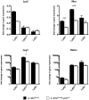
IL-4Rαflox/ΔLysMCre mice (open bars) and IL-4Rαflox/Δ littermate controls (solid bars) were infected percutaneously with 35 cercariae. From mice infected for 9 weeks, CD45+ SiglecF- CD11b+ Ly6G- F4/80+ CD64+ liver leukocytes were sorted and separated based on Ly6C expression with a flow cytometer. Gene expression was measured by qPCR (n = 3; *p<0.05, ***p<0.001). Fold change is displayed relative to gene expression from CD45+ SiglecF- CD11b+ Ly6G- F4/80+ CD64+ Ly6C+ cells sorted from naïve IL-4Rαflox/Δ littermate control livers. Data shown are mean ±SEM and represent at least two independent experiments.
Figure 8
Figure 8. Distinct populations of alternatively activated macrophages control inflammation and fibrosis.
Diagram showing the roles of various subsets of alternatively activated macrophages in the pathogenesis of schistosomiasis. In IL-4Rαflox/ΔLysMCre mice, LysMCre results in the elimination of only mature IL-4Rα+Arg1+Lyz2hi AAMs. The loss of this subset of AAMs results in the failure to downmodulate granulomatous inflammation in acute and chronic schistosomiasis. However, a substantial population of inflammatory IL-4Rα+Lyz2lo AAMs is preserved in these mice that expresses Arg1 in response in response to IL-4 and IL-13 and slows the progression of fibrosis. In Arg1flox/floxTie2Cre mice, both populations of AAMs are defective because of the loss of Arg1 activity, leading to exacerbation of both egg-induced inflammation and fibrosis at both the acute and chronic stage of infection (far right panel). In comparison, Arg1flox/ΔLysMCre primarily deletes Arg1 in the mature Lyz2hi population, leading to a much more modest increase in fibrosis and granulomatous inflammation, which only reaches significance at the chronic stage of infection with S. mansoni. Wild-type IL-4Rαflox/Δmice have both mature resident and inflammatory AAMs expressing IL-4Rα and Arg1 so both forms of pathology (inflammation and fibrosis) are substantially controlled in these mice (far left panel).

Similar articles

Cited by

References

    1. Mosser DM, Edwards JP (2008) Exploring the full spectrum of macrophage activation. Nat Rev Immunol 8: 958–969. - PMC - PubMed
    1. Murray PJ, Allen JE, Biswas SK, Fisher EA, Gilroy DW, et al. (2014) Macrophage Activation and Polarization: Nomenclature and Experimental Guidelines. Immunity 41: 14–20. - PMC - PubMed
    1. Gordon S (2003) Alternative activation of macrophages. Nat Rev Immunol 3: 23–35. - PubMed
    1. Wynn TA (2004) Fibrotic disease and the T(H)1/T(H)2 paradigm. Nat Rev Immunol 4: 583–594. - PMC - PubMed
    1. Duffield JS, Lupher M, Thannickal VJ, Wynn TA (2013) Host responses in tissue repair and fibrosis. Annu Rev Pathol 8: 241–276. - PMC - PubMed

Publication types

MeSH terms

LinkOut - more resources