. 2014 Oct 1;193(7):3600-12.
doi: 10.4049/jimmunol.1401088. Epub 2014 Sep 3.

Type I IFN induces IL-10 production in an IL-27-independent manner and blocks responsiveness to IFN-γ for production of IL-12 and bacterial killing in Mycobacterium tuberculosis-infected macrophages

Affiliations

Type I IFN induces IL-10 production in an IL-27-independent manner and blocks responsiveness to IFN-γ for production of IL-12 and bacterial killing in Mycobacterium tuberculosis-infected macrophages

Finlay W McNab et al. J Immunol. .

Abstract

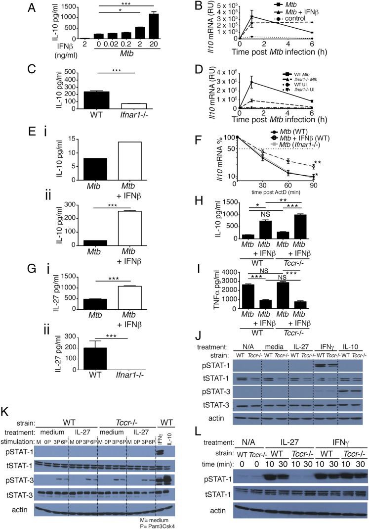
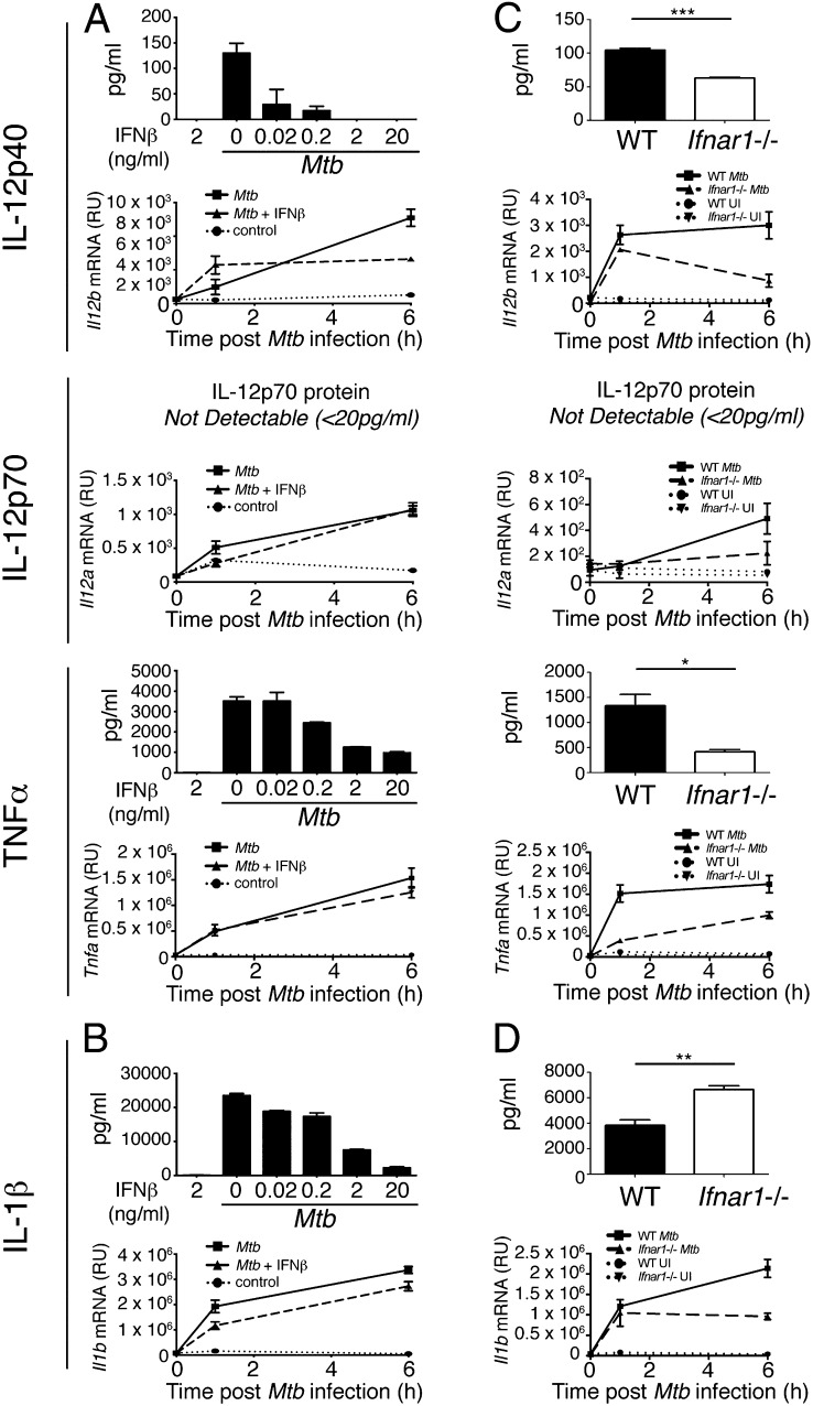
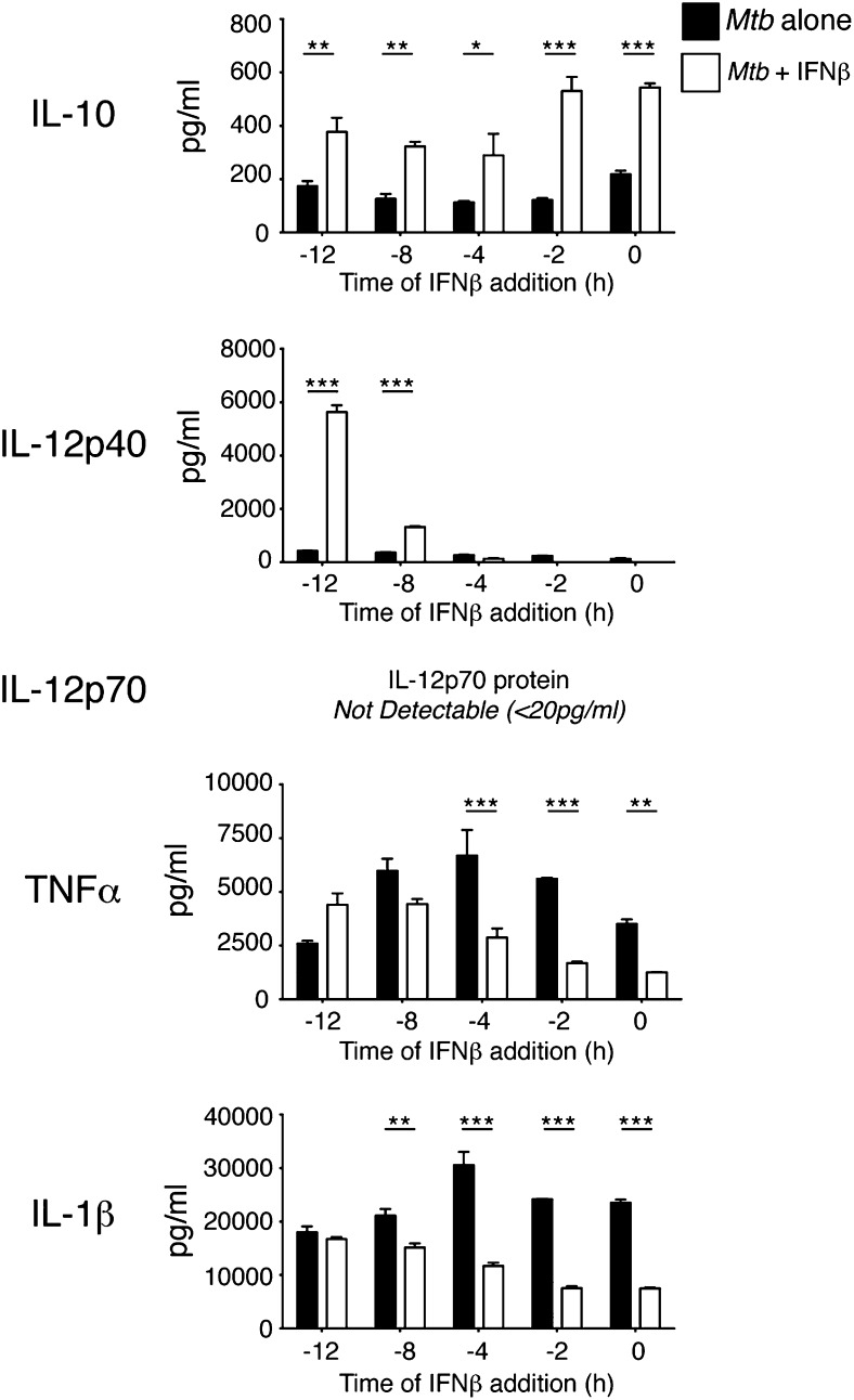
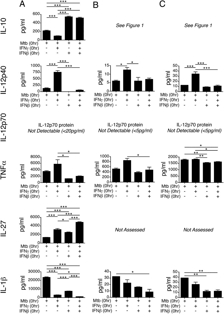
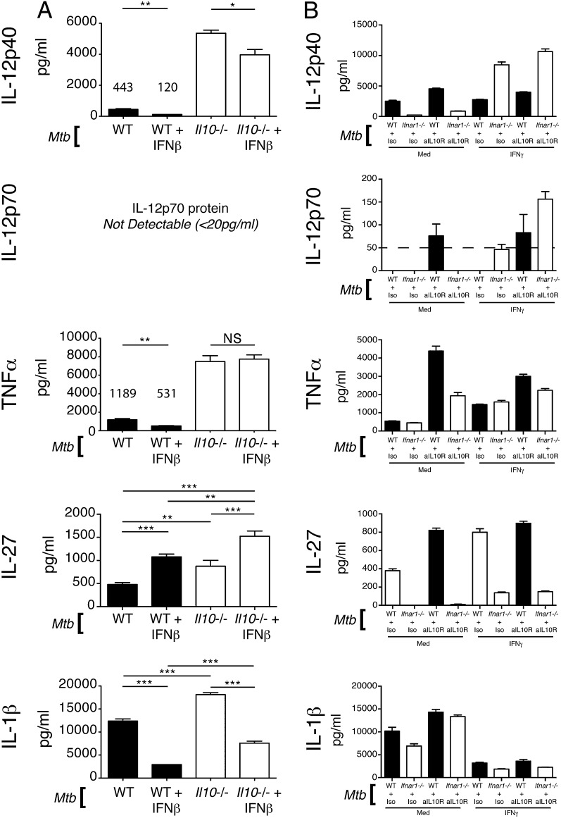
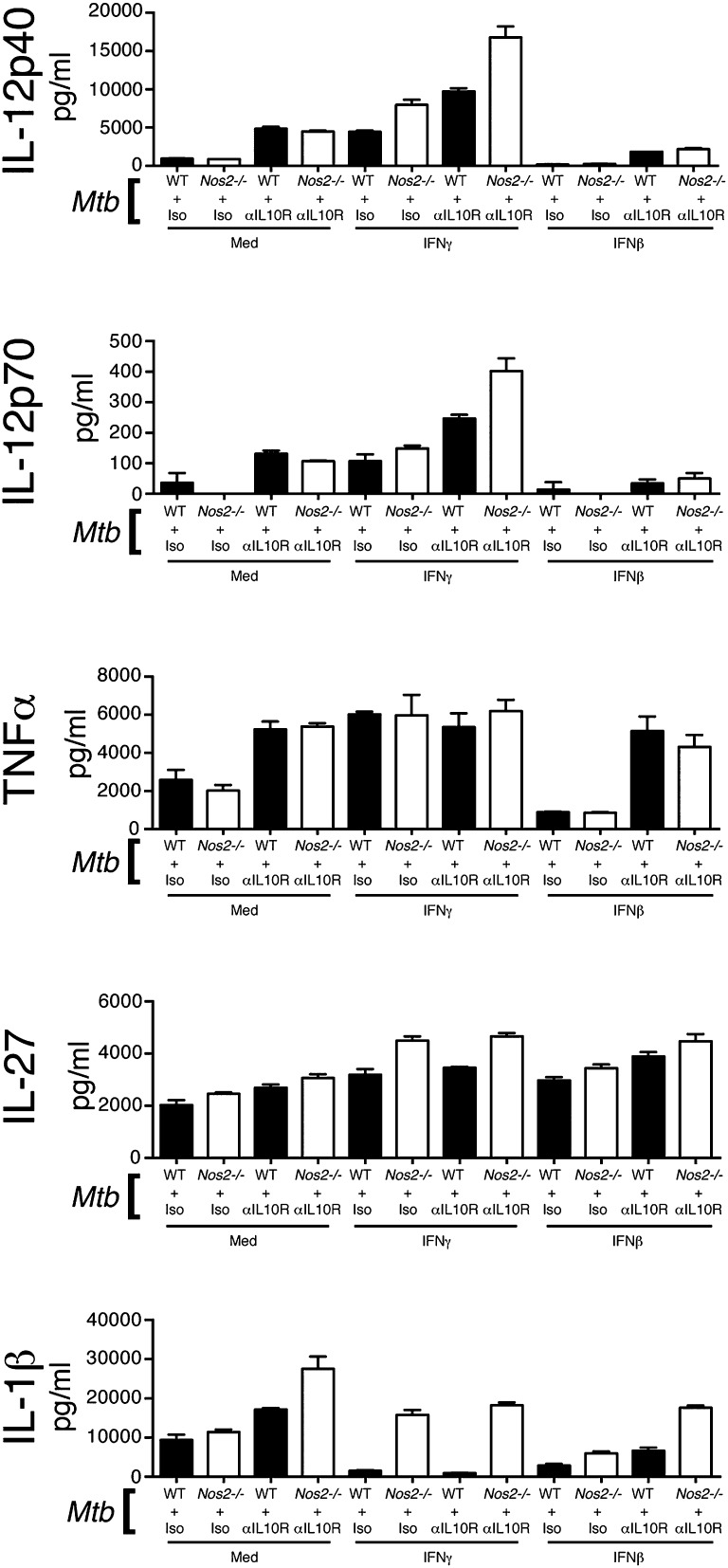
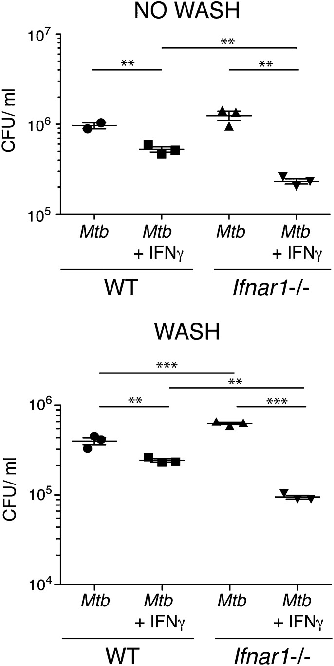
Tuberculosis, caused by the intracellular bacterium Mycobacterium tuberculosis, currently causes ∼1.4 million deaths per year, and it therefore remains a leading global health problem. The immune response during tuberculosis remains incompletely understood, particularly regarding immune factors that are harmful rather than protective to the host. Overproduction of the type I IFN family of cytokines is associated with exacerbated tuberculosis in both mouse models and in humans, although the mechanisms by which type I IFN promotes disease are not well understood. We have investigated the effect of type I IFN on M. tuberculosis-infected macrophages and found that production of host-protective cytokines such as TNF-α, IL-12, and IL-1β is inhibited by exogenous type I IFN, whereas production of immunosuppressive IL-10 is promoted in an IL-27-independent manner. Furthermore, much of the ability of type I IFN to inhibit cytokine production was mediated by IL-10. Additionally, type I IFN compromised macrophage activation by the lymphoid immune response through severely disrupting responsiveness to IFN-γ, including M. tuberculosis killing. These findings describe important mechanisms by which type I IFN inhibits the immune response during tuberculosis.


Figures

FIGURE 1.
FIGURE 1.
Type I IFN regulates IL-10 production in M. tuberculosis–infected macrophages independently of IL-27 signaling. (A) WT macrophages were infected with M. tuberculosis in the presence of increasing concentrations of IFN-β, added at the time of infection, and levels of IL-10 in culture supernatant were determined by ELISA at 24 h postinfection. (B) WT macrophages were infected with M. tuberculosis in the presence or absence of 2 ng/ml IFN-β, added at the time of infection, and levels of Il10 mRNA were determined by quantitative RT-PCR (qRT-PCR) at the time points indicated after infection. (C) WT and Ifnar1−/− macrophages were infected with M. tuberculosis and levels of IL-10 in culture supernatant were determined by ELISA at 24 h postinfection. (D) WT and Ifnar1−/− macrophages were infected with M. tuberculosis and levels of Il10 mRNA determined by qRT-PCR at the time points indicated after infection. (E) WT myeloid cells taken ex vivo from lungs (i) and BM (ii) were infected with M. tuberculosis in the presence or absence of 2 ng/ml IFN-β, added at the time of infection, and levels of IL-10 in culture supernatant were determined by Luminex bead array at 24 h postinfection. (F) WT, Ifnar1−/−, and WT treated with IFN-β macrophages were infected with M. tuberculosis, and at 1 h postinfection ActD was added. mRNA was then taken at the time points indicated and Il10 mRNA levels were determined by qRT-PCR. (G) WT macrophages treated (or not) with 2 ng/ml IFN-β at the time of infection (i) and WT and Ifnar1−/− macrophages (ii) were infected with M. tuberculosis, and levels of IL-27 in culture supernatant were determined by ELISA at 24 h postinfection. (H and I) WT and Tccr−/− (IL-27Rα−/−) macrophages were infected with M. tuberculosis in the presence or absence of 2 ng/ml IFN-β, added at the time of infection, and levels of IL-10 (H) or TNF-α (I) in culture supernatant were determined by ELISA at 24 h postinfection. (J) WT and Tccr−/− (IL-27Rα−/−) macrophages were treated for 20 min with rIL-27 (50 ng/ml), rIFN-γ (10 ng/ml), or rIL-10 (10 ng/ml) and whole-cell extracts were then analyzed by immunoblotting with the indicated Abs. (K) WT and Tccr−/− (IL-27Rα−/−) macrophages were stimulated for 0, 3, or 6 h with Pam3CSK4 (200 ng/ml) and then treated (or not) for 20 min with rIL-27 (50 ng/ml). WT macrophages treated with IFN-γ (10 ng/ml) or IL-10 (10 ng/ml) were included as positive controls for STAT-1 and STAT-3 phosphorylation, respectively. Whole-cell extracts were then analyzed by immunoblotting with the indicated Abs. (L) WT and Tccr−/− splenocytes were treated for the indicated times with rIL-27 (50 ng/ml) or rIFN-γ (10 ng/ml). Whole-cell extracts were then analyzed by immunoblotting with the indicated Abs. Graphs show means ± SEM of triplicate samples, except for (E), which shows duplicates. For ELISA and Luminex bead array results, uninfected control samples were below the detection limit (20 and 5 pg/ml, respectively) for the cytokines measured (data not shown). Significance was determined using an unpaired t test (A, C, E, and G), a one-way ANOVA with a Bonferroni post hoc test (H and I), or a two-way ANOVA, with significance relative to WT (F). Data are representative of at least two independent experiments. *p < 0.05, **p < 0.01, ***p < 0.001.
FIGURE 2.
FIGURE 2.
Opposing effects of exogenous IFN-β treatment and autocrine type I IFN signaling on IL-12 and TNF-α production in M. tuberculosis–infected macrophages. (A) WT macrophages were infected with M. tuberculosis in the presence of increasing concentrations of IFN-β (for protein) or 2 ng/ml IFN-β (for mRNA), added at the time of infection, and levels of IL-12p40, IL-12p70, and TNF-α protein in supernatant and Il12b, Il12a, and Tnfa mRNA from cells were determined by ELISA at 24 h postinfection and qRT-PCR at indicated times after infection. (B) IL-1β protein and Il1b mRNA from macrophages in (A) were measured by ELISA at 24 h postinfection and qRT-PCR at indicated times after infection. (C) WT and Ifnar1−/− macrophages were infected with M. tuberculosis and IL-12p40, IL-12p70, and TNF-α protein in supernatant and Il12b, Il12a, and Tnfa mRNA from cells were determined by ELISA at 24 h postinfection and qRT-PCR at indicated times after infection. (D) IL-1β protein and Il1b mRNA from macrophages in (C) were measured by ELISA at 24 h postinfection and qRT-PCR at indicated times after infection. Graphs show means ± SEM of triplicate samples. For ELISA results, uninfected control samples were below the detection limit (20 pg/ml) for the cytokines measured (data not shown). Significance was determined using an unpaired t test. Data are representative of at least three independent experiments. *p < 0.05, **p < 0.01, ***p < 0.001.
FIGURE 3.
FIGURE 3.
IFN-β pretreatment of M. tuberculosis–infected macrophages enhances IL-12 and TNF-α production. WT macrophages were infected with M. tuberculosis alone or in the presence of IFN-β (2 ng/ml), added at the indicated times prior to infection. Cytokine levels in culture supernatants were determined by ELISA at 24 h postinfection. Uninfected control samples were below the detection limit (20 pg/ml) for the cytokines measured (data not shown). Graphs show means ± SEM of triplicate samples. Data are representative of two independent experiments. Significance was determined using a two-way ANOVA with a Bonferroni post hoc test. *p < 0.05, **p < 0.01, ***p < 0.001.
FIGURE 4.
FIGURE 4.
Exogenous IFN-β inhibits M. tuberculosis–infected macrophage responsiveness to concomitant IFN-γ addition. (A) WT macrophages were infected with M. tuberculosis alone or with M. tuberculosis and IFN-β (2 ng/ml), M. tuberculosis and IFN-γ (5 ng/ml), or M. tuberculosis and both IFN-β (2 ng/ml) and IFN-γ (5 ng/ml) together, added at the time of infection. Cytokine levels in culture supernatants were determined at 24 h postinfection. Uninfected control samples were below the detection limit (20 pg/ml) for the cytokines measured (data not shown). Graphs show means ± SEM. Significance was determined using a one-way ANOVA with a Bonferroni post hoc test. Data are representative of three independent experiments. (B) Myeloid cells (LinLy6c+Ly6G) were sorted from the lungs of WT mice and infected with M. tuberculosis and treated with IFN as in (A). Cytokine levels in culture supernatant were determined at 24 h postinfection using a Luminex bead array. Uninfected control samples were below the detection limit (5 pg/ml) for the cytokines measured (data not shown). Graphs show means ± SEM. Data are representative of two independent experiments. (C) Myeloid cells (LinLy6c+Ly6G) were sorted from the BM of WT mice and infected with M. tuberculosis and treated with IFN as in (A) and (B). Cytokine levels in culture supernatant were determined at 24 h postinfection using a Luminex bead array. Uninfected control samples were below the detection limit for the cytokines measured (data not shown). Graphs show means ± SEM. Data are representative of three independent experiments. *p < 0.05, **p < 0.01, ***p < 0.001.
FIGURE 5.
FIGURE 5.
Exogenous IFN-β and endogenous type I IFN signaling affect proinflammatory cytokine production and responsiveness to IFN-γ in M. tuberculosis–infected macrophages through IL-10–dependent and –independent mechanisms. (A) WT and Il10−/− macrophages were infected with M. tuberculosis or with M. tuberculosis and IFN-β (2 ng/ml), added at the time of infection. Levels of the indicated cytokines in culture supernatant were determined at 24 h postinfection by ELISA. Graphs show means ± SEM of triplicate samples. Significance was determined using a one-way ANOVA with a Bonferroni post hoc test. (B) WT and Ifnar1−/− macrophages were infected with M. tuberculosis, treated with anti–IL-10R or isotype control Abs and with IFN-γ (5 ng/ml) or not, added at the time of infection. Levels of the indicated cytokines in culture supernatant were determined at 24 h postinfection by ELISA. Uninfected control samples were below the detection limit (20 pg/ml) for the cytokines measured (data not shown). Graphs show means ± SEM of triplicate samples. Data are representative of three independent experiments. *p < 0.05, **p < 0.01, ***p < 0.001.
FIGURE 6.
FIGURE 6.
Type I IFN regulation of IL-1β production is dependent on iNOS and IL-10. WT and Nos2−/− macrophages were infected with M. tuberculosis, treated with anti–IL-10R or isotype control Abs and with either IFN-γ (5 ng/ml) or IFN-β (2 ng/ml) or media, added at the time of infection. Levels of the indicated cytokines in culture supernatant were determined at 24 h postinfection by ELISA. Uninfected control samples were below the detection limit (20 pg/ml) for the cytokines measured (data not shown). Graphs show means ± SEM of triplicate samples. Data are representative of at least three independent experiments.
FIGURE 7.
FIGURE 7.
Type I IFN signaling inhibits macrophage restriction of M. tuberculosis growth and killing in response to IFN-γ. WT and Ifnar1−/− macrophages were infected with M. tuberculosis in the presence or absence of IFN-γ (5 ng/ml), added at the time of infection. At 4 h postinfection media containing M. tuberculosis was removed, cells were washed in PBS, and fresh media (with no M. tuberculosis) were replaced (bottom panel) or not (top panel). At 96 h postinfection cells were washed in PBS, lysed in 0.2% saponin, and bacterial loads were enumerated via serial dilution and plating. Line and bars show means ± SEM. Significance was determined using an unpaired t test. Data are representative of at least two independent experiments for each of the two systems (i.e., wash or no wash). **p < 0.01, ***p < 0.001.

Similar articles

Cited by

References

    1. Cooper A. M. 2009. Cell-mediated immune responses in tuberculosis. Annu. Rev. Immunol. 27: 393–422 - PMC - PubMed
    1. Flynn J. L., Chan J.. 2001. Immunology of tuberculosis. Annu. Rev. Immunol. 19: 93–129 - PubMed
    1. North R. J., Jung Y. J.. 2004. Immunity to tuberculosis. Annu. Rev. Immunol. 22: 599–623 - PubMed
    1. Chan J., Xing Y., Magliozzo R. S., Bloom B. R.. 1992. Killing of virulent Mycobacterium tuberculosis by reactive nitrogen intermediates produced by activated murine macrophages. J. Exp. Med. 175: 1111–1122 - PMC - PubMed
    1. MacMicking J. D., North R. J., LaCourse R., Mudgett J. S., Shah S. K., Nathan C. F.. 1997. Identification of nitric oxide synthase as a protective locus against tuberculosis. Proc. Natl. Acad. Sci. USA 94: 5243–5248 - PMC - PubMed

Publication types

MeSH terms