RNA-binding protein HuR promotes growth of small intestinal mucosa by activating the Wnt signaling pathway
- PMID: 25165135
- PMCID: PMC4214778
- DOI: 10.1091/mbc.E14-03-0853
RNA-binding protein HuR promotes growth of small intestinal mucosa by activating the Wnt signaling pathway
Abstract
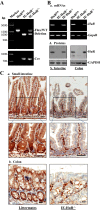
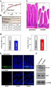
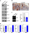
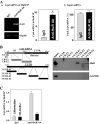
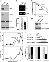
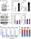
Inhibition of growth of the intestinal epithelium, a rapidly self-renewing tissue, is commonly found in various critical disorders. The RNA-binding protein HuR is highly expressed in the gut mucosa and modulates the stability and translation of target mRNAs, but its exact biological function in the intestinal epithelium remains unclear. Here, we investigated the role of HuR in intestinal homeostasis using a genetic model and further defined its target mRNAs. Targeted deletion of HuR in intestinal epithelial cells caused significant mucosal atrophy in the small intestine, as indicated by decreased cell proliferation within the crypts and subsequent shrinkages of crypts and villi. In addition, the HuR-deficient intestinal epithelium also displayed decreased regenerative potential of crypt progenitors after exposure to irradiation. HuR deficiency decreased expression of the Wnt coreceptor LDL receptor-related protein 6 (LRP6) in the mucosal tissues. At the molecular level, HuR was found to bind the Lrp6 mRNA via its 3'-untranslated region and enhanced LRP6 expression by stabilizing Lrp6 mRNA and stimulating its translation. These results indicate that HuR is essential for normal mucosal growth in the small intestine by altering Wnt signals through up-regulation of LRP6 expression and highlight a novel role of HuR deficiency in the pathogenesis of intestinal mucosal atrophy under pathological conditions.
© 2014 Liu, Christodoulou-Vafeiadou, et al. This article is distributed by The American Society for Cell Biology under license from the author(s). Two months after publication it is available to the public under an Attribution–Noncommercial–Share Alike 3.0 Unported Creative Commons License (http://creativecommons .org/licenses/by-nc-sa/3.0).
Figures

Similar articles
-
Post-transcriptional regulation of Wnt co-receptor LRP6 and RNA-binding protein HuR by miR-29b in intestinal epithelial cells.Biochem J. 2016 Jun 1;473(11):1641-9. doi: 10.1042/BCJ20160057. Epub 2016 Apr 18. Biochem J. 2016. PMID: 27089893 Free PMC article.
-
HuR Enhances Early Restitution of the Intestinal Epithelium by Increasing Cdc42 Translation.Mol Cell Biol. 2017 Mar 17;37(7):e00574-16. doi: 10.1128/MCB.00574-16. Print 2017 Apr 1. Mol Cell Biol. 2017. PMID: 28031329 Free PMC article.
-
Jnk2 deletion disrupts intestinal mucosal homeostasis and maturation by differentially modulating RNA-binding proteins HuR and CUGBP1.Am J Physiol Cell Physiol. 2014 Jun 15;306(12):C1167-75. doi: 10.1152/ajpcell.00093.2014. Epub 2014 Apr 16. Am J Physiol Cell Physiol. 2014. PMID: 24740539 Free PMC article.
-
HuR and Its Interactions with Noncoding RNAs in Gut Epithelium Homeostasis and Diseases.Front Biosci (Landmark Ed). 2023 Oct 25;28(10):262. doi: 10.31083/j.fbl2810262. Front Biosci (Landmark Ed). 2023. PMID: 37919092 Review.
-
RNA-binding proteins and microRNAs in gastrointestinal epithelial homeostasis and diseases.Curr Opin Pharmacol. 2014 Dec;19:46-53. doi: 10.1016/j.coph.2014.07.006. Epub 2014 Jul 24. Curr Opin Pharmacol. 2014. PMID: 25063919 Free PMC article. Review.
Cited by
-
Embryonic lethal abnormal vision proteins and adenine and uridine-rich element mRNAs after global cerebral ischemia and reperfusion in the rat.J Cereb Blood Flow Metab. 2017 Apr;37(4):1494-1507. doi: 10.1177/0271678X16657572. Epub 2016 Jan 1. J Cereb Blood Flow Metab. 2017. PMID: 27381823 Free PMC article.
-
Cooperative Repression of Insulin-Like Growth Factor Type 2 Receptor Translation by MicroRNA 195 and RNA-Binding Protein CUGBP1.Mol Cell Biol. 2017 Sep 12;37(19):e00225-17. doi: 10.1128/MCB.00225-17. Print 2017 Oct 1. Mol Cell Biol. 2017. PMID: 28716948 Free PMC article.
-
Polyamines in Gut Epithelial Renewal and Barrier Function.Physiology (Bethesda). 2020 Sep 1;35(5):328-337. doi: 10.1152/physiol.00011.2020. Physiology (Bethesda). 2020. PMID: 32783609 Free PMC article. Review.
-
Circular RNA CircHIPK3 Promotes Homeostasis of the Intestinal Epithelium by Reducing MicroRNA 29b Function.Gastroenterology. 2021 Oct;161(4):1303-1317.e3. doi: 10.1053/j.gastro.2021.05.060. Epub 2021 Jun 9. Gastroenterology. 2021. PMID: 34116030 Free PMC article.
-
Inhibition of Caspase-2 Translation by the mRNA Binding Protein HuR: A Novel Path of Therapy Resistance in Colon Carcinoma Cells?Cells. 2019 Jul 30;8(8):797. doi: 10.3390/cells8080797. Cells. 2019. PMID: 31366165 Free PMC article. Review.
References
-
- Clevers H. Wnt/b-catenin signaling in development and disease. Cell. 2006;127:469–480. - PubMed
Publication types
MeSH terms
Substances
Grants and funding
LinkOut - more resources
Full Text Sources
Other Literature Sources
Molecular Biology Databases
Miscellaneous

