. 2014 Aug 21;41(2):244-56.
doi: 10.1016/j.immuni.2014.06.017.

Transcription factor T-bet regulates intraepithelial lymphocyte functional maturation

Affiliations

Transcription factor T-bet regulates intraepithelial lymphocyte functional maturation

Bernardo S Reis et al. Immunity. .

Abstract

The intestinal epithelium harbors large populations of activated and memory lymphocytes, yet these cells do not cause tissue damage in the steady state. We investigated how intestinal T cell effector differentiation is regulated upon migration to the intestinal epithelium. Using gene loss- and gain-of-function strategies, as well as reporter approaches, we showed that cooperation between the transcription factors T-bet and Runx3 resulted in suppression of conventional CD4(+) T helper functions and induction of an intraepithelial lymphocyte (IEL) program that included expression of IEL markers such as CD8αα homodimers. Interferon-γ sensing and T-bet expression by CD4(+) T cells were both required for this program, which was distinct from conventional T helper differentiation but shared by other IEL populations, including TCRαβ(+)CD8αα(+) IELs. We conclude that the gut environment provides cues for IEL maturation through the interplay between T-bet and Runx3, allowing tissue-specific adaptation of mature T lymphocytes.


Figures

Figure 1
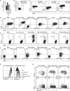
Figure 1. T-bet Upregulation Is Linked to IEL Differentiation
(A) Expression of Tbx21 by sorted small intestine IEL populations, TCRαβ+CD4+, TCRαβ+CD8αα+ (CD4, CD8β), TCRαβ+CD8β+CD8αα+ (CD4), TCRγδ+CD8αα+ (CD4, CD8β), and naive splenic CD4+ T cells from WT mice. (B) Expression Tbx21 by sorted small intestine IEL populations, TCRβ+CD4+CD8β ThPOKlo and ThPOKhi from naive Zbtb7b -GFP reporter mice. (C) CD8α, T-bet, 2B4, and CD103 expression by TCRβ+CD4+CD8β small intestine IELs of naive WT mice. (D–H) In vitro CD4+CD8αα+ IEL induction. Sorted, naive Va2+CD4+ T cells isolated from WT reporter OTII mice (OTII Zbtb7b -GFP) (D–G) or OTII Runx3-YFP (G) were cocultured with DCs, OVA peptide, and indicated cytokines. (D) T-bet and IFN-γ, (E) Foxp3, and (F) CD8α and CD103 expression by cultured OTII cells. (G) Histograms of ThPOK (left) or Runx3 (right) expression by OTII cells from different culture conditions. (H) CD103, CD8α, and ThPOK expression by gated CD4+CD8β OTII cells. (B) Error bars represent SEM of triplicates. All plots are representative of at least three independent experiments.
Figure 2
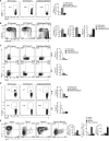
Figure 2. T-bet Regulates IEL Development
Sorted naive Va2+CD4+ T cells isolated from WT (OTII), Tbx21–/– (OTII[DTbx21]) or Runx3 conditional knockout (OTII[DRunx3]) mice were cocultured with DCs, OVA peptide, and TGF-β+RA+IFN-γ (A and B). (A) CD8α (contour plot) and (B) CD103 (histogram) expression by gated CD4+CD8βT cells. (C) Sorted naive polyclonal CD4+ T cells isolated from WT, DTbx21 or Cd4(DRunx3) Zbtb7b -GFP reporter mice were cultured with plate-bound α-CD3 and soluble α-CD28 in the presence of TGF-β+RA+IFN-γ. Histogram of ThPOK expression by CD4+CD8βT cells is shown. (D–I) Ex vivo analysis of small intestinal IELs from 10- to 14-week-old (D–H) or 25- to 30-week-old (I) WT and Tbx21–/– mice. (D and I) TL-tetramer (CD8αα) and CD8β expression by gated TCRαβ+CD4 IEL. (E) Ratio TCRγδ+ to TCRαβ+ among CD45+ cells. (F) CD8αα quantification in natural IELs TCRγδ+ (left) and TCRαβ+CD8βCD4 (right) T cells right. (G) CD8α and CD103 expression by TCRαβ+CD4+CD8β IELs. (H) CD8αα quantification in induced IELs TCRαβ+CD4+ (left) and TCRαβ+CD8β+ (right). (J and K) Ex vivo analysis of small intestine IELs from 10- to 14-week-old WT and Tbx21–/– Zbtb7b -GFP reporter mice. (J) ThPOK and CD103 expression by gated TCRαβ+CD4+CD8β cells; (K) IL-17 and CD8α expression by gated TCRαβ+CD4+CD8β cells, stimulated with PMA and ionomycin. Plots are representative of at least three independent experiments. Statistical significance analyzed by Student's t test of pooled experiments. Error bars represent SEM. *p < 0.05; **p < 0.01; ***p < 0.001.
Figure 3
Figure 3. Cell-Intrinsic Effects of T-bet on CD4-IEL Maturation
(A–G) Sorted naive CD4+GFPhi T cells from WT and Tbx21–/– Zbtb7b -GFP reporter mice were adoptively transferred to Rag1–/– recipient mice and analyzed 40 to 50 days later. (A) CD8α and CD103 expression, (B) ThPOK loss and CD8αα expression by gated TCRβ+CD4+CD8β small intestine IELs of recipient mice. (C–F) Analysis of spleen, mesenteric lymph node (mLN), and large intestine IEL and LPL cells from recipient mice after PMA and ionomycin stimulation. (C) Plots for IL-17 and IFN-γ expression. (D–G) Frequency of (D) IL-17 single producer (SP), (E) IL-17-IFN-γ double producer (DP), (F) IFN-γ single producer, and (G) Foxp3 positive TCRβ+CD4+CD8β cells from two pooled independent experiments. Error bars represent SEM. *p < 0.05; **p < 0.01. Plots are representative of three independent experiments.
Figure 4
Figure 4. Runx3 and T-bet Play Both Synergistic and Complementary Roles in Functional Differentiation of Intestinal T Cells
(A–D) Sorted naive CD4+GFPhi T cells isolated from WT or Cd4DRunx3 Zbtb7b -GFP mice were adoptively transferred to Rag1–/– recipients, and analyzed 40 to 50 days later. Prior to transfer cells were transduced with Tbx21-expressing or empty control (mock) retrovirus. (A) CD8α, (B) CD103, and ThPOK expression by gated TCRβ+CD4+CD8β small intestine IELs of recipient mice. (C) RORgt, T-bet, and (D) Foxp3 expression by TCRβ+CD4+CD8β cells isolated from the mesenteric lymph node (mLN) and small intestine IEL compartment of recipient mice. Plots are representative of two independent experiments. (E) IL-17 and IFN-γ expression by sorted naive CD4+ T cells isolated from Tbx21–/– mice and infected with Tbx21-Thy1.1 (Tbx21), Runx3-GFP (Runx3), both (Tbx21+Runx3), or empty control (mock) retroviruses. Transduced cells were cultured in vitro for 4.5 days under Th17-polarizing differentiation conditions (TGF-β+IL-6+IL1β+IL-23). Plots are representative of two independent experiments. Error bars represent SEM.
Figure 5
Figure 5. Hierarchical Roles for Runx3 and T-bet in IEL Differentiation
(A) Histogram and mean fluorescence index (MFI) of CD103 expression by WT CD4+ T cells transduced with Runx1, Runx3, or an empty retrovirus (mock). (B) Histogram of Runx3-YFP expression by small intestinal IEL populations from WT or Tbx21–/–Tbx21) Runx3-YFP reporter mice. (C) Runx3 expression by sorted small intestinal IEL populations from WT and Tbx21–/– mice. (D and E) Anti-T-bet ChIP-qPCR for the −1 kb region of Up1, the −34 kb CNS region of Ifng, the −39 kb and the −17 kb Runx3 regulatory regions (D) or the Zbtb7b RBS 1 and 2 (E) in sorted peripheral CD4+ and CD8+ T cells from WT mice, Cd4(DRunx3) and Tbx21–/– mice. In (D), the regions −39 kb and −17 kb of Runx3 were only analyzed in cells from WT and Tbx21–/– mice. Error bars represent SD from pooled independent ChIP-qPCR experiments with biological replicates. (F and G) Anti-T-bet ChIP-qPCR (as in D and E) in sorted naive Vα2+CD4+ T cells isolated from WT OTII mice cocultured with DCs, OVA peptide, and IL-12 + α−IL-4-(Th1) or TGF-β+RA+IFN-γ (CD4CD8αα). Error bars represent SD from pooled independent ChIP-qPCR amplification analysis.
Figure 6
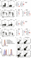
Figure 6. Retinoic Acid and T-bet-Inducing Cytokines Play Specific Roles in IEL Differentiation
(A–J) Ex vivo analysis of small intestinal IELs from 12- to 16-week-old Ifngr1–/– mice and Ifngr1+/– littermate and cage mate controls (A–E) or from 8- to 10-week-old Il27ra –/– mice and Il27ra+/– littermate and cage mate controls (F–J). (A and F) CD8α and CD103 expression by gated TCRαβ+CD4+CD8β IELs. (B and G) TL-tetramer (CD8αα) quantification in induced IELs, TCRαβ+CD4+CD8β (left), and TCRαβ+CD8β+ (right). (C and H) CD8αα and CD8β expression by gated TCRβ+CD4 IELs. (D and I) Ratio of TCRγδ+ to TCRαβ+ among CD45+ cells. (E and J) CD8αα quantification in natural IELs, TCRγδ+ (left), and TCRαβ+CD4CD8β (right). (K–M) Sorted naive Va2+CD4+ T cells isolated from WT and (OTII) dominant-negative RA receptor (OTII [dnRara]) Zbtb7b -GFP reporter mice were cocultured with DCs, OVA peptide, and indicated cytokines. (K) Histograms of T-bet expression by OTII cells from WT (OTII) or (OTII[dnRara]). (L) CD8β and CD8α expression by CD4+ T cells from OTII (left) or OTII (dnRara) (right) mice. (M) Histogram of ThPOK (left) and CD103 (right) expression by gated CD4+CD8β OTII cells. Plots are representative of three independent experiments. Statistical significance analyzed by Student's t test of pooled experiments. Error bars represent SEM. *p < 0.05; **p < 0.01; ***p < 0.001.

Comment in

Similar articles

Cited by

References

    1. Cheroutre H, Lambolez F. Doubting the TCR coreceptor function of CD8alphaalpha. Immunity. 2008;28:149–159. - PubMed
    1. Cheroutre H, Lambolez F, Mucida D. The light and dark sides of intestinal intraepithelial lymphocytes. Nat. Rev. Immunol. 2011;11:445–456. - PMC - PubMed
    1. Cruz-Guilloty F, Pipkin ME, Djuretic IM, Levanon D, Lotem J, Lichtenheld MG, Groner Y, Rao A. Runx3 and T-box proteins cooperate to establish the transcriptional program of effector CTLs. J. Exp. Med. 2009;206:51–59. - PMC - PubMed
    1. Das G, Augustine MM, Das J, Bottomly K, Ray P, Ray A. An important regulatory role for CD4+CD8 alpha alpha T cells in the intestinal epithelial layer in the prevention of inflammatory bowel disease. Proc. Natl. Acad. Sci. USA. 2003;100:5324–5329. - PMC - PubMed
    1. Denning TL, Granger SW, Mucida D, Graddy R, Leclercq G, Zhang W, Honey K, Rasmussen JP, Cheroutre H, Rudensky AY, Kronenberg M. Mouse TCRalphabeta+CD8alphaalpha intraepithelial lymphocytes express genes that down-regulate their antigen reactivity and suppress immune responses. J. Immunol. 2007;178:4230–4239. - PubMed

Publication types

MeSH terms