. 2014 Aug 14;158(4):764-777.
doi: 10.1016/j.cell.2014.06.023.

Stem-loop recognition by DDX17 facilitates miRNA processing and antiviral defense

Affiliations

Stem-loop recognition by DDX17 facilitates miRNA processing and antiviral defense

Ryan H Moy et al. Cell. .

Abstract

DEAD-box helicases play essential roles in RNA metabolism across species, but emerging data suggest that they have additional functions in immunity. Through RNAi screening, we identify an evolutionarily conserved and interferon-independent role for the DEAD-box helicase DDX17 in restricting Rift Valley fever virus (RVFV), a mosquito-transmitted virus in the bunyavirus family that causes severe morbidity and mortality in humans and livestock. Loss of Drosophila DDX17 (Rm62) in cells and flies enhanced RVFV infection. Similarly, depletion of DDX17 but not the related helicase DDX5 increased RVFV replication in human cells. Using crosslinking immunoprecipitation high-throughput sequencing (CLIP-seq), we show that DDX17 binds the stem loops of host pri-miRNA to facilitate their processing and also an essential stem loop in bunyaviral RNA to restrict infection. Thus, DDX17 has dual roles in the recognition of stem loops: in the nucleus for endogenous microRNA (miRNA) biogenesis and in the cytoplasm for surveillance against structured non-self-elements.


Figures

Figure 1
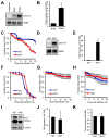
Figure 1. DEAD-box helicase screen identifies RVFV restriction factors
A. Drosophila cells were treated with the indicated dsRNA, infected with RVFV (MOI=0.04) and processed for immunofluorescence (IF) and automated microscopy 30 hpi. Average fold increase in percent infection compared to control dsRNA-treated cells is shown. Mean±SEM. B. Average fold change in total nuclei from (A). C. Representative IF image of RVFV-infected Drosophila cells treated with the indicated dsRNA. D. Immunoblot for RVFV protein (Gn) from infected cells treated with the indicated dsRNA 30 hpi. E. Average fold increase in RVFV RNA in cells treated with the indicated dsRNA as quantified by Northern. Mean±SEM; *p<0.05, Student’s t-test. All data represent three independent experiments. See also Figure S1 and Table S1.
Figure 2
Figure 2. Rm62 restricts RVFV infection in adult flies
A. Rm62-silenced flies (Hs-Gal4> UAS-Rm62 IR) or sibling controls were infected with RVFV and monitored for survival. Mean±SEM; p<0.05, Log-rank test. B. Northern for RVFV RNA from infected flies at d6 and d9 post-infection. C. Fold increase in RVFV S segment RNA from Rm62-silenced flies 6 dpi. Mean±SEM; *p<0.05, Student’s t-test. D. Survival of Rm62 mutant flies (Rm62CB02119/Rm6201086; MUT) or sibling control flies (Rm62CB02119/+; HET) infected with RVFV. Mean±SEM; p<0.001, Log-rank test. E. Representative RNA blot for RVFV RNA from RVFV-infected flies 6 dpi. F. Fold increase in RVFV S segment RNA in Rm62 mutant flies quantified by RNA blot. Mean±SEM; *p<0.05, Student’s t-test. All data represent at least three independent experiments. See also Figure S2.
Figure 3
Figure 3. Rm62 specifically restricts bunyavirus infection in flies
A. Drosophila cells were treated with the indicated dsRNA and infected with LACV (MOI=1). Viral RNA (LACV N) was monitored by RNA blot 36 hpi. B. Fold increase in LACV N RNA levels. Mean±SEM; *p<0.05, Student’s t-test. C. Survival of LACV- infected Rm62 mutant (Rm62CB02119/Rm6201086; MUT) or control flies (Rm62CB02119/+; HET). Mean±SEM; p<0.005, Log-rank test. D. Northern for LACV N mRNA from LACV-infected flies 6 dpi. E. Fold increase in LACV N in Rm62 mutant flies. Mean±SEM; *p<0.05, Student’s t-test. F. Survival of DCV-infected flies. G. Survival of VSV-GFP-infected flies. H. Survival of SINV-GFP-infected flies. I. Immunoblot for DCV capsid from DCV-infected flies 4 dpi. J. Fold increase in viral RNA (GFP) from VSV-GFP infected flies 6 dpi quantified by Northern. Mean±SEM. K. Fold increase in viral RNA (GFP) from SINV-GFP infected flies 6 dpi quantified by Northern. Mean±SEM. All data represent three independent experiments. See also Figure S3.
Figure 4
Figure 4. DDX17 controls RVFV and LACV replication in human cells
A. Representative IF image of RVFV-infected U2OS cells (MOI=0.3) transfected with the indicated siRNAs 16 hpi. B. Relative percent RVFV infection as quantified by automated image analysis. Mean±SEM; *p<0.05, Student’s t-test. C. Representative RNA blot from siRNA-transfected U2OS cells infected with RVFV 16 hpi. D. Fold increase in RVFV N mRNA compared to control siRNA-treated U2OS cells as quantified by Northern. Mean±SEM; *p<0.05, Student’s t-test. E. Representative IF image of LACV-infected U2OS cells (MOI=0.3) transfected with the indicated siRNAs 16 hpi. F. Relative percent LACV infection as quantified by automated image analysis. Mean±SEM; *p<0.05, Student’s t-test. G. Representative Northern for LACV N mRNA from siRNA-transfected U2OS cells infected with LACV 16 hpi. H. U2OS cells were transfected with the indicated siRNAs and infected with RVFV for 16h. Viral protein (Gn) was monitored by immunoblot. I. Immunoblot for viral protein (GFP) from U2OS cells treated with the indicated siRNAs and infected with VSV-GFP at 16 hpi (MOI=1). J. Ifit1 mRNA expression from uninfected or VSV-infected U2OS cells treated with the indicated siRNAs at 16 hpi by qRT-PCR, normalized to uninfected siCON-treated cells. Mean±SEM. K. Ifit1 mRNA expression from uninfected or RVFV-infected U2OS cells treated with the indicated siRNAs at 16 hpi by qRT-PCR, normalized to uninfected siCON-treated cells. Mean±SEM. All data represent three independent experiments. See also Figure S4.
Figure 5
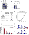
Figure 5. CLIP-seq analysis of DDX17-bound RNAs from uninfected and RVFV-infected U2OS cells
A. Immunoblot from uninfected or RVFV-infected U2OS cells with immunoprecipitation (IP) using anti-DDX17 or anti-FLAG (control). Input, IP and unbound fractions are shown, with high efficiency of DDX17 IP. B. Autoradiograph of immunopurified and 32P-labeled DDX17-RNA complexes transferred to nitrocellulose membrane. Immunoprecipitation with anti-FLAG as a control shows high specificity of the DDX17-RNA signal. C. Flowchart of CLIP-seq alignment and processing pipeline, resulting in alignment-clusters. D. Alignment clusters overlapping annotated regions of the genome (refSeq) were further searched for significant peaks and the overlap between infected and uninfected DDX17 significant CLIP-seq peaks (FDR < 0.001) in protein-coding genes from refSeq at increasing peak height is plotted. R2= 0.88. E. Percentage of total nucleotides under significant CLIP-seq peaks within refSeq protein-coding genes broken down into transcript feature types extracted from refSeq. F. Composite motif logo of the multiple sequence alignment of the 20 most enriched hexamers under significant CLIP-seq peaks within protein-coding genes as identified by z-score, comparing hexamer frequencies to 100 permutations of binding site locations within bound transcripts for uninfected (top) or infected (bottom) cells. See also Figure S5 and Table S2.
Figure 6
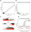
Figure 6. DDX17 directly binds miRNA stem loops in human U2OS cells
A. Normalized CLIP-seq signal (TPKM, tags per kilobase of pre-miRNA per million CLIP-seq reads) in pre-miRNA hairpin loci with CLIP signal extracted from miRBase. Linear regression of infected TPKM on uninfected TPKM is plotted, R2=0.79. B. Scatterplot of miRNAs that are bound; normalized pre-miRNA expression (RPKM) from small RNA-seq and the mean of normalized CLIP-seq signal (TPKM) between infected and uninfected U2OS cells are plotted, R2=0.001. C. Alignment clusters overlapping miRBase pre-miRNA hairpin loci on the UCSC genome browser with uninfected cells colored black, and infected cells colored red. D. RNA map of DDX17 CLIP signal in pre-miRNA hairpins. Fraction of 160 hairpins bound is plotted at single-nucleotide resolution relative to the center of the stem-loop. See also Tables S3 and S4.
Figure 7
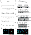
Figure 7. DDX17 binds RVFV RNA to restrict viral infection
A. DDX17 CLIP-seq clusters aligned to the RVFV tripartite genome, plotted 3′ to 5′ (genome orientation) along the x-axis. Binding sites that map to the genome are below and to the antigenome are above the line. CLIP-seq signal intensity (black) is measured in total overlapping reads at each nucleotide position. B. Predicted secondary structure of a 75-nt RNA from DDX17 CLIP peak on the RVFV S segment between N and NSs as determined by RNAfold (asterisk in A). C. The 75-nt DDX17 CLIP peak RNA from (B) was synthesized by T7 in vitro transcription and biotinylated. Biotinylated RVFV RNA was incubated with U2OS cell protein lysates and immunoprecipitated, and DDX17-RVFV RNA complexes were analyzed by immunoblot. D. RNA-protein interaction assays were performed as in (C) using the biotinylated RVFV stem loop and non-specific control RNA from RVFV not bound in the DDX17 CLIP-seq dataset. E. Representative immunoblot of U2OS cells transfected with the indicated siRNAs and infected with wild type SINV (WT) or SINV encoding the RVFV hairpin (SINV-hp) 8 hpi. F. Representative immunoblot of Drosophila cells treated with control (β-gal) or Rm62 dsRNA and infected with SINV WT or SINV-hp 24 hpi (MOI=0.3). G. Representative IF images of DDX17 and RVFV N from uninfected or infected U2OS cells 12 hpi. H. Representative IF images of DDX5 and RVFV N from uninfected or infected U2OS cells 12 hpi. (Helicase, green; RVFV-N, red; Nuclei, blue). See also Figure S6.

Comment in

Similar articles

Cited by

References

    1. Bick MJ, Carroll JW, Gao G, Goff SP, Rice CM, MacDonald MR. Expression of the zinc-finger antiviral protein inhibits alphavirus replication. Journal of virology. 2003;77:11555–11562. - PMC - PubMed
    1. Boeke J, Bag I, Ramaiah MJ, Vetter I, Kremmer E, Pal-Bhadra M, Bhadra U, Imhof A. The RNA helicase Rm62 cooperates with SU(VAR)3–9 to re-silence active transcription in Drosophila melanogaster. PloS one. 2011;6:e20761. - PMC - PubMed
    1. Bortz E, Westera L, Maamary J, Steel J, Albrecht RA, Manicassamy B, Chase G, Martinez-Sobrido L, Schwemmle M, Garcia-Sastre A. Host- and strain-specific regulation of influenza virus polymerase activity by interacting cellular proteins. mBio. 2011;2 - PMC - PubMed
    1. Boutros M, Kiger AA, Armknecht S, Kerr K, Hild M, Koch B, Haas SA, Paro R, Perrimon N. Genome-wide RNAi analysis of growth and viability in Drosophila cells. Science. 2004;303:832–835. - PubMed
    1. Bowie AG, Unterholzner L. Viral evasion and subversion of pattern-recognition receptor signalling. Nature reviews Immunology. 2008;8:911–922. - PMC - PubMed

Publication types