. 2014 Nov;88(21):12276-95.
doi: 10.1128/JVI.00970-14. Epub 2014 Aug 13.

Modulation of hepatitis C virus genome replication by glycosphingolipids and four-phosphate adaptor protein 2

Affiliations

Modulation of hepatitis C virus genome replication by glycosphingolipids and four-phosphate adaptor protein 2

Irfan Khan et al. J Virol. 2014 Nov.

Abstract

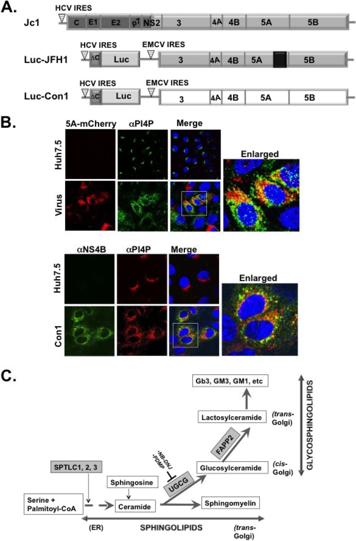
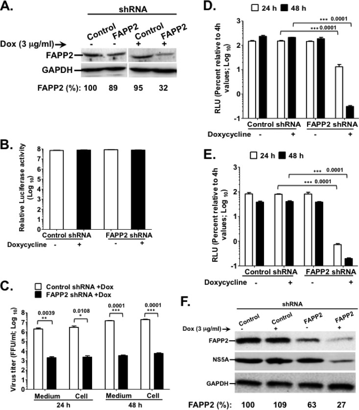
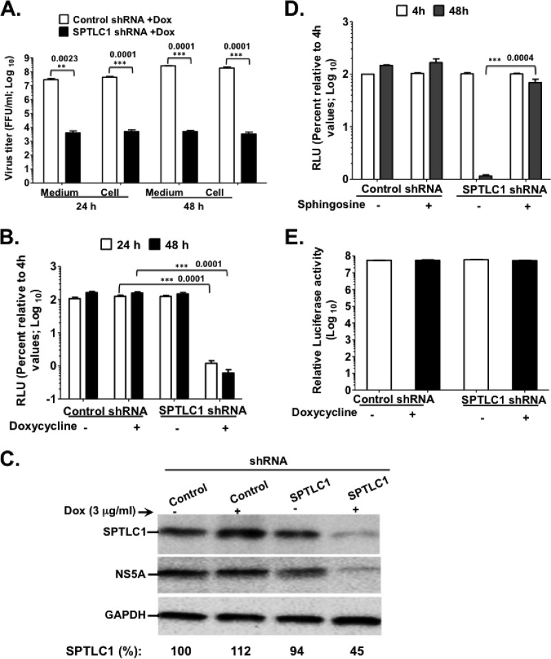
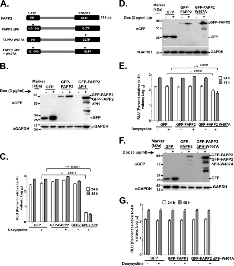
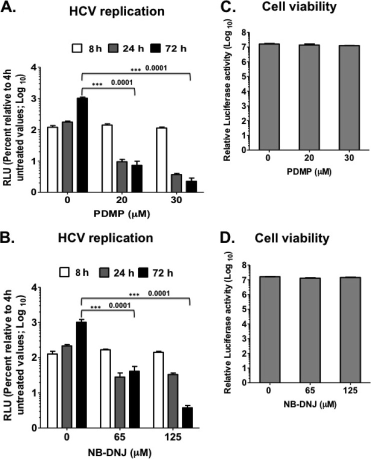
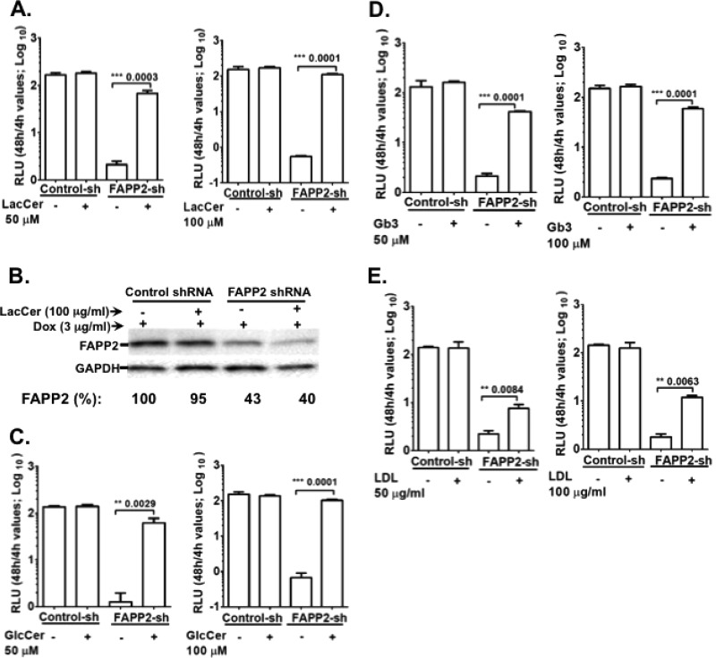
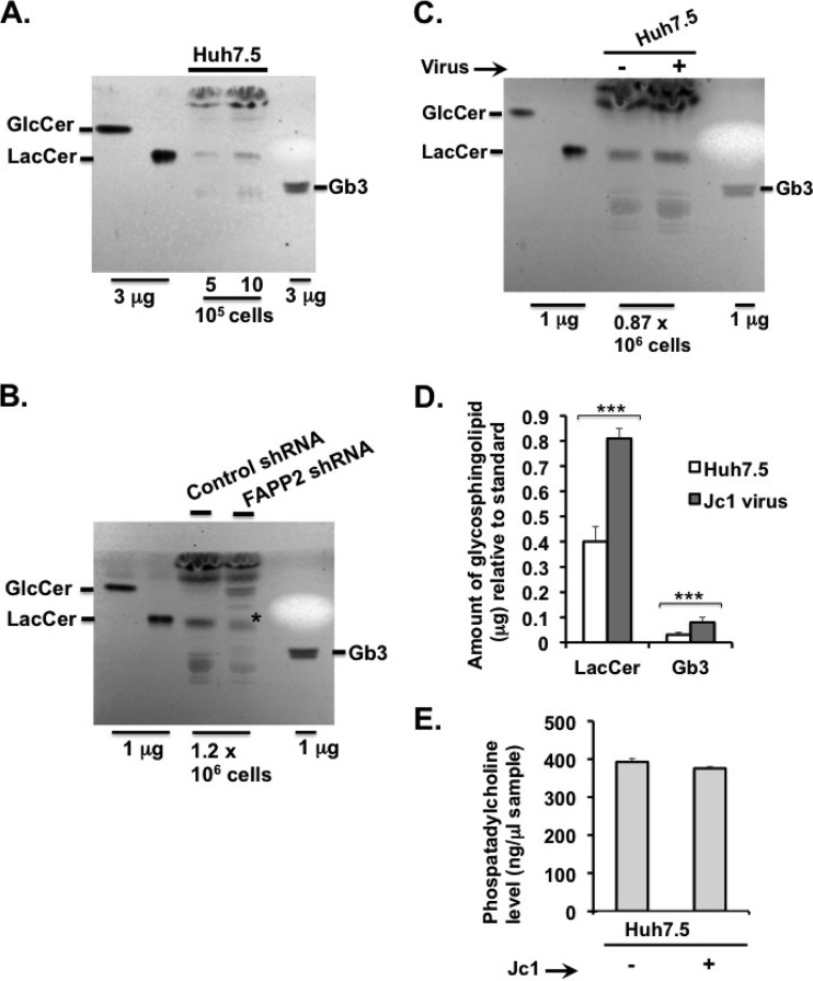
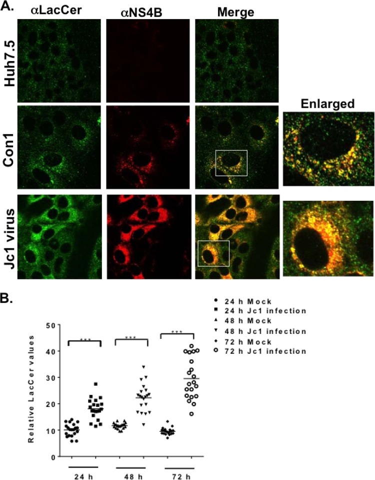
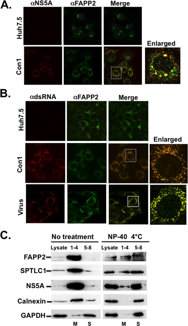
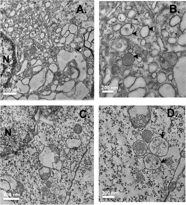
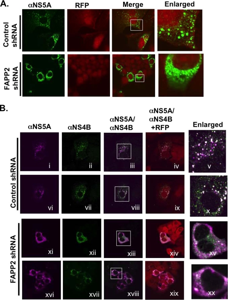
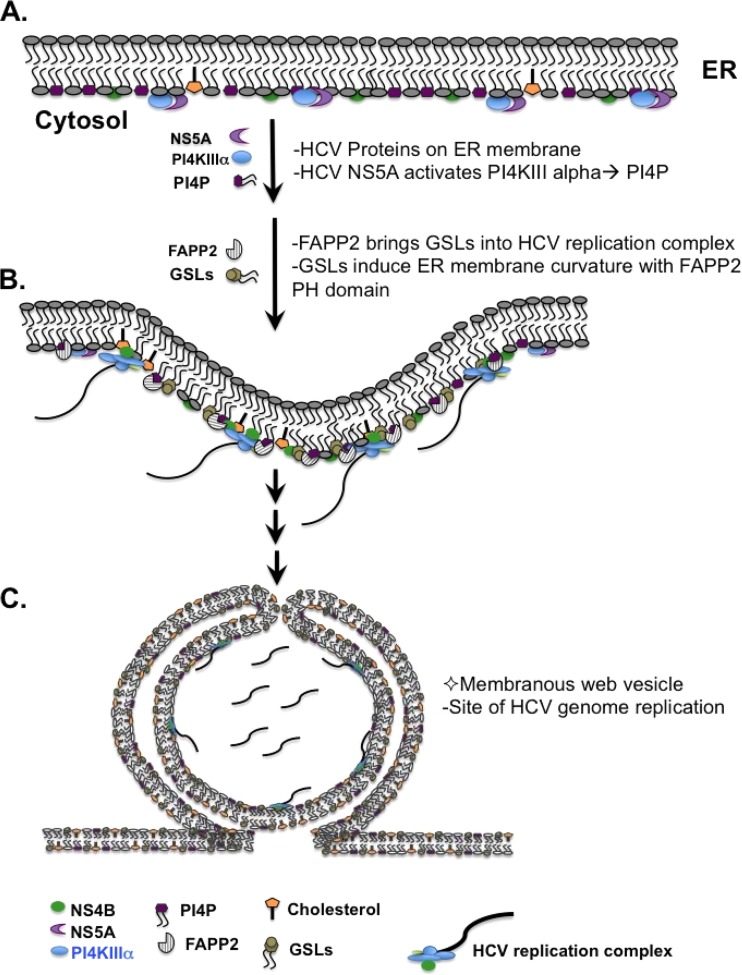
Hepatitis C virus (HCV) assembles its replication complex on cytosolic membrane vesicles often clustered in a membranous web (MW). During infection, HCV NS5A protein activates PI4KIIIα enzyme, causing massive production and redistribution of phosphatidylinositol 4-phosphate (PI4P) lipid to the replication complex. However, the role of PI4P in the HCV life cycle is not well understood. We postulated that PI4P recruits host effectors to modulate HCV genome replication or virus particle production. To test this hypothesis, we generated cell lines for doxycycline-inducible expression of short hairpin RNAs (shRNAs) targeting the PI4P effector, four-phosphate adaptor protein 2 (FAPP2). FAPP2 depletion attenuated HCV infectivity and impeded HCV RNA synthesis. Indeed, FAPP2 has two functional lipid-binding domains specific for PI4P and glycosphingolipids. While expression of the PI4P-binding mutant protein was expected to inhibit HCV replication, a marked drop in replication efficiency was observed unexpectedly with the glycosphingolipid-binding mutant protein. These data suggest that both domains are crucial for the role of FAPP2 in HCV genome replication. We also found that HCV significantly increases the level of some glycosphingolipids, whereas adding these lipids to FAPP2-depleted cells partially rescued replication, further arguing for the importance of glycosphingolipids in HCV RNA synthesis. Interestingly, FAPP2 is redistributed to the replication complex (RC) characterized by HCV NS5A, NS4B, or double-stranded RNA (dsRNA) foci. Additionally, FAPP2 depletion disrupts the RC and alters the colocalization of HCV replicase proteins. Altogether, our study implies that HCV coopts FAPP2 for virus genome replication via PI4P binding and glycosphingolipid transport to the HCV RC.

Importance: Like most viruses with a positive-sense RNA genome, HCV replicates its RNA on remodeled host membranes composed of lipids hijacked from various internal membrane compartments. During infection, HCV induces massive production and retargeting of the PI4P lipid to its replication complex. However, the role of PI4P in HCV replication is not well understood. In this study, we have shown that FAPP2, a PI4P effector and glycosphingolipid-binding protein, is recruited to the HCV replication complex and is required for HCV genome replication and replication complex formation. More importantly, this study demonstrates, for the first time, the crucial role of glycosphingolipids in the HCV life cycle and suggests a link between PI4P and glycosphingolipids in HCV genome replication.


Figures

FIG 1
FIG 1
(A) Schematic of the Jc1 (genotype 2a) virus and luciferase reporter replicons (Luc-JFH1 [genotype 2a] and Luc-Con1 [genotype 1b]) used to determine the role of the glycosphingolipid machinery in HCV replication. Note that NS5A has a C-terminal mCherry fusion (in Luc-JFH1 and virus used for panel B) as reported by Gottwein et al. (36). (B) Huh7.5 cells were mock infected or infected with HCV J6/JFH1 (MOI of 0.1) with a C-terminal mCherry fusion to NS5A as described for panel A (36). At 48 h postinfection, the cells were processed for confocal microscopy with mouse monoclonal αPI4P antibody (green). NS5A was detected via mCherry fluorescence. Alternatively, HCV Con1 replicon cells were grown for 48 h and stained with mouse monoclonal α-PI4P antibody (red) and rabbit polyclonal antibody against NS4B (green). The boxed areas are a magnified view for colocalization (yellow) of HCV NS5A or NS4B protein with PI4P. (C) Diagram of the de novo biosynthetic pathway leading to sphingolipids (e.g., ceramide and sphingomyelin) and glycosphingolipids (e.g., glucosylceramide and lactosylceramide) production. The SPTLC1, 2, 3 complex encodes the subunit of SPT (highlighted in gray), the first enzyme in the pathway leading to ceramide production. The SPTLC1 subunit interacts with SPTLC2 or SPTLC3 to form two distinct enzymatic functional complexes. Notice that ceramide is an intermediate product for generating both sphingolipids and glycosphingolipids. UGCG is highlighted in gray and codes for glucosylceramide synthase, a rate-limiting enzyme in glycosphingolipid synthesis. NB-DNJ and PDMP (54–59) are two pharmacological inhibitors of UGCGC. FAPP2 is highlighted in gray and carries glucosylceramide from the cis-Golgi to the trans-Golgi network for conversion into lactosylceramide and other glycosphingolipids. CoA, coenzyme A.
FIG 2
FIG 2
FAPP2 function is required for HCV genome replication. (A) Control and FAPP2 shRNA-expressing cells were treated with 3 μg/ml doxycycline or left untreated. Forty-eight h posttreatment, cell lysates were separated by SDS-PAGE, followed by immunoblotting with αFAPP2 (1:1,000) and αGAPDH (1:8,000) antibodies. (B) Control and FAPP2 shRNA cells were treated as described for panel A. At 48 h posttreatment, cell viability was determined using the CellTiter-Glo luminescent cell viability assay. (C) Control and FAPP2 shRNA cells were induced with 3 μg/ml doxycycline. At 48 h postinduction, the cells were electroporated with 10 μg of HCV Jc1 RNA. At 24 h and 48 h posttransfection, cell-associated (Cell) and extracellular (Medium) viruses were collected. Virus titers were measured using a limiting-dilution assay (16, 20), and the results are expressed as focus-forming units (FFU)/ml. (D and E) Control and FAPP2 shRNA cells were grown for 48 h with or without doxycycline, followed by transfection with 10 μg of Luc-JFH1 (D) or Luc-Con1 (E) replicon RNA. At 4 h, 24 h, and 48 h posttransfection, cell lysates were collected and HCV replication efficiency was measured by luciferase reporter activity as reported previously (16, 20). RLU, relative light units. The values represent percent luciferase activity relative to 4-h values. (F) The cell extracts also were collected at 48 h posttransfection (D) for immunoblotting with FAPP2 (1:1,000)-, NS5A (1:8,000)-, or GAPDH (1:8,000)-specific antibody. The data are representative of at least two independent experiments with triplicate samples for panels B to E. *, P < 0.05 (statistically significant); **, P < 0.01 (very significant); ***, P < 0.001 (extremely significant).
FIG 3
FIG 3
SPTLC1 knockdown impedes HCV genome replication. (A) Control and SPTLC1 shRNA-expressing cells were electroporated with 10 μg of HCV Jc1 RNA as described in the legend to Fig. 2B. At 24 h and 48 h posttransfection, virus titers were determined as described for Fig. 2C. (B and C) Control and SPTLC1 shRNA cells were grown for 48 h with or without doxycycline, followed by transfection with 10 μg of Luc-JFH1 replicon RNA. At 4 h, 24 h, 48 h, and 72 h posttransfection, HCV replication efficiency (B) was measured as described for Fig. 2C. (C) The cell extracts also were collected at 48 h posttransfection for immunoblotting with αSPTLC1 (1:500)-, αNS5A (1:8,000)-, or αGAPDH (1:8,000)-specific antibody. (D) Control and SPTLC1 shRNA cells were transfected with Luc-JFH1 replicon RNA as described for panel B. At 4 h posttransfection, 300 μg/ml sphingosine (49) was added to transfected cells, and HCV replication efficiency was measured at 48 h posttransfection. (E) Control and SPTLC1 shRNA cells were left untreated or were treated with doxycycline. At 48 h posttreatment, cell viability was determined as described for Fig. 2B. The data are representative of at least two independent experiments with triplicate samples for panels B to D and F. *, P < 0.05 (statistically significant); **, P < 0.01 (very significant); ***, P < 0.001 (extremely significant).
FIG 4
FIG 4
FAPP2 domains are required for HCV genome replication. (A) Diagram of FAPP2 protein (519 amino acids [aa]) and its functional domains. The PH domain (aa 1 to 112) and the GLTP domain (aa 320 to 519) bind to PI4P and glucosylceramide, respectively. The mutation in the PH domain (FAPP2 ΔPH; aa 1 to 100) or GLTP domain (FAPP2 W407A) is indicated by an asterisk. (B) Stable Huh7.5 cells expressing control GFP, WT GFP-FAPP2, or GFP-FAPP2 ΔPH were grown for 48 h with or without doxycycline, followed by immunoblotting with αGFP (1:2,000) or αGAPDH (1:8,000) antibody. Note that these cells express no shRNA. (C) The stable cells shown in panel B were transfected with 10 μg of Luc-JFH1 replicon RNA, and HCV replication efficiency was determined as described for Fig. 2C. (D and E) Stable cells, with FAPP2 W407A mutation or controls, were treated and processed for immunoblotting (D) and HCV replication efficiency assay (E), respectively. (F and G) Stable cells, with FAPP2 ΔPH-W407A mutations or controls, were treated and processed for immunoblotting (F) and HCV replication efficiency assay (G), respectively. The data are representative of at least two independent experiments with triplicate samples for panels C and D. *, P < 0.05 (statistically significant); **, P < 0.01 (very significant); ***, P < 0.001 (extremely significant).
FIG 5
FIG 5
Glucosylceramide synthase (UGCG) inhibitors impede HCV replication efficiency. (A and B) Huh7.5 cells were transfected with 10 μg of JFH1-Luc replicon RNA and were left untreated or treated with various concentrations of PDMP or NB-DNJ. HCV replication efficiency was determined at 8 h, 24 h, and 72 h posttransfection. Note that the cells were treated at 4 h postelectroporation. (C and D) Impact of PDMP and NB-DNJ on cell viability. The cells were treated with 20 and 30 μM PDMP or 63 and 125 μM NB-DNJ for 72 h, followed by cell viability measurement as described in Materials and Methods. The results are representative of three independent experiments with triplicate samples for data from panels A to D. *, P < 0.05 (statistically significant); **, P < 0.01 (very significant); ***, P < 0.001 (extremely significant).
FIG 6
FIG 6
HCV replication is restored to near completion by some glycosphingolipids in FAPP2 knockdown cells. (A and B) Dox-induced control and FAPP2 shRNA cells were transfected with 10 μg of Luc-JFH1 replicon RNA as described in Materials and Methods. (A) At 4 h posttransfection, 0, 50, or 100 μM LacCer was added to transfected cells, and HCV replication efficiency was determined at 4 h and 48 h posttransfection. (B) Additionally, cell lysates were subjected to immunoblotting with αFAPP2 (1:1,000) or αGAPDH (1:8000) antibody. (C) Control and FAPP2 shRNA cells were transfected with replicon RNA as described for panel A but treated with 0, 50, or 100 μM GlcCer. HCV replication efficiency was determined as described for panel A. (D and E) Control and FAPP2 shRNA cells were transfected with replicon RNA as described for panel A but treated with various concentrations of Gb3 (D) or cholesterol as a low-density lipoprotein (LDL) (E). HCV replication efficiency was determined as described for panel A. The data are representative of three independent experiments with triplicate samples for panels A, C, D, and E. *, P < 0.05 (statistically significant); **, P < 0.01 (very significant); ***, P < 0.001 (extremely significant).
FIG 7
FIG 7
Changes in glycosphingolipid levels in various HCV-expressing cells. (A) Huh7.5 cells were grown for 48 h, followed by glycosphingolipid extraction from 107 cells, as discussed in Materials and Methods. Extracted lipids, from 5 × 10⁵ to 10 × 105 cells, were separated on TLC plates in a chloroform-methanol-water (60:35:8) solvent system and visualized by spraying with orcinol-sulfuric acid reagent. (B) Control and FAPP2 shRNA cells were treated with 3 μg/ml Dox for 48 h. Glycosphingolipids were extracted from 107 cells, and aliquots that correspond to 1.2 × 10⁶ cells were separated on TLC plates and processed as described for panel A. (C) Huh7.5 cells (9 × 10⁶) were mock infected or infected with HCV Jc1 at an MOI of 30 to ensure at least 95% infection efficiency. At 48 h postinfection, glycosphingolipids were extracted from 7 × 10⁶ cells, and aliquots that correspond to 0.87 × 10⁶ cells were separated on TLC plates as described for panel A. (D) The amount of glycosphingolipids relative to the standard (1 μg of LacCer or Gb3), in mock-infected and Jc1 virus-infected Huh7.5 cells shown in panel C, was determined with ImageJ software. ***, P < 0.001 (extremely significant). (E) Two million mock-infected and Jc1 virus-infected cells from panel C were processed for phosphatidylcholine assay as described in Materials and Methods. The results shown in panels D and E are representative of at least two independent experiments, each containing data from 3 TLC runs.
FIG 8
FIG 8
Lactosylceramide is associated with HCV NS4B protein. (A) Parental Huh7.5 and HCV Con1 (genotype 1b) replicon cells were grown for 48 h and processed for confocal microscopy with mouse monoclonal αLacCer antibody (1:500; green) and rabbit polyclonal αNS4B antibody (1:150; red). Huh7.5 cells also were infected with HCV Jc1 (MOI of 1) and processed for confocal microscopy as described above. The boxed areas represent a magnified view for colocalization (yellow) of lactosylceramide and HCV NS4B protein. (B) Huh7.5 cells were infected with HCV Jc1 (MOI of 1) as described for panel A and processed for confocal microscopy 24 h, 48 h, and 72 h postinfection. For each infection time point, confocal images were taken of 20 representative cells. The intensity of lactosylceramide pixels was calculated with the JACoP plugin in ImageJ software. Each filled circle or square (24 h; mock or Jc1 infected), upper or lower triangle (48 h; mock or Jc1 infected), diamond (72 h; mock), or open circle (72 h; Jc1 infected) represents one cell.
FIG 9
FIG 9
FAPP2 colocalizes with HCV NS5A and viral dsRNA. (A) Parental Huh7.5 and HCV Con1 (genotype 1b) replicon cells were grown for 48 h and processed for confocal microscopy with mouse monoclonal αNS5A antibody (1:1,000; red) and rabbit polyclonal αFAPP2 antibody (1:100; green). (B) Huh7.5, Con1 replicon, or Jc1 (34) virus-infected cells were grown as described for panel A and processed for confocal microscopy with mouse monoclonal antibody against dsRNA (1: 200; red) and rabbit polyclonal αFAPP2 antibody (1:100; green). The boxed areas indicate the magnified view for colocalization (yellow color) of HCV NS5A (A) or dsRNA (B) with FAPP2 protein. (C) FAPP2 cofractionates with HCV replicase NS5A protein in the detergent-resistant membrane fraction. Con1 replicon cell lysates were left untreated or were treated with 1% NP-40 on ice and subjected to membrane floatation. Proteins from pooled fractions (1 to 4 and 5 to 9) were separated by SDS-PAGE, followed by immunoblotting with antibodies against FAPP2, SPTLC1, NS5A, calnexin, or GAPDH. Numbers 1 to 4 refer to membrane (M) fractions, and numbers 5 to 8 refer to soluble (S) fractions.
FIG 10
FIG 10
Ultrastructural analysis of shRNA cells infected with Jc1 virus. Control (A and B) and FAPP2 (C and D) shRNA cells were treated with Dox as described in the legend to Fig. 7, followed by Jc1 virus infection as described in Materials and Methods. The cells were processed for electron microscopy at 48 h postinfection. (B and D) Magnified images of panels A and C, respectively. (B) Arrowheads indicate vesicles of 150 to 200 nm in size. (D) Arrows show vesicles larger than 250 nm in size. Scale bars indicate the magnification for each image.
FIG 11
FIG 11
FAPP2 knockdown disrupts HCV NS4B and NS5A focus formation. (A) Control and FAPP2 shRNA cells were treated with 3 μg/ml doxycycline as described in the legend to Fig. 7, followed by transfection with pIRES vector expressing HCV NS3-4A-4B-5A-5B polyprotein in the presence of doxycycline. At 48 h posttransfection, the cells were fixed and processed for confocal microscopy with mouse monoclonal αNS5A antibody (1:1,000; green). Red fluorescent protein (RFP) indicates control and FAPP2 shRNA cells. Magnified areas, with NS5A subcellular distribution, are shown by rectangles. (B) Control (i to x) and FAPP2 (xi to xx) shRNA cells were treated as described for panel A and processed for confocal microscopy with mouse monoclonal αNS5A antibody (1:1,000; magenta) and rabbit polyclonal antibody against NS4B (1:25; green). RFP indicates shRNA-expressing cells as described for panel A. Magnified areas, with putative NS4B and NS5A colocalization (white), are indicated by rectangles.
FIG 12
FIG 12
Proposed model for the role of FAPP2 in HCV genome replication. (A) After translation and processing on endoplasmic reticulum (ER) membranes, HCV NS5A activates PI4KIIIα to produce PI4P. (B) FAPP2 brings glycosphingolipids (GSLs) into the HCV RC in part via interaction with PI4P. GSLs and the FAPP2 PH domain regulate ER membrane curvature to form the MW vesicles. Alternatively, GSLs control the size of the MW vesicles or the local concentration of the replicase proteins. (C) Schematic of the double-membrane vesicles, as depicted by Paul et al. (79); these vesicles are clustered into an MW structure.

Similar articles

Cited by

References

    1. Averhoff FM, Glass N, Holtzman D. 2012. Global burden of hepatitis C: considerations for healthcare providers in the United States. Clin. Infect. Dis. 55(Suppl 1):S10–S15. 10.1093/cid/cis361. - DOI - PubMed
    1. Lavanchy D. 2011. Evolving epidemiology of hepatitis C virus. Clin. Microbiol. Infect. 17:107–115. 10.1111/j.1469-0691.2010.03432.x. - DOI - PubMed
    1. Miller RH, Purcell RH. 1990. Hepatitis C virus shares amino acid sequence similarity with pestiviruses and flaviviruses as well as members of two plant virus supergroups. Proc. Natl. Acad. Sci. U. S. A. 87:2057–2061. 10.1073/pnas.87.6.2057. - DOI - PMC - PubMed
    1. Simmonds P, Bukh J, Combet C, Deleage G, Enomoto N, Feinstone S, Halfon P, Inchauspe G, Kuiken C, Maertens G, Mizokami M, Murphy DG, Okamoto H, Pawlotsky JM, Penin F, Sablon E, Shin IT, Stuyver LJ, Thiel HJ, Viazov S, Weiner AJ, Widell A. 2005. Consensus proposals for a unified system of nomenclature of hepatitis C virus genotypes. Hepatology 42:962–973. 10.1002/hep.20819. - DOI - PubMed
    1. Bartenschlager R, Lohmann V. 2000. Replication of hepatitis C virus. J. Gen. Virol. 81(Part 7):1631–1648 http://vir.sgmjournals.org/content/81/7/1631.long. - PubMed

Publication types

MeSH terms

Substances