. 2014 Nov;4(11):1310-25.
doi: 10.1158/2159-8290.CD-13-1010. Epub 2014 Aug 13.

The androgen-regulated protease TMPRSS2 activates a proteolytic cascade involving components of the tumor microenvironment and promotes prostate cancer metastasis

Affiliations

The androgen-regulated protease TMPRSS2 activates a proteolytic cascade involving components of the tumor microenvironment and promotes prostate cancer metastasis

Jared M Lucas et al. Cancer Discov. 2014 Nov.

Abstract

TMPRSS2 is an androgen-regulated cell-surface serine protease expressed predominantly in prostate epithelium. TMPRSS2 is expressed highly in localized high-grade prostate cancers and in the majority of human prostate cancer metastases. Through the generation of mouse models with a targeted deletion of Tmprss2, we demonstrate that the activity of this protease regulates cancer cell invasion and metastasis to distant organs. By screening combinatorial peptide libraries, we identified a spectrum of TMPRSS2 substrates that include pro-hepatocyte growth factor (HGF). HGF activated by TMPRSS2 promoted c-MET receptor tyrosine kinase signaling, and initiated a proinvasive epithelial-to-mesenchymal transition phenotype. Chemical library screens identified a potent bioavailable TMPRSS2 inhibitor that suppressed prostate cancer metastasis in vivo. Together, these findings provide a mechanistic link between androgen-regulated signaling programs and prostate cancer metastasis that operate via context-dependent interactions with extracellular constituents of the tumor microenvironment.

Significance: The vast majority of prostate cancer deaths are due to metastasis. Loss of TMPRSS2 activity dramatically attenuated the metastatic phenotype through mechanisms involving the HGF-c-MET axis. Therapeutic approaches directed toward inhibiting TMPRSS2 may reduce the incidence or progression of metastasis in patients with prostate cancer.


Conflict of interest statement

Conflicts of Interest: None of the authors have any relevant conflicts of interest pertaining to the studies and data in this manuscript.

Figures

Figure 1
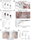
Figure 1. Expression of TMPRSS2 in prostate carcinoma
(A, C) qRT-PCR measurements of TMPRSS2, hepsin and matriptase mRNA from microdissected primary prostate carcinoma and metastasis from men with advanced CRPC. (B) Immunohistochemical localization of TMPRSS2 protein in benign prostate epithelium (white arrows) and prostate carcinoma (black arrows). TMPRSS2 in benign epithelium is oriented to the ductal lumen with little protein in contact with stromal constituents. In contrast, cancer cells expressing TMPRSS2 are in direct contact with stroma. (D) TMPRSS2 proteinin prostate cancer metastasis. Lymph node stroma and osteoid are denoted by an asterix. The consistency of TMPRSS2 protein in multiple different metastases from 44 patients with advanced prostate cancer demonstrates that most metastatic foci express TMPRSS2 with a general concordance in multiple tumors from the same individual. Each data point represents an individual tumor focus and black (odd numbered patients) and gray (even numbered patients) data points alternate for clarity. (E) TMPRSS2 protein staining intensity in ERG positive versus ERG negative primary prostate cancers. (F) Representative example of TMPRSS2 protein expression in primary prostate cancers with or without ERG expression (G) Expression of TMPRSS2 in human prostate epithelium is attenuated following treatment with androgen-suppressing therapeutics, estradiol or the LHRH agonist leuprolide, compared to untreated controls.
Figure 2
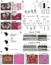
Figure 2. Tmprss2 influences prostate cancer growth and the development of metastasis
(A) Representative anatomical and histological images of prostate glands from wild type (WT; WT) and TRAMP mice with (Tmprss2+/+; TRAMP) and without (Tmprss2−/−; TRAMP) Tmprss2 protease activity. Both TRAMP genotypes exhibited a spectrum of well- to poorly-differentiated carcinomas. P, prostate gland; SV, seminal vesicle. Prostate glands are circled. (B) Weights of the genitourinary tracts (GU-SV) excised from TRAMP mice with (Tmprss2+/+; TRAMP) or without (Tmprss2−/−; TRAMP) Tmprss2 activity. At 32 weeks of age, prostate tumors in Tmprss2−/−; TRAMP mice were substantially larger than those tumors excised from Tmprss2+/+; TRAMP animals (P=0.01). (C) The frequency of lymph node metastasis in Tmprss2−/−; TRAMP versus Tmprss2+/+; TRAMP genotypes was similar. The frequency of metastasis to solid organs in TRAMP mice with inactive Tmprss2 was significantly reduced compared to TRAMP mice with wild-type Tmprss2 (P=0.02). (D) Basement membrane invasion assays demonstrating enhanced invasion of primary cells from Tmprss2WT; TRAMP tumors relative to benign epithelium or primary tumor cells deficient in Tmprss2 activity (Tmprss2−/−; TRAMP). (E) Soft-agar colony formation assay demonstrating enhanced anchorage-independent growth of DU145 prostate cancer cells expressing wild-type active human TMPRSS2 (TMPRSS2WT) versus a vector control or protease-dead TMPRSS2 mutant (TMPRSS2PM). (F) Schematic of the metastasis assay: primary tumors developing in Tmprss2−/−; TRAMP or Tmprss2+/+; TRAMP were resected, cells dissociated, and injected into tail veins of recipient hosts. (G) Detection of S40T antigen by PCR in blood (b), liver (l) or lung (lu) of mice at designated time points following tail-vein injections with primary prostate tumor cells from Tmprss2+/+; TRAMP or Tmprss2−/−; TRAMP mice. TCR, T-cell receptor.(H) Representative anatomical and histological images of livers from mice receiving vascular injections of cells from Tmprss2−/−; TRAMP andTmprss2+/+; TRAMP tumors.
Figure 3
Figure 3. Identification of TMPRSS2 protease substrates
(A) Results of positional scanning of synthetic combinatorial peptide libraries (PS-SCL). The amino acid cleavage preferences are: P1 (R); P2 (T, F, W, A, V); P3 (E, M, Q); P4 (G, I, M). (B) pro-HGF is a substrate for TMPRSS2. The arrow denotes the single-chain (pro-) HGF and is not visualized following incubation with active TMPRSS2. The inactive TMPRSS2 protease-dead mutant (TMPRSS2PM) is incapable of catalyzing HGF proteolysis. TMPRSS2 does not degrade the BSA carrier protein (dominant bands). Western blot analysis using a polyclonal anti-HGF antibody that recognizes multiple epitopes in the α and β chains of HGF demonstrates the loss of single chain (pro) HGF following incubation with active TMPRSS2 (lane 2), but not protease-dead TMPRSS2 (lane 3). (C) pro-HGF proteolyzed by TMPRSS2 activates c-Met as determined by immunodetection of phosphorylated c-Met (p-cMet) in DU145 prostate cancer cells. Control reactions with matriptase also demonstrate c-Met activation. Pro-HGF alone and the addition of anti-HGF neutralizing antibody abolishes c-Met phosphorylation.
Figure 4
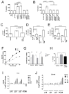
Figure 4. TMPRSS2-activated HGF consistently promotes invasion but differentially enhances or suppresses proliferation
(A) Exposure of DU145 prostate cancer cells to pro-HGF activated by either TMPRSS2 or matriptase increases cellular invasion. (B) Exposure of DU145 prostate cancer cells to prostate fibroblast conditioned medium and TMPRSS2 increases cellular invasion, which is not observed with an inactive TMPRSS2 protease mutant (TMPRSS2PM) and abolished with the addition of anti-HGF neutralizing antibody or the cMet inhibitor SU11274. The pro-invasive phenotype associated with TMPRSS2 expression in LNCaP (C) PC3 (D) and DU145 (E) cells is promoted by exogenous paracrine-acting HGF. SF is serum-free medium. (F, G) TMPRSS2-activated HGF enhances the proliferation of DU145 cells and suppresses the proliferation of BPH1 and TRAMP C2 cells (TC2). Cell numbers were determined at 96 hours and represent a mean of 3 replicates (asterix = p<0.01). (H) Invasion of TRAMP C2 cells is promoted by TMPRSS2-activated HGF and attenuated with anti-HGF neutralizing antibody (asterix = p<0.01). Differential effects of activated HGF on cell cycle regulators p21 and p27 in TC2 (I) and DU145 (J) cells.
Figure 5
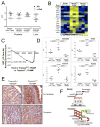
Figure 5. TMPRSS2 associates with a gene expression program involved in epithelial to mesenchymal transition (EMT)
(A) AR and c-Met transcript levels in primary and metastatic TRAMP tumors determined by qRT-PCR.(B) Transcript profiling of mRNAs from microdissected prostate epithelium. Genes associated with EMT are elevated in Tmprss2WT; TRAMP tumors (yellow) and decreased in Tmprss2−/−; TRAMP tumors (blue). (C) Gene set enrichment analysis (GSEA) confirms a significant enrichment in EMT-associated genes in Tmprss2WT; TRAMP tumors (p<0.001). (D) qRT-PCR quantitation of gene expression from benign and neoplastic epithelium microdissected from TRAMP mouse strains with and without Tmprss2 activity. (E) Immunohistochemical staining of TRAMP tumors for EMT-associated proteins vimentin and N-cadherin. (F) Schematic view of the proteolytic cascades influenced by TMPRSS2 in the context of normal physiology in benign epithelium and in neoplasia where substrates normally confined to the stroma are available for interactions with the TMPRSS2 protease. SC, secretory epithelial cell; BC, basal epithelial cell; BM, basement membrane; CC, cancerous epithelial cell, EMT, epithelial to mesenchymal transition.
Figure 6
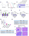
Figure 6. TMPRSS2 chemical inhibitors suppress prostate tumor growth and metastasis in vivo
(A) Chemical structure of the identified TMPRSS2 inhibitor bromhexine (BHH).(B) BHH exhibits substantially greater inhibitory activity toward TMPRSS2 relative to hepsin or matriptase. Optimal peptide substrates were used for each enzyme and substrate concentrations were determined after 30 minutes.(C) Suppression of TMPRSS2-induced cleavage of the TMPRSS2 substrate pro-PLAT by the chemical TMPRSS2 inhibitors identified through screening chemical libraries. T2 is active TMPRSS2. BHH is bromhexine. (D) BHH exposure does not induce cell death or substantially suppress the growth of DU145 cells. BHH suppresses the serum- and HGF-mediated invasion of (E) DU145 and (F) PC3 cells expressing TMPRSS2. (G) In vivo treatment with BHH increases the size of primary TRAMP tumors compared to TRAMP mice treated with DMSO vehicle. Treatment was initiated at age 15 weeks and tumor weights were determined after 20 weeks of treatment.(H) TRAMP mice treated with BHH have substantially fewer spontaneous metastasis to lung or liver. (I) BHH treatment substantially reduced the frequency of distant metastasis following tail vein injections of tumor cells harvested from primary TRAMP tumors. (J) Representative histology of murine liver 8 weeks following the intravenous injection of TRAMP tumor cells showing liver metastasis in mice treated with DMSO vehicle and normal liver histology without metastasis in BHH treated mice.

Comment in

Similar articles

Cited by

References

    1. Netzel-Arnett S, Hooper JD, Szabo R, Madison EL, Quigley JP, Bugge TH, et al. Membrane anchored serine proteases: a rapidly expanding group of cell surface proteolytic enzymes with potential roles in cancer. Cancer Metastasis Rev. 2003;22:237–58. - PubMed
    1. Hooper JD, Clements JA, Quigley JP, Antalis TM. Type II transmembrane serine proteases. Insights into an emerging class of cell surface proteolytic enzymes. J Biol Chem. 2001;276:857–60. - PubMed
    1. Szabo R, Bugge TH. Type II transmembrane serine proteases in development and disease. Int J Biochem Cell Biol. 2008;40:1297–316. - PubMed
    1. Szabo R, Wu Q, Dickson RB, Netzel-Arnett S, Antalis TM, Bugge TH. Type II transmembrane serine proteases. Thromb Haemost. 2003;90:185–93. - PubMed
    1. List K, Bugge TH, Szabo R. Matriptase: potent proteolysis on the cell surface. Mol Med. 2006;12:1–7. - PMC - PubMed

Publication types