. 2014 Jun 25:3:e29080.
doi: 10.4161/onci.29080. eCollection 2014.

Bone marrow-derived cells are implicated as a source of lymphatic endothelial progenitors in human breast cancer

Affiliations

Bone marrow-derived cells are implicated as a source of lymphatic endothelial progenitors in human breast cancer

Eveline Faes Van't Hull et al. Oncoimmunology. .

Abstract

Bone marrow-derived endothelial progenitor cells (EPCs) infiltrate into sites of neovascularization in adult tissues and mature into functional blood endothelial cells (BECs) during a process called vasculogenesis. Human marrow-derived EPCs have recently been reported to display a mixed myeloid and lymphatic endothelial cell (LEC) phenotype during inflammation-induced angiogenesis; however, their role in cancer remains poorly understood. We report the in vitro differentiation of human cord blood CD133+CD34+ progenitors into podoplanin+ cells expressing both myeloid markers (CD11b, CD14) and the canonical LEC markers vascular endothelium growth factor receptor 3 (VEGFR-3), lymphatic vessel endothelial hyaluronan receptor 1 (LYVE-1), and prospero homeobox 1 (PROX-1). These podoplanin+ cells displayed sprouting behavior comparable to that of LECs in vitro and a dual hemangiogenic and lymphangiogenic activity in vivo in an endothelial cell sprouting assay and corneal vascularization assay, respectively. Furthermore, these cells expressed vascular endothelium growth factor (VEGF) family members A, -C, and -D. Thus, bone-marrow derived EPCs stimulate hemangiogenesis and lymphangiogenesis through their ability to differentiate into LECs and to produce angiogenic factors. Importantly, plasma from patients with breast cancer induced differentiation of CD34+ cord blood progenitors into hemangiogenic and lymphangiogenic CD11b+ myeloid cells, whereas plasma from healthy women did not have this effect. Consistent with these findings, circulating CD11b+ cells from breast cancer patients, but not from healthy women, displayed a similar dual angiogenic activity. Taken together, our results show that marrow-derived EPCs become hemangiogenic and lymphangiogenic upon exposure to cancer plasma. These newly identified functions of bone-marrow derived EPCs are expected to influence the diagnosis and treatment of breast cancer.

Keywords: angiogenesis; bone-marrow derived cells; breast cancer; lymphangiogenesis; lymphatic endothelial cells; podoplanin; vasculogenesis.


Figures

None
Figure 1. Increased hemangiogenic and lymphangiogenic activities of CD11b+ cells differentiated in vitro from CD34+ progenitors in the presence of plasma from breast cancer patients. (A–D) The hemangiogenic and lymphangiogenic activities of CD11b+ cells were assessed in vitro by measuring their ability to induce sprouting of human umbilical vein endothelial cells (HUVECs) and lymphatic endothelial cells (LECs) grown on microcarrier beads and embedded in a 3D fibrin gel. (A) Cumulative sprout length for HUVEC and LEC beads following exposure for 10 d to purified CD11b+ cells differentiated in vitro from CD34+ progenitors in the presence of plasma from healthy (H) individuals or breast cancer (BC) patients (n = 10–12). (B) Cumulative sprout length for CD11b+ cells isolated from peripheral blood of healthy individuals (H) and breast cancer patients (BC). (C–D) CD11b+ cells were differentiated in vitro from CD34+ progenitors following exposure to healthy plasma and the kinetics of hemangiogenic and lymphangiogenic activities (C) or differentiation into podoplanin+ cells (D) were measured. Background sprouting measured with coated HUVEC and LEC beads alone is shown (none). Data in (C) show cumulated sprout length normalized to that measured in the absence of bone marrow-derived cells (BMDCs) and in the presence of 50 ng/mL VEGF-A. Statistical significance was determined by Student’s t test and differences are indicated for all panels (*P < 0.05; **P < 0.005; ***P < 0.001).
None
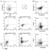
Figure 2. In vitro differentiation of CD34+ cord blood precursors into podoplanin+ cells. (A–B) CD34+ hematopoietic progenitors from cord blood were isolated by immunomagnetic selection and cultured in vitro. Resultant cells were characterized by immunostaining and cytofluorimetric analysis 15 to 35 d after the isolation of CD34+ cord blood precursors. Representative flow cytometry dot plots of the expression profiles of podoplanin, CD31, CD34, and CD45 (panel A) and podoplanin, CD133, and neuropilin-1 (panel B) are shown from 10 distinct cultures.
<b>Figure 3</b>.
Figure 3.
In vitro differentiated podoplanin+ cells display markers of lymphatic endothelial cells and myeloid cells.(A–C)CD34+ hematopoietic progenitors from cord blood were isolated by immunomagnetic selection and cultured in vitro. Expression of the lymphatic endothelial cell (LEC) markers vascular endothelium growth factor receptors (VEGFRs), neuropilins, prospero-related homeobox 1 (PROX-1), and lymphatic vessel endothelial hyaluronan receptor 1 (LYVE-1) and of VE-cadherin was assessed by immunostaining and fluorescence cytometry (A) and confocal microscopy (B) in podoplanin+ and podoplanin cell populations. A. Flow cytometry histograms of the expression level of the indicated marker in each of the two cell types, with filled histograms depicting labeling with isotype control antibodies. (B) All podoplanin+ cells (arrows) expressed LYVE-1 and most (> 80%) showed intermediate PROX-1 expression relative to human umbilical vein endothelial cells (HUVECs) and human dermal lymphatic microvascular endothelial cells (DLECs). (C) Podoplanin+ cells expression of the myeloid cell markers CD11b and CD14. Representative images from three to five experiments are shown. Cell characterizations were performed in 22- to 35-d-old cultures. Bar, 25 µm.
None
Figure 4. Podoplanin+ cells and LECs show similar abilities to form sprouts in vitro in response to vascular endothelium growth factor (VEGF). In vitro sprouting of endothelial cells cultured 25 d using podoplanin+ cells positively selected using anti–podoplanin-PE antibody and immunomagnetic separation. Podoplanin+ cells were labeled with carboxyfluorescein diacetate succinimidyl ester (CSFE) and cultured on collagen-coated microcarrier beads embedded in a 3D fibrin gel, as described below. (A) Epifluorescence microscopy image of CFSE-labeled sprouts (dashed line) of podoplanin+ cells after 5 d of stimulation with vascular endothelium growth factor (VEGF)-A and -C. (B) Confocal microscopy images of sprouts of podoplanin+ cells in 3D fibrin gel 3 d post-stimulation. Green, CD31; blue, podoplanin; red, VEGF receptor (VEGFR). Bar, 75 µm. (C–F) Sprouting capacity of endothelial cells in response to VEGF was evaluated in vitro by measuring the mean cumulative sprout length. (C) Beads were coated exclusively with human umbilical vein endothelial cells (HUVECs) or lymphatic microvascular endothelial cells (LECs) (D–F) Chimeric beads were coated with both HUVECs and LECs (D), HUVEC and podoplanin+ cells (E), or LECs and podoplanin+ cells in a 2:1 ratio (F). Sprouts of lymphatic origin were identified by incorporation of CFSE into LECs or podoplanin+ cells. Cumulative data from a minimum of three experiments are shown. Background sprouting (96.8 ± 60 pixels) measured with coated beads alone was subtracted from all data. Significant differences between cell types in response to the same treatment were measured by Student’s t test; *P < 0.05 in panels C–F; in panel C, significant differences among the treatments for HUVECs and LECs are indicated by blue and red asterisks, respectively.
None
Figure 5. Podoplanin+ cells are hemangiogenic and lymphangiogenic in vitro and in vivo. The hemangiogenic and lymphangiogenic activities of podoplanin+ cells (derived from cultured cord blood CD34+ precursor cells) and microvascular lymphatic endothelial cells (LECs) were assessed in vitro by measuring their aptitude to induce sprouting of human umbilical vein endothelial cells (HUVECs) and LECs grown on microcarrier beads and embedded in a 3D fibrin gel. (A) Cumulated sprout length for HUVEC (left panel) and LEC (right panel) beads following exposure to podoplanin+ cells or LECs. Background sprouting (71.4 ± 57 pixels) measured with HUVEC and LEC beads alone was subtracted. Significant differences were determined by Student’s t test, *P < 0.05; **P < 0.005). (B) Corneal vascularization assay to assess the in vivo hemangiogenic and lymphangiogenic activity of podoplanin+ cells was performed by transplantation of 30 000 podoplanin+ cells into the corneal stroma of anesthetized NOD-SCID/IL2Rγnull mice. Fluorescent microscopy images of sagittal sections of mouse eyes using antibodies specific for CD31 and lymphatic vessel endothelial hyaluronan receptor 1 (LYVE-1) revealed de novo formation of blood and lymphatic vessels emerging from the peripheral limbal vasculature (arrow heads) and expanding into the cornea (vertical double-headed arrow). Single arrow depicts the iris. Bar, 100 μm. Cumulative data of three independent experiments are shown in (A) and a representative experiment among three is shown in (B). (C) Expression of vascular endothelium growth factor (VEGF)-A and -C in the podoplanin+ cell fraction detected by confocal microscopy. Bar, 12 µm.

Similar articles

Cited by

References

    1. Choi I, Lee S, Hong YK. The new era of the lymphatic system: no longer secondary to the blood vascular system. Cold Spring Harb Perspect Med. 2012;2:a006445. doi: 10.1101/cshperspect.a006445. - DOI - PMC - PubMed
    1. Olsson AK, Dimberg A, Kreuger J, Claesson-Welsh L. VEGF receptor signalling - in control of vascular function. Nat Rev Mol Cell Biol. 2006;7:359–71. doi: 10.1038/nrm1911. - DOI - PubMed
    1. Zumsteg A, Christofori G. Myeloid cells and lymphangiogenesis. Cold Spring Harb Perspect Med. 2012;2:a006494. doi: 10.1101/cshperspect.a006494. - DOI - PMC - PubMed
    1. Asahara T, Kawamoto A. Endothelial progenitor cells for postnatal vasculogenesis. Am J Physiol Cell Physiol. 2004;287:C572–9. doi: 10.1152/ajpcell.00330.2003. - DOI - PubMed
    1. Velazquez OC. Angiogenesis and vasculogenesis: inducing the growth of new blood vessels and wound healing by stimulation of bone marrow-derived progenitor cell mobilization and homing. J Vasc Surg. 2007;45(Suppl A):A39–47. doi: 10.1016/j.jvs.2007.02.068. - DOI - PMC - PubMed

Publication types