Review
. 2014 Aug 1;6(8):a018747.
doi: 10.1101/cshperspect.a018747.

Structural and functional coordination of DNA and histone methylation

Affiliations
Review

Structural and functional coordination of DNA and histone methylation

Xiaodong Cheng. Cold Spring Harb Perspect Biol. .

Abstract

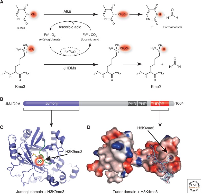
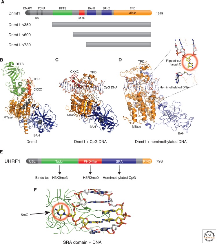
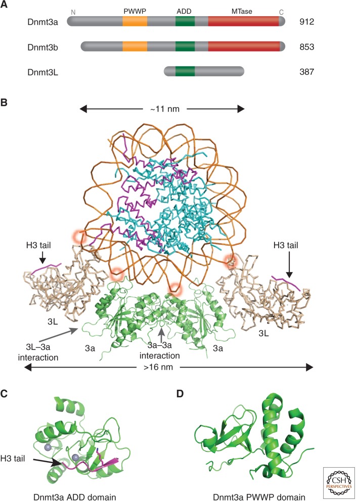
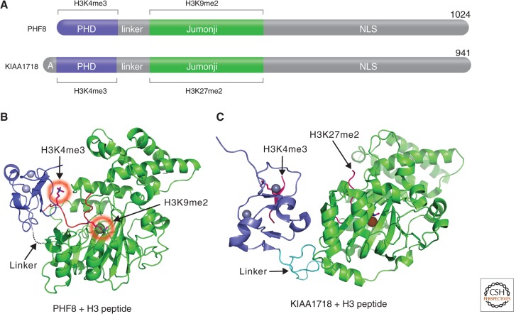
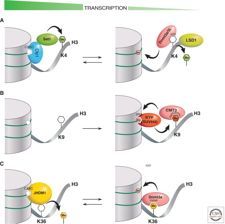
One of the most fundamental questions in the control of gene expression in mammals is how epigenetic methylation patterns of DNA and histones are established, erased, and recognized. This central process in controlling gene expression includes coordinated covalent modifications of DNA and its associated histones. This article focuses on structural aspects of enzymatic activities of histone (arginine and lysine) methylation and demethylation and functional links between the methylation status of the DNA and histones. An interconnected network of methyltransferases, demethylases, and accessory proteins is responsible for changing or maintaining the modification status of specific regions of chromatin.


Figures

Figure 1.
Figure 1.
Structural features of SUV39H1 and SUV39H2. (A) Ribbon diagram of amino-terminal chromodomain of SUV39H1 (PDB 3MTS). (B) Carboxyl-terminal SET domain structure of SUV39H2 (PDB 2R3A). (C) Formation of the pseudoknot by motifs III and IV. (D) Formation of the active site showing the methyl donor (S-adenosyl-l-methionine [AdoMet]), target H3K9 lysine, catalytic Y280 residue, and F370 Phe/Tyr Switch (Collins et al. 2005). AdoMet-dependent methyltransferases (including HKMTs) share a reaction mechanism in which the nucleophile acceptor (NH2) attacks the electrophilic carbon of AdoMet in an SN2 displacement reaction. (E) SUV39H1 and H2 have a pre-SET segment containing nine invariant cysteines, the SET region containing four signature motifs, and the post-SET region containing three invariant cysteines. An enlargement of the pre-SET Zn3Cys9 triangular zinc cluster structure is illustrated on the bottom left and the post-SET zinc center on the bottom right panel.
Figure 2.
Figure 2.
Dot1p family (non-SET HKMTs). (A) Schematic representation of Dot1 homologs from yeast and human, indicating the conserved methyltransferase core regions. (B) Superimposition of the conserved core regions of yeast Dot1p (residues 176-567; PDB 1U2Z), colored in green and brown, and human Dot1L (residues 5-332; PDB 1NW3), colored in cyan. The amino-terminal helical domains are shown on the left side of the panel in green for yDot1p or cyan for hDot1L. The carboxyl-terminal catalytic domain is shown on the right side of the panel with the bound methyl donor, AdoMet, as spheres (circled in red with an arrow). (C) A model of yDot1p docked with a nucleosome, adapted from Sawada et al. (2004). The structure of the nucleosome core particle is shown as ribbons (red, H3; green, H4; magenta, H2A; yellow, H2B; gray lines, DNA). The model was put together by aligning the target H3K79, located on the nucleosome disk surface, with the active site pocket of Dot1p.
Figure 3.
Figure 3.
PRMT family. (A) Reactions catalyzed by the two major types of protein arginine methylation. (B) Representatives of PRMT members (type I: PRMT1 and PRMT4; type II: PRMT5). The conserved methyltransferase (MTase) domain is in green and the unique β-barrel domain in yellow. (C) Dimeric structures of PRMT1 (PDB 1OR8) and PRMT4 (PDB 3B3F). Dimerization arms are indicated by red circles. (D) Type II PRMT5-MEP50 tetramer complex (DPB 4GQB), formed by the stacking of two dimers. The second dimer is faded in the background. MEP50 (colored in brown) interacts with the amino-terminal domain (gray) of PRMT5. Bound H4 peptide is colored in red. (E) An example of three coactivators acting synergistically for p53-mediated transcription.
Figure 4.
Figure 4.
Histone demethylation by oxidation. (A) Schematic representation of human LSD1 domain organization: the amino-terminal putative nuclear localization signal, followed by a SWIRM (Swi3p, Rsc8p, and Moira) domain, and the catalytic oxidase domain. The oxidase domain contains an atypical insertion of the Tower domain not found in other oxidases. (B) Scheme of the demethylation reaction catalyzed by LSD1. (C) Crystal structure of LSD1-CoREST in complex with the SNAIL1 peptide (PDB 2Y48). LSD1 includes residues 171–836 in red, blue, and magenta. CoREST shows residues 308–440 in orange. The SNAIL1 peptide is in green, and the FAD cofactor is shown as a yellow ball-and-stick. (D) Superposition between SNAIL1 (orange) and histone H3 (gray) peptides. (Adapted from Baron et al. 2011).
Figure 5.
Figure 5.
Demethylation by hydroxylation. (A) Mechanisms of demethylation of 3-methylthymine by AlkB (top) and of methyllysine by Jumonji-domain proteins (bottom). (B) Schematic representation of JMJD2A domain organization. (C) Structure of the amino-terminal Jumonji (ribbons) in complex with H3K9me3 (PDB 2OX0). (D) Structure of the carboxyl-terminal double Tudor domain (surface representation) in complex with H3K4me3 (PDB 2GFA). (Adapted, with permission, from Huang et al. 2006.)
Figure 6.
Figure 6.
Structures of maintenance Dnmt1 and UHRF1. (A) Schematic representation of mouse Dnmt1 domain organization and available Dnmt1 amino-terminal deletion mutants. An amino-terminal region interacts with Dnmt1-associated protein(s) (DMAP1; Rountree et al. 2000). Then an adjacent lysine and serine are subject to a methylation and phosphorylation switch that determines Dnmt1 stability (Esteve et al. 2011), a PCNA (proliferating cell nuclear antigen) interacting sequence (Chuang et al. 1997), and an RFTS (Leonhardt et al. 1992) that interacts with the SET- and RING-associated (SRA) domain of UHRF1 (Achour et al. 2008). This is followed by a CpG-interacting CXXC domain (Song et al. 2011), a tandem BAH (bromo-adjacent homology) domain (Callebaut et al. 1999), and the catalytic DNA methyltransferase domain that includes the target-recognizing domain (Lauster et al. 1989) at the carboxyl terminus. (B) Structure of Dnmt1 in the absence of DNA (PDB 3AV4). (C) Structure of Dnmt1 in the presence of unmethylated CpG (PDB 3PT6). (D) Structure of Dnmt1 with hemimethylated CpG DNA oligonucleotides (PDB 4DA4). (E) UHRF1 harbors at least five recognizable functional domains: an ubiquitin-like domain at the amino terminus, followed by a tandem tudor domain recognizing H3K9me3 (Rothbart et al. 2012), a plant homeodomain (PHD) recognizing H3R2me0 (Rajakumara et al. 2011), an SRA domain recognizing hemimethylated CpG, and really interesting new gene (RING) domain at the carboxyl terminus that may endow UHRF1 with E3 ubiquitin ligase activity to histones (Citterio et al. 2004). (F) Structure of SRA-DNA complex illustrates 5mC flipped out from the DNA helix and bound in a cage-like pocket (circled in red; PDB 2ZO1). (Adapted from Hashimoto et al. 2009).
Figure 7.
Figure 7.
Structures of the de novo Dnmt3a-Dnmt3L complex. (A) Domain architecture of Dnmt3a, 3b, and 3L. (B) A nucleosome is shown docked to a Dnmt3L-3a-3a-3L tetramer (Dnmt3a is colored in green and Dnmt3L in gray; PDB 2QRV). (Adapted from Cheng and Blumenthal 2008). The position of the histone H3 amino-terminal tail (purple) bound to Dnmt3L is shown, taken from a cocrystal structure (PDB 2PVC). By wrapping the Dnmt3a/3L tetramer around the nucleosome, the two Dnmt3L molecules are able to bind both histone tails emanating from one nucleosome. The ∼10 bp periodicity of binding to the major DNA groove is indicated by red circles. (C) Structure of the Dnmt3a ADD domain (PDB 3A1B) possibly interacting with histone tails from neighboring nucleosomes. (D) Structure of the Dnmt3a PWWP domain (PDB 3L1R).
Figure 8.
Figure 8.
CpG-interacting proteins including MLL1. (A) Domain architecture of CXXC domain-containing proteins. (BD) The structures of three isolated MLL1 domains in complex with interacting histones or DNA: (B) the amino-terminal CXXC domain in complex with CpG DNA (PDB 2KKF; Cierpicki et al. 2010), (C) the central PHD-bromodomain in complex with H3K4me3 peptide (PDB 3LQJ; Wang et al. 2010), and (D) the carboxyl-terminal SET domain (PDB 2W5Z; Southall et al. 2009).
Figure 9.
Figure 9.
Coordinated methyllysine erasure between a Jumonji and a PHD. (A) Schematic representations of PHF8 and KIAA1718 domain structure. (B) A bent conformation of PHF8 bound to a histone H3 peptide (in red) containing K4me3 and K9me2 (circled in red; PDB 3KV4). (C) An extended conformation of KIAA1718 (PDB 3KV6). The position of the histone H3 peptide is taken from a cocrystal structure of Caenorhabditis elegans KIAA1718 (PDB 3N9P).
Figure 10.
Figure 10.
Cartoons of interactions that regulate DNA methylation and associated histone H3 modifications. There are three examples of PTM cross talk between histone and DNA methylation. Chromatin on the left represents transcriptionally active states, whereas chromatin on the right represents transcriptionally repressive states. The “Me”-labeled filled hexagons indicate one or more methyl groups in DNA (pink) or protein lysine residues (K). The catalytic action of methylation writers and erasers are indicated by curved black arrows. Methylation readers interact via specific labeled domains that fit in a lock-and-key fashion to methylated (filled hexagons) or unmethylated CpGs or lysines (unfilled hexagons). (A) Enzymatic reactions by the Dnmt3a-Dnmt3L complex and the SET1/CFP1 complex regulate the inverse correlation of DNA methylation and H3K4 methylation. (B) In plants, enzymatic reactions by CMT3 and KYP reinforce the correlation between DNA CHG methylation and H3K9 methylation. (C) Enzymatic reactions by Dnmt3a and JHDM1 positively regulate the association of DNA methylation with H3K36 methylation.

Similar articles

Cited by

References

    1. Achour M, Jacq X, Ronde P, Alhosin M, Charlot C, Chataigneau T, Jeanblanc M, Macaluso M, Giordano A, Hughes AD, et al. 2008. The interaction of the SRA domain of ICBP90 with a novel domain of DNMT1 is involved in the regulation of VEGF gene expression. Oncogene 27: 2187–2197 - PubMed
    1. Allis CD, Jenuwein T, Reinberg D 2014. Overview and concepts. Cold Spring Harb Perspect Biol 10.1101/cshperspect.a018739 - DOI
    1. An W, Kim J, Roeder RG 2004. Ordered cooperative functions of PRMT1, p300, and CARM1 in transcriptional activation by p53. Cell 117: 735–748 - PubMed
    1. Ansari KI, Mandal SS 2010. Mixed lineage leukemia: Roles in gene expression, hormone signaling and mRNA processing. FEBS J 277: 1790–1804 - PubMed
    1. Antonysamy S, Bonday Z, Campbell RM, Doyle B, Druzina Z, Gheyi T, Han B, Jungheim LN, Qian Y, Rauch C, et al. 2012. Crystal structure of the human PRMT5:MEP50 complex. Proc Natl Acad Sci 109: 17960–17965 - PMC - PubMed

Publication types

LinkOut - more resources