. 2015 Jan;87(1):74-84.
doi: 10.1038/ki.2014.271. Epub 2014 Jul 30.

Nlrp3-inflammasome activation in non-myeloid-derived cells aggravates diabetic nephropathy

Affiliations
Free PMC article

Nlrp3-inflammasome activation in non-myeloid-derived cells aggravates diabetic nephropathy

Khurrum Shahzad et al. Kidney Int. 2015 Jan.
Free PMC article

Abstract

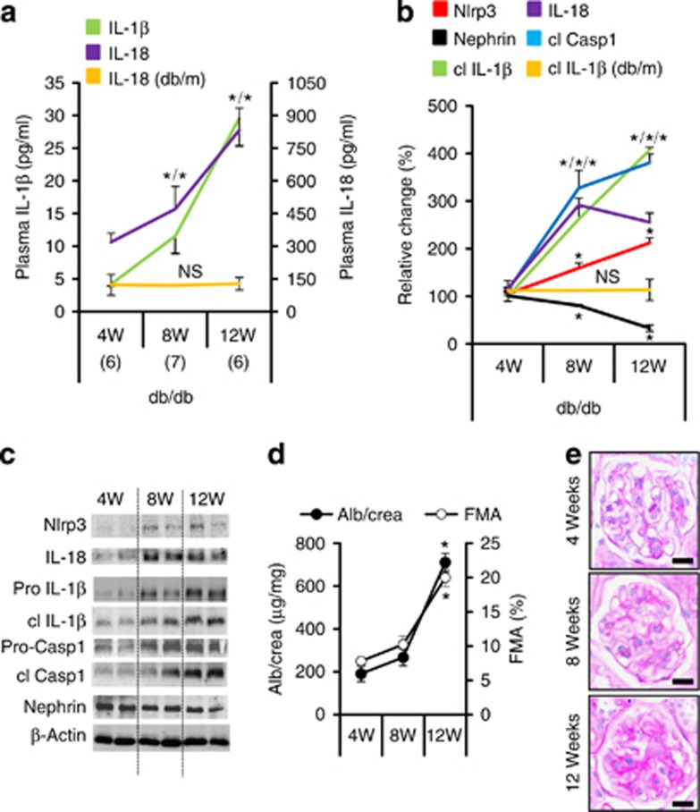
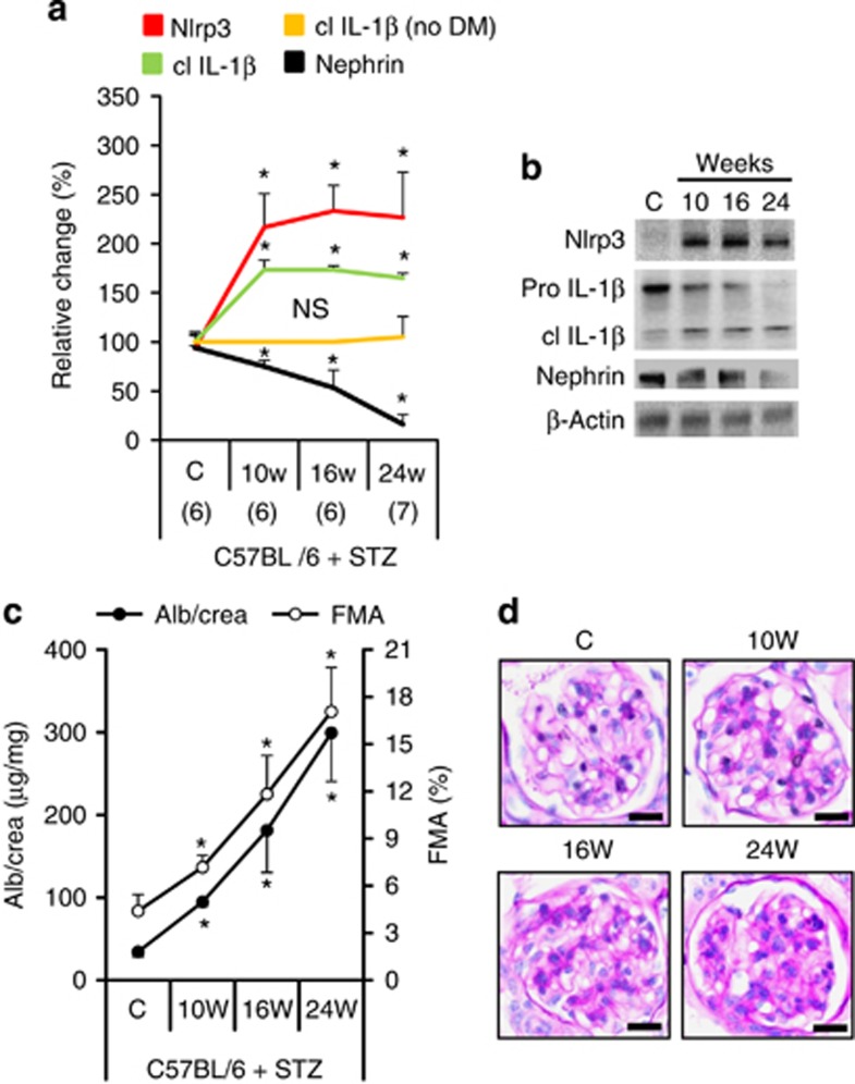
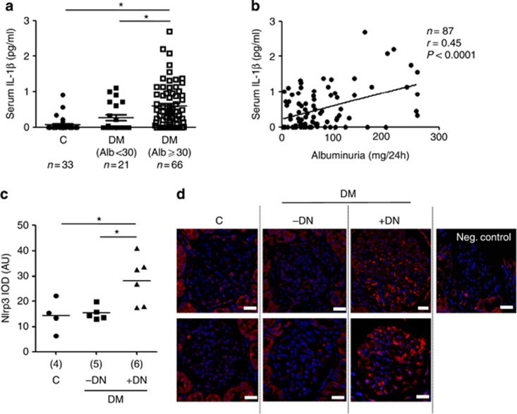
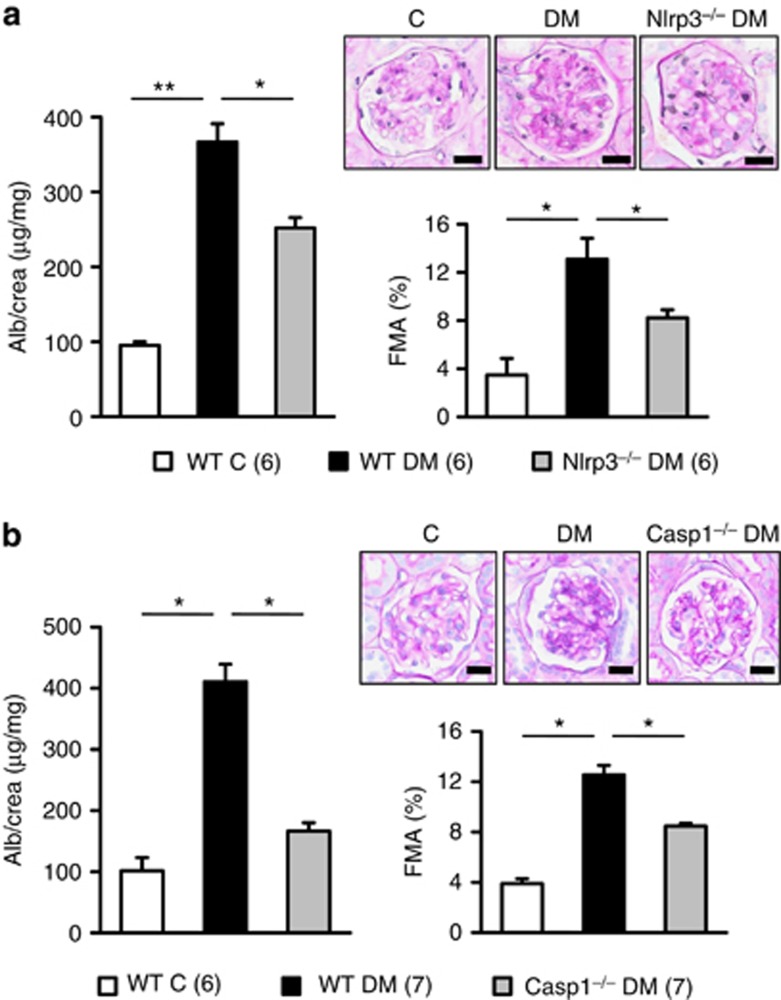
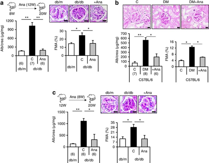
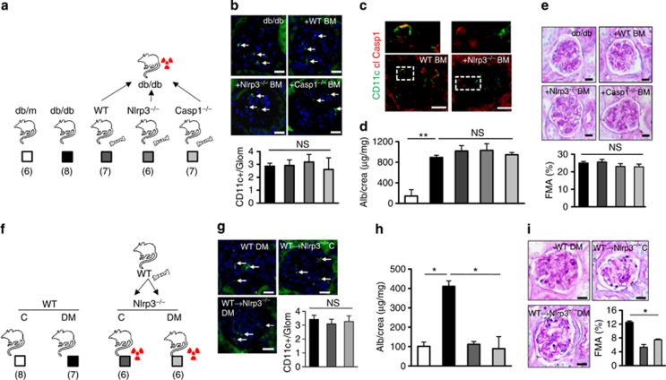
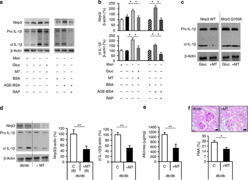
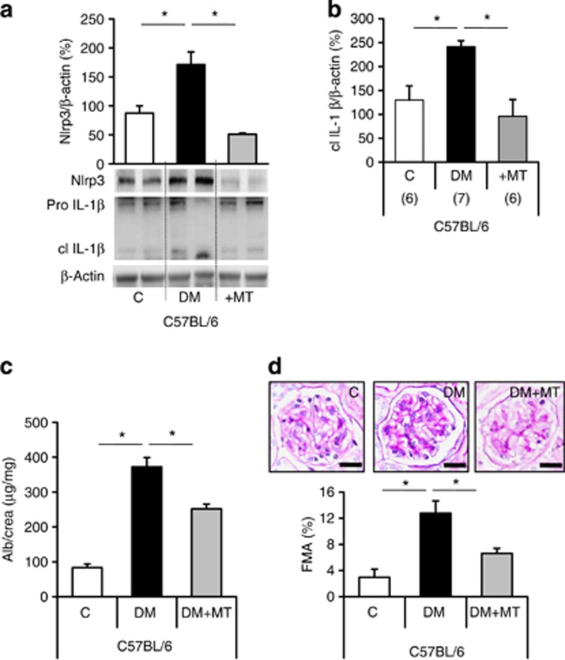
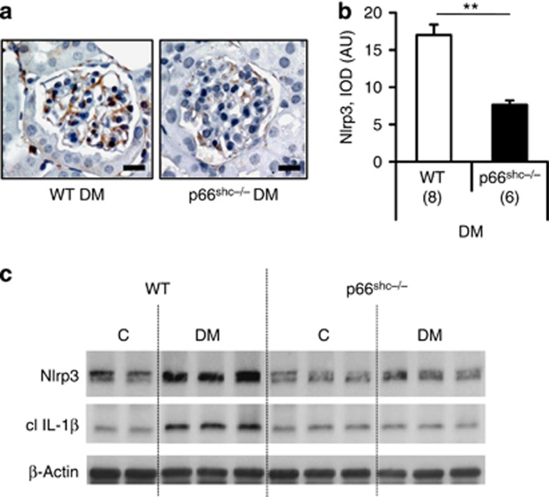
Diabetic nephropathy is a growing health concern with characteristic sterile inflammation. As the underlying mechanisms of this inflammation remain poorly defined, specific therapies targeting sterile inflammation in diabetic nephropathy are lacking. Intriguingly, an association of diabetic nephropathy with inflammasome activation has recently been shown, but the pathophysiological relevance of this finding remains unknown. Within glomeruli, inflammasome activation was detected in endothelial cells and podocytes in diabetic humans and mice and in glucose-stressed glomerular endothelial cells and podocytes in vitro. Abolishing Nlrp3 or caspase-1 expression in bone marrow-derived cells fails to protect mice against diabetic nephropathy. Conversely, Nlrp3-deficient mice are protected against diabetic nephropathy despite transplantation of wild-type bone marrow. Pharmacological IL-1R antagonism prevented or even reversed diabetic nephropathy in mice. Mitochondrial reactive oxygen species (ROS) activate the Nlrp3 inflammasome in glucose or advanced glycation end product stressed podocytes. Inhibition of mitochondrial ROS prevents glomerular inflammasome activation and nephropathy in diabetic mice. Thus, mitochondrial ROS and Nlrp3-inflammasome activation in non-myeloid-derived cells aggravate diabetic nephropathy. Targeting the inflammasome may be a potential therapeutic approach to diabetic nephropathy.


Figures

Figure 1
Figure 1
Inflammasome activation is associated with the onset of diabetic nephropathy in db/db mice. IL-1β and IL-18 plasma levels (a) and tissue levels of Nlrp3 (nucleotide-binding domain and leucine-rich repeat pyrin 3 domain) and cleaved IL-1β (cl IL-1β) in renal cortex extracts (b, c) are increased in db/db mice at 8 weeks of age (compared with 4 weeks of age) and are further increased at age 12 weeks. Tissue levels of cleaved caspase-1 (cl Casp1) and IL-18 increase between age 4 and 8 weeks and then remain elevated (b, c). Nephrin expression is reduced at 8 and decreases further at 12 weeks of age (b, c). These changes are associated with a significant increase in albuminuria (Alb) and the fractional mesangial area (FMA) in 12- but not in 8-week-old db/db mice (d, e). Markers of inflammasome activation (plasma IL-18, a; cleaved IL-1β, b) remain normal in nondiabetic db/m control mice. Mean value±s.e.m. Number of mice (ae) in each group is shown in parentheses in a; representative immunoblots in (c) and representative periodic acid–Schiff–stained glomeruli in e, scale bar=20 μm; *P<0.05. FMA, fractional mesangial area; IL, interleukin; NS, not significant.
Figure 2
Figure 2
Inflammasome activation is associated with the onset of diabetic nephropathy in streptozotocin (STZ)-treated C57BL/6 mice. Tissue levels of Nlrp3 (nucleotide-binding domain and leucine-rich repeat pyrin 3 domain) and cleaved interleukin-1β (cl IL-1β) increase, whereas expression of nephrin declines in renal cortex extracts of diabetic C57BL/6 mice (STZ model; a, b). Inflammasome activation is paralleled by an increase in albuminuria and fractional mesangial area (FMA, c, d; overviews for d in Supplementary Figure S2a online). Cleaved IL-1β remains normal in nondiabetic C57BL/6 control mice (no DM, a). Mean value±s.e.m. Number of mice (ad) in each group is shown in parentheses in a; representative immunoblots in b and periodic acid Schiff-stained glomeruli in d, scale bar=20 μm; *P<0.05. DM, diabetic mice; NS, not significant.
Figure 3
Figure 3
Inflammasome activation in human diabetic patients. Serum interleukin (IL)-1β levels are increased in albuminuric diabetic patients when compared to nondiabetic controls and non-albuminuric diabetic patients (a) and serum IL-1β levels in diabetic patients correlate with albuminuria (b). Nlrp3 (nucleotide-binding domain and leucine-rich repeat pyrin 3 domain) expression is increased in glomeruli of diabetic patients with diabetic nephropathy (DM+DN) as compared to nondiabetic controls (C) or diabetic patients without diabetic nephropathy (DM−DN) (c, d). Scatter diagram (c) and two representative images each (d) of glomeruli from indicated groups; immunofluorescence labeling for Nlrp3 (red), 4,6-diamidino-2-phenylindole counterstain (blue), and a negative control using a nonspecific primary antibody (Neg. control); scale bar=20 μm. Scatter diagrams representing individual data points and mean (horizontal line (a, c); *P<0.05. AU, arbitrary units; DM, diabetic mice; IOD, integrated optical density.
Figure 4
Figure 4
Nlrp3 (nucleotide-binding domain and leucine-rich repeat pyrin 3 domain) or caspase-1-deficiency ameliorates diabetic nephropathy (DN). Albuminuria and fractional mesangial area (FMA) are significantly reduced in Nlrp3−/− (a) or caspase-1−/− (b) diabetic mice in comparison with wild-type diabetic mice (WT DM). Mean value±s.e.m. Number of mice (a, b) in each group is shown in parentheses in a; representative periodic acid Schiff-stained glomeruli; scale bar (a, b)=20 μm; *P<0.05, **P<0.01.
Figure 5
Figure 5
Inhibition of the inflammasome with anakinra (Ana) ameliorates or reverses diabetic nephropathy. Initiating anakinra (interleukin (IL)-1 receptor antagonist) treatment in 8-week-old db/db mice (a) or diabetic (streptozotocin (STZ) model) uninephrectomized C57BL/6 mice (b) prevents albuminuria (Alb) and extracellular matrix accumulation (fractional mesangial area, FMA) over a period of 12 weeks (db/db) or 8 weeks (C57BL/6 + STZ), respectively. Initiating anakinra treatment in 12–week-old db/db mice and hence after establishment of experimental diabetic nephropathy anakinra normalizes albuminuria and FMA (c). Mean value±s.e.m. Number of mice in each group is shown in parentheses in ac; representative periodic acid–Schiff–stained glomeruli; scale bar (ac)=20 μm. *P<0.05, **P<0.01. DM, diabetic mice.
Figure 6
Figure 6
Inflammasome activation in resident glomerular cells in diabetic nephropathy. Nlrp3 (nucleotide-binding domain and leucine-rich repeat pyrin 3 domain; red) colocalizes with markers for podocytes (synaptopodin, green, left) and endothelial cells (PECAM, green, right) in glomeruli of microalbuminuric diabetic patients (a), scale bar=20 μm. In glomeruli of 20-week-old db/db mice, cleaved caspase-1 (red) colocalizes with markers for podocyte (synaptopodin, green, left) and endothelial cell (CD34, green, right; b, scale bar=20 μm). Representative images of glomeruli obtained by confocal microscopy (Zeiss LSM 4 Pascal microscope, Carl Zeiss, Jena, Germany) and higher magnification of areas indicated by white dotted lines; yellow reflects colocalization (a, b). In mouse glomerular endothelial cells (GENC) and human podocytes (hPodo) glucose (Gluc, 25 mmol/l, 24 h) induces Nlrp3 and cleaved interleukin-1β (cl IL-1β) in comparison to control (C, phosphate-buffered saline) or mannitol (M, 25 mmol/l mannitol)-treated cells (c); representative immunoblots, β-actin as loading control. In primary podocytes isolated from C57BL/6 wild-type (WT), but not from caspase-1–deficient (casp1−/−) mice, cl IL-1β can be readily detected (d). Glucose (G, 25 mmol/l), but not mannitol (M, 25 mmol/l), induces cl IL-1β in WT, but not in caspapse-1–deficient podocytes (d); representative immunoblots, β-actin as loading control.
Figure 7
Figure 7
Non-myeloid-derived cells are sufficient to promote diabetic nephropathy. Following transplantation of Nlrp3−/− or Casp1−/− bone marrow into db/db mice (a: experimental approach) the frequency of CD11c+ cells in glomeruli is similar to that in control db/db mice (b), immunofluorescence, CD11c: green; 4,6-diamidino-2-phenylindole (DAPI) counterstain, but no cleaved caspase-1 (cl Casp1) can be detected in CD11c+ cells (c), representative immunofluorescence following transplantation of Nlrp3−/− bone marrow; cleaved caspase-1: red; CD11c: green; colocalization: yellow; white dotted lines indicate areas shown at higher magnification above. Albuminuria and FMA remain high in db/db mice despite transplantation with Nlrp3−/− or caspase-1−/− bone marrow (d, e). Conversely, diabetic and uninephrectomized Nlrp3−/− mice are protected from nephropathy despite transplantation of Nlrp3+/+ bone marrow (f: experimental approach). The frequency of CD11c+ cells in glomeruli is similar in experimental groups (g); CD11c: green; DAPI counterstain, but albuminuria and FMA remain normal in diabetic Nlrp3−/− mice despite reconstitution with Nlrp3+/+ bone marrow (h, i). Mean value±s.e.m. Number of mice (ae and fi) in each group is shown in parentheses in a and f; representative periodic acid–Schiff–stained glomeruli in e and i; scale bar=20 μm (b, c, e, g, and i); white arrows in b and g indicate CD11c+ cells; *P<0.05; **P<0.01. BM, bone marrow; DM, diabetic mice; FMA, fractional mesangial area; IL, interleukin; Nlrp3, nucleotide-binding domain and leucine-rich repeat pyrin 3 domain; NS, not significant; WT, wild type.
Figure 8
Figure 8
Mitochondrial reactive oxygen species cause inflammasome activation and promote diabetic nephropathy in db/db mice. Glucose (Gluc, 25 mmol/l for 24 h) and AGE-BSA (200 μg/ml) induce Nlrp3 expression and IL-1β cleavage in mouse podocytes (a, b). Inhibition of the receptor RAGE using RAP (RAGE antagonist peptide, 10 μmol/l for 24 h) was sufficient to prevent the AGE-BSA-dependent induction of Nlrp3 and IL-1β cleavage (a, b). The mitochondrial-targeted antioxidant MitoTempo (MT) reduces glucose-induced Nlrp3 expression and IL-1β cleavage in mouse (a, b) and human (c) podocytes, respectively (ac). Following transfection of human podocytes with a constitutive active human Nlrp3 mutant (Q705K), MitoTempo (10 μm) fails to suppress IL-1β cleavage (b); representative immunoblots of at least three independent repeat experiments (a, c) and bar graph (b) summarizing densitometric results. Treatment of 8-week-old db/db mice with MitoTempo (MT for 8 weeks) reduces Nlrp3 expression and IL-1β cleavage in renal cortex tissue extracts (d); representative immunoblots of Nlrp3, IL-1β, and β-actin and bar graph summarizing results, as well as albuminuria (e) and FMA (f) in comparison with phosphate-buffered saline-treated db/db controls (C: controls); mean value±s.e.m. Number of mice (df) in each group is shown in parentheses in d; representative periodic acid–Schiff–stained glomeruli in f; scale bar (e)=20 μm; *P<0.05; **P<0.01. FMA, fractional mesangial area; IL, interleukin; Nlrp3, nucleotide-binding domain and leucine-rich repeat pyrin 3 domain.
Figure 9
Figure 9
MitoTempo protects against diabetic nephropathy in diabetic C57BL/6 mice. Treatment of diabetic uninephrectomized (DM, STZ model) C57BL/6 mice (DM) with MitoTempo (+MT, for 8 weeks) reduces levels of Nlrp3 and cleaved IL-1β in renal cortex tissue extracts (a): bottom: representative immunoblots of Nlrp3, IL-1β, and β-actin; a and b: bar graphs summarizing results, albuminuria (Alb) (c), and FMA (d) in comparison with phosphate-buffered saline-treated diabetic controls (DM). Nondiabetic C57BL/6 mice served as controls (C); mean value±s.e.m. Number of mice (ad) in each group is shown in parentheses in b; representative periodic acid–Schiff–stained glomeruli in d; scale bar=20 μm; *P<0.05. DM, diabetic mice; FMA, fractional mesangial area; IL, interleukin; Nlrp3, nucleotide-binding domain and leucine-rich repeat pyrin 3 domain.
Figure 10
Figure 10
Deficiency of the redox-protein p66Shc reduces inflammasome activation in diabetic nephropathy. In diabetic mice lacking the redox-protein p66Shc levels of Nlrp3 (ac) and cleaved IL-1β (c) are reduced in comparison with diabetic wild-type (WT) mice (streptozotocin model, 24 weeks of hyperglycemia). Immunohistochemical detection of Nlrp3 in glomeruli of diabetic WT and p66Shc−/− mice (a), Nlrp3-positive cells detected by HRP-DAB reaction, brown; hematoxylin counterstain, blue; scale bar=20 μm; bar graph summarizing results of integrated optical density (IOD) of positive DAB stain (b); mean value±s.e.m. Number of mice (ac) in each group is shown in parentheses in b, and representative immunoblots in c; **P<0.01. AU, arbitrary units; DM, diabetic mice; IL, interleukin; Nlrp3, nucleotide-binding domain and leucine-rich repeat pyrin 3 domain.

Comment in

Similar articles

Cited by

References

    1. Chow F, Ozols E, Nikolic-Paterson DJ, et al. Macrophages in mouse type 2 diabetic nephropathy: correlation with diabetic state and progressive renal injury. Kidney Int. 2004;65:116–128. - PubMed
    1. Nguyen D, Ping F, Mu W, et al. Macrophage accumulation in human progressive diabetic nephropathy. Nephrology (Carlton) 2006;11:226–231. - PubMed
    1. Lim AK, Ma FY, Nikolic-Paterson DJ, et al. Lymphocytes promote albuminuria, but not renal dysfunction or histological damage in a mouse model of diabetic renal injury. Diabetologia. 2010;53:1772–1782. - PubMed
    1. Moriya R, Manivel JC, Mauer M. Juxtaglomerular apparatus T-cell infiltration affects glomerular structure in Type 1 diabetic patients. Diabetologia. 2004;47:82–88. - PubMed
    1. Awad AS, Kinsey GR, Khutsishvili K, et al. Monocyte/macrophage chemokine receptor CCR2 mediates diabetic renal injury. Am J Physiol Renal Physiol. 2011;301:F1358–F1366. - PMC - PubMed

Publication types