. 2015 Jan;60(1):86-100.
doi: 10.1007/s10620-014-3307-z. Epub 2014 Jul 29.

Krüpple-like factor 5 is required for proper maintenance of adult intestinal crypt cellular proliferation

Affiliations

Krüpple-like factor 5 is required for proper maintenance of adult intestinal crypt cellular proliferation

Kristin N Bell et al. Dig Dis Sci. 2015 Jan.

Abstract

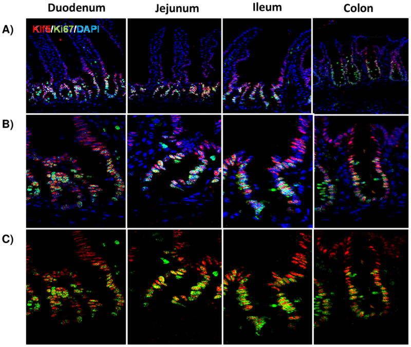
Background: Krüpple-like factor 5 (KLF5) is a transcription factor that is highly expressed in the proliferative compartment of the intestinal crypt. There, it is thought to regulate epithelial turnover and homeostasis.

Aim: In this study, we sought to determine the role for Klf5 in the maintenance of cellular proliferation, cytodifferentiation, and morphology of the crypt-villus axis.

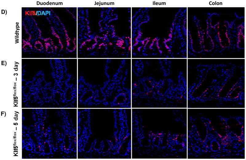
Methods: Tamoxifen-induced recombination directed by the epithelial-specific Villin promoter (in Villin-CreERT2 transgenic mice) was used to delete Klf5 (in Klf5 (loxP/loxP) mice) from the adult mouse intestine and analyzed by immunostaining and RT-qPCR. Control mice were tamoxifen-treated Klf5 (loxP/loxP) mice lacking Villin-CreERT2.

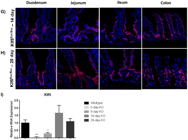
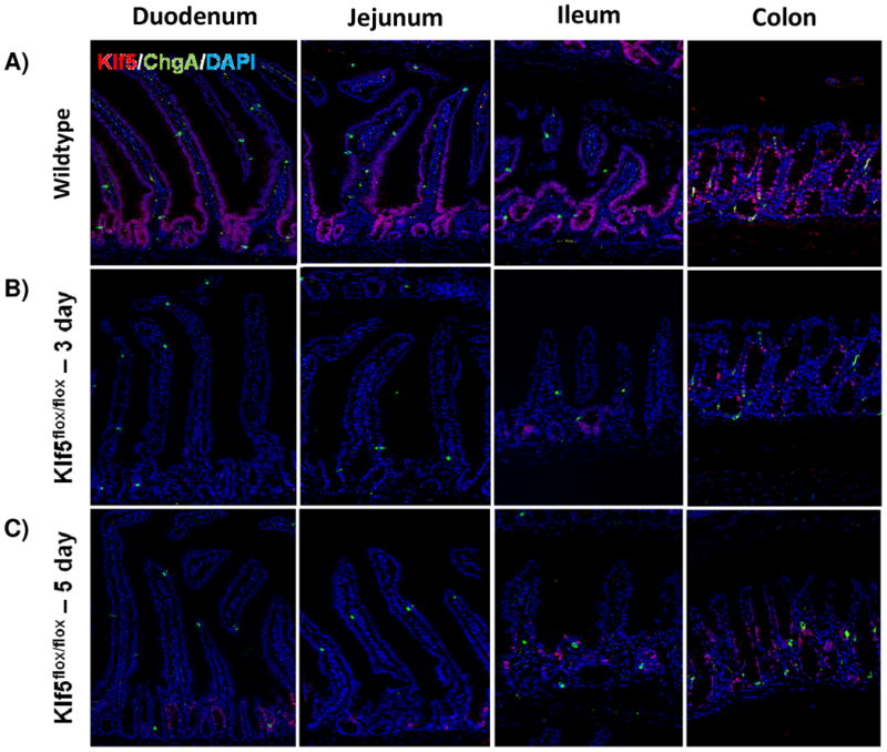
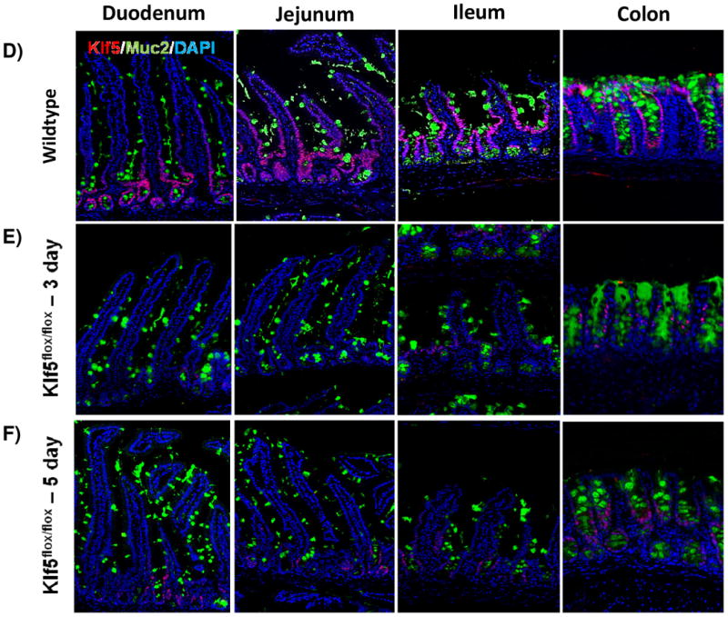
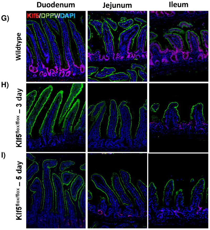
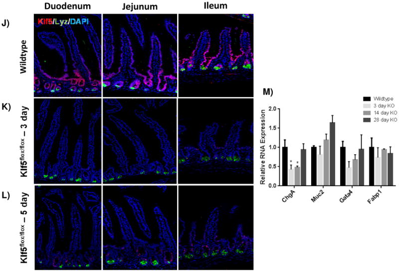
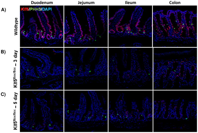
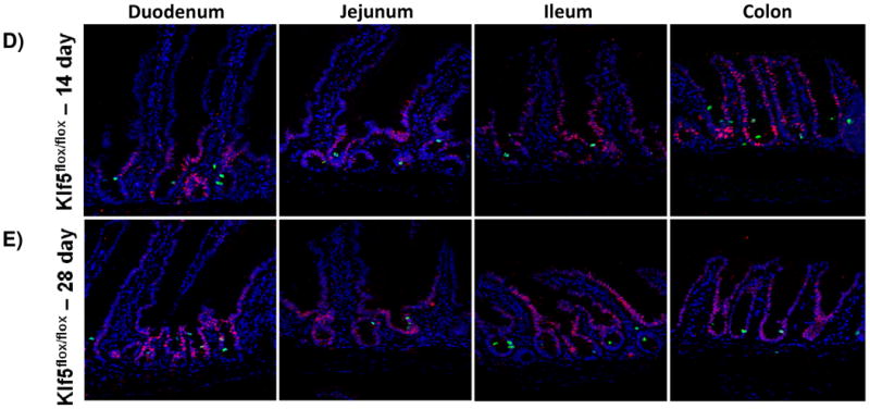
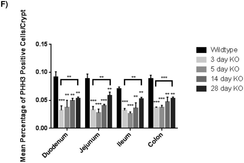
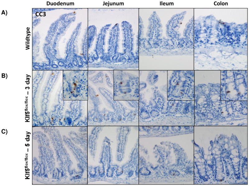
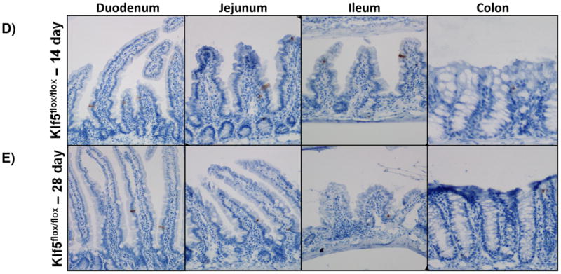
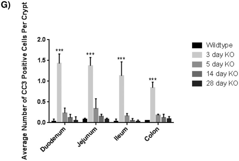
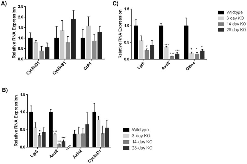
Results: Three days after tamoxifen-induced recombination, the mitosis marker phospho-histone H3 was significantly reduced within the Klf5-mutant crypt epithelium, coincident with increased expression of the apoptosis marker cleaved-caspase 3 within the crypt where cell death rarely occurs normally. We also observed a reduction in Chromagranin A expressing enteroendocrine cells, though no significant change was seen in other secretory or absorptive cell types. To examine the long-term repercussions of Klf5 loss, we killed mice 5, 14, and 28 days post recombination and found reemerging expression of KLF5. Furthermore, we observed restoration of cellular proliferation, though not to levels seen wildtype intestinal crypts. Reduction of apoptosis to levels comparable to the wildtype intestinal crypt was also observed at later time points. Analysis of cell cycle machinery indicated no significant perturbation upon deletion of Klf5; however, a reduction of stem cell markers Ascl2, Lgr5, and Olfm4 was observed at all time points following Klf5 deletion.

Conclusions: These results indicate that Klf5 is necessary to maintain adult intestinal crypt proliferation and proper cellular differentiation. Rapid replacement of Klf5-mutant crypts with wildtype cells and reduction of stem cell markers suggests further that Klf5 is required for self renewal of intestinal stem cells.


Figures

Figure 1
Figure 1. KLF5 is expressed in proliferative crypts of the small and large intestine
(A) Immunofluorescence staining for coexpression of KLF5 (red) and Ki67 (green). DAPI (blue) was utilized as a nuclear stain.(B) Immunofluorescence staining of the crypt with DAPI (B) and without DAPI (C) show regions of Klf5 and Ki67 expression overlap. Colocalization between these two proteins is seen in yellow. (D-H) Regional histological analysis of tamoxifen-induced recombination in Klf5loxP/loxP; VilWT control mice (D) and Klf5loxP/loxP; VilCreERT2 experimental mice 3 (E), 5 (F), 14 (G), and 28 days (H). (I) Comparison of Klf5 mRNA expression analysis by RT-qPCR from jejunal segments of the small intestine. Student's t-test was performed to determine p-values; expression was normalized to Actb. (Mean ± SEM: P ≤ 0.05, **P ≤ 0.01, ***P ≤ 0.001, n = 3-6).
Figure 2
Figure 2. Loss of KLF5 affects cytodifferentiation
Histological comparison of Klf5loxP/loxP; VilWT (WT) and Klf5loxP/loxP; VilCreERT2 (Klf5-mutant) intestinal tissues for cellular differentiation utilizing immunofluorescent staining for chromagranin A (green; enteroendocrine cells) (A-C),mucin2 (green; goblet cells) (D-F), lysozyme (green; Paneth cells) (G-I), and dipeptidyl peptidase-4 (green; enterocytes) (J-L). Nuclear stain is DAPI (blue). (M) Comparison of ChgA, Muc2, Gata4, and Fabp1 mRNA expression analysis by RT-qPCR from jejunal segments of the small intestine. Student's t-test was performed to determine p-values; expression was normalized to Gapdh. (Mean ± SEM: P ≤ 0.05, **P ≤ 0.01, ***P ≤ 0.001, n = 3-6).
Figure 3
Figure 3. Loss of KLF5 attenuates cellular proliferation in the adult intestine
(A) Histological analysis for the comparison of cellular proliferation utilizing immunofluorescent staining for mitotic marker, phosphohistoneH3 (green) in wildtype and Klf5 loss-of-function mice (3, 5, 14, and 28 days post tamoxifen-induced recombination). Nuclear stain is DAPI (blue). (B) Quantification of average percentage of phosphohistoneH3 positive cells per crypt. Significance determined between groups of two (wildtype vs. loss of Klf5 for 3 days, wildtype vs. loss of Klf5 for 5 days, wildtype vs. loss of Klf5 for 14 days, wildtype vs. loss of Klf5 for 28 days, and loss of Klf5 for 3 vs. 28 days). (Mean ± SD, T-test: * P ≤ 0.05, **P ≤ 0.01, ***P ≤ 0.001, n = 3)
Figure 4
Figure 4. Intestinal crypt cell death is increased early after loss of KLF5, but disappears over time
(A-E) Immunohistochemical analysis of apoposis is shown using cleaved-caspase 3 (brown) counterstained with hemotoxylin (blue) in wildtype and Klf5 loss-of-function mice (3, 5, 14, 28 days post tamoxifen-induced recombination). (F) Quantification of the number of cleaved-caspase 3 positive cells per crypt. Significance determined between groups of two (wildtype vs. loss of Klf5 for 3 days, wildtype vs. loss of Klf5 for 5 days, wildtype vs. loss of Klf5 for 14 days, wildtype vs. loss of Klf5 for 28 days, and loss of Klf5 for 3 vs. 28 days). (Mean ± SD, T-test: * P ≤ 0.05, **P ≤ 0.01, ***P ≤ 0.001, n = 3)
Figure 5
Figure 5. Loss of Klf5 results in loss of active stem cell marker expression in the small intestine
Results from of mRNA of expression for (A) cell cycle proteins ( CyclinD1, CyclinB1, and Cdk1) , (B) downstream targets of canonical Wnt signaling (Lgr5, Ascl2, Axin2, and CyclinD1), and (C) active intestinal stem cell markers (Lgr5, Ascl2, Axin2, CyclinD1) by RT-qPCR from jejunal segments of the small intestine. Student's t-test was performed to determine p-values; expression was normalized to Gapdh (SYBR) or Actb (Taqman). (Mean ± SEM: P ≤ 0.05, **P ≤ 0.01, ***P ≤ 0.001, n = 3-6). Note that some qRT-PCR results for relative RNA expression appear on two graphs for comparison purposes.

Similar articles

Cited by

References

    1. Buddington RK, Diamond JM. Ontogenetic development of intestinal nutrient transporters. Annual Review of Physiology. 1989;1989(51):601–619. - PubMed
    1. Deplancke B, Gaskins HR. Microbial modulation of innate defense: goblet cells and the intestinal mucus layer. American Journal of Clincal Nutrition. 2001;73:1131S–1141S. - PubMed
    1. Shimizu M. Modulation of intestinal functions by food substances. Die Nahrung. 1999;43(3):154–158. - PubMed
    1. Strader AD, Woods SC. Gastrointestinal hormones and food intake. Gastroenterology. 2005;128(1):175–191. - PubMed
    1. Cheng H, Leblond CP. Origin, differentiation and renewal of the four main epithelial cell types in the mouse small intestine. V. Unitarian Theory of the origin of the four epithelial cell types. The American Journal of Anatomy. 1974;141(4):537–561. - PubMed

Publication types

MeSH terms