

TRPV4 channel inhibits TGF-β1-induced proliferation of hepatic stellate cells
- PMID: 25013893
- PMCID: PMC4094468
- DOI: 10.1371/journal.pone.0101179
TRPV4 channel inhibits TGF-β1-induced proliferation of hepatic stellate cells
Abstract
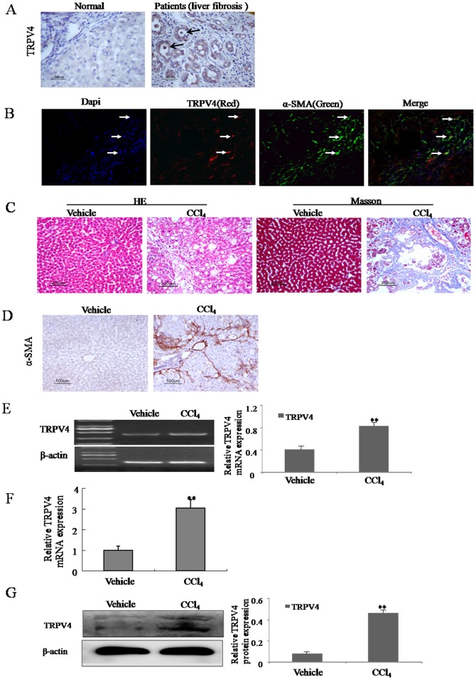
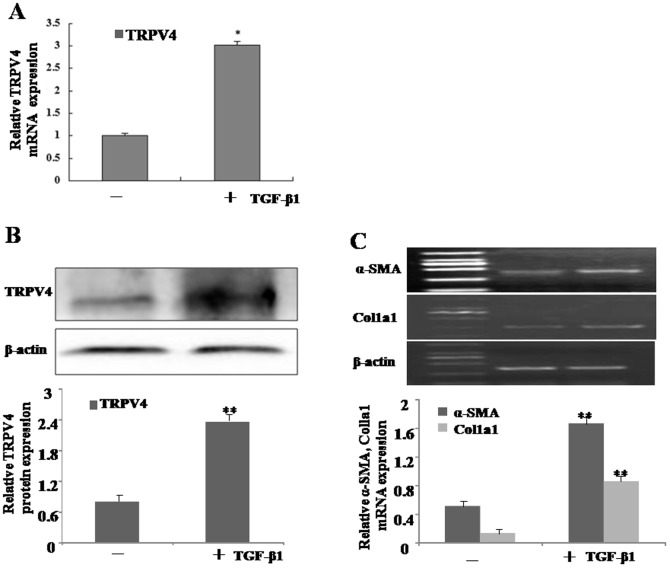
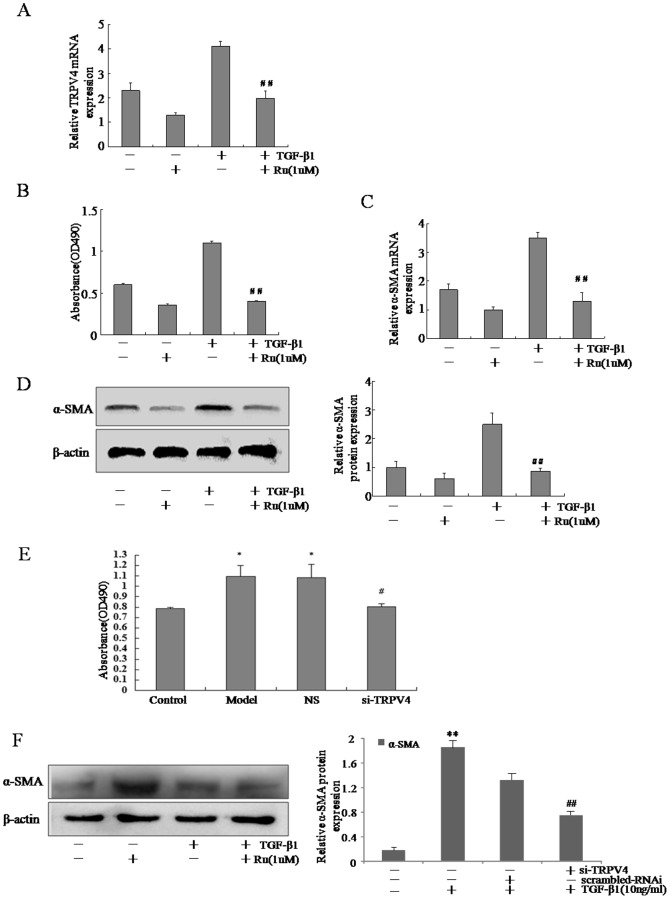
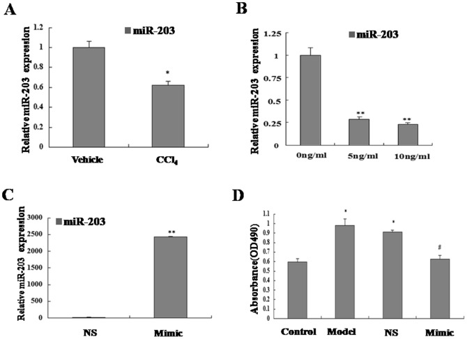
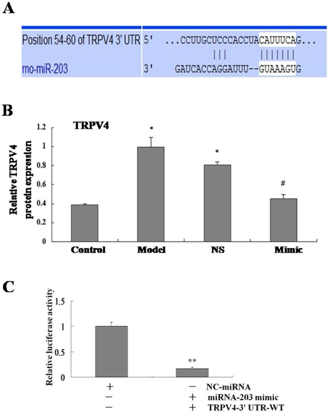
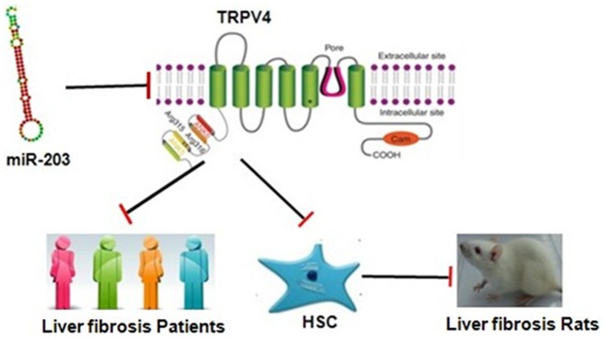
TRPV4, one of the TRP channels, is implicated in diverse physiological and pathological processes including cell proliferation. However, the role of TRPV4 in liver fibrosis is largely unknown. Here, we characterized the role of TRPV4 in regulating HSC-T6 cell proliferation. TRPV4 mRNA and protein were measured by RT-PCR and Western blot in patients and rat model of liver fibrosis in vivo and TGF-β1-activated HSC-T6 cells in vitro. Both mRNA and protein of TRPV4 were dramatically increased in liver fibrotic tissues of both patients and CCl4-treated rats. Stimulation of HSC-T6 cells with TGF-β1 resulted in increase of TRPV4 mRNA and protein. However, TGF-β1-induced HSC-T6 cell proliferation was inhibited by Ruthenium Red (Ru) or synthetic siRNA targeting TRPV4, and this was accompanied by downregulation of myofibroblast markers including α-SMA and Col1α1. Moreover, our study revealed that miR-203 was downregulated in liver fibrotic tissues and TGF-β1-treated HSC-T6 cell. Bioinformatics analyses predict that TRPV4 is the potential target of miR-203. In addition, overexpression of miR-203 in TGF-β1-induced HSC significantly reduced TRPV4 expression, indicating TRPV4, which was regulated by miR-203, may function as a novel regulator to modulate TGF-β1-induced HSC-T6 proliferation.
Conflict of interest statement
Figures
Similar articles
-
MicroRNA-146a modulates TGF-beta1-induced hepatic stellate cell proliferation by targeting SMAD4.Cell Signal. 2012 Oct;24(10):1923-30. doi: 10.1016/j.cellsig.2012.06.003. Epub 2012 Jun 24. Cell Signal. 2012. PMID: 22735812
-
TRPM7 channel regulates PDGF-BB-induced proliferation of hepatic stellate cells via PI3K and ERK pathways.Toxicol Appl Pharmacol. 2013 Nov 1;272(3):713-25. doi: 10.1016/j.taap.2013.08.009. Epub 2013 Aug 16. Toxicol Appl Pharmacol. 2013. PMID: 23958495
-
TGF-β1-elevated TRPM7 channel regulates collagen expression in hepatic stellate cells via TGF-β1/Smad pathway.Toxicol Appl Pharmacol. 2014 Oct 15;280(2):335-44. doi: 10.1016/j.taap.2014.08.006. Epub 2014 Aug 19. Toxicol Appl Pharmacol. 2014. PMID: 25150141
-
Blockade of YAP alleviates hepatic fibrosis through accelerating apoptosis and reversion of activated hepatic stellate cells.Mol Immunol. 2019 Mar;107:29-40. doi: 10.1016/j.molimm.2019.01.004. Epub 2019 Jan 11. Mol Immunol. 2019. PMID: 30639476
-
Therapeutic implications of targeting autophagy and TGF-β crosstalk for the treatment of liver fibrosis.Life Sci. 2023 Sep 15;329:121894. doi: 10.1016/j.lfs.2023.121894. Epub 2023 Jun 26. Life Sci. 2023. PMID: 37380126 Review.
Cited by
-
Role of Transient Receptor Potential Vanilloid 4 in Vascular Function.Front Mol Biosci. 2021 Apr 26;8:677661. doi: 10.3389/fmolb.2021.677661. eCollection 2021. Front Mol Biosci. 2021. PMID: 33981725 Free PMC article. Review.
-
CTGF siRNA ameliorates tubular cell apoptosis and tubulointerstitial fibrosis in obstructed mouse kidneys in a Sirt1-independent manner.Drug Des Devel Ther. 2015 Jul 31;9:4155-71. doi: 10.2147/DDDT.S86748. eCollection 2015. Drug Des Devel Ther. 2015. PMID: 26257513 Free PMC article.
-
Transient receptor potential ion-channel subfamily V member 4: a potential target for cancer treatment.Cell Death Dis. 2019 Jun 24;10(7):497. doi: 10.1038/s41419-019-1708-9. Cell Death Dis. 2019. PMID: 31235786 Free PMC article. Review.
-
Hydrogel cultures reveal Transient Receptor Potential Vanilloid 4 regulation of myofibroblast activation and proliferation in valvular interstitial cells.FASEB J. 2022 May;36(5):e22306. doi: 10.1096/fj.202101863R. FASEB J. 2022. PMID: 35385164 Free PMC article.
-
PI3-kinase/Akt pathway-regulated membrane transportation of acid-sensing ion channel 1a/Calcium ion influx/endoplasmic reticulum stress activation on PDGF-induced HSC Activation.J Cell Mol Med. 2019 Jun;23(6):3940-3950. doi: 10.1111/jcmm.14275. Epub 2019 Apr 2. J Cell Mol Med. 2019. PMID: 30938088 Free PMC article.
References
-
- Hernandez-Gea V, Friedman SL (2011) Pathogenesis of liver fibrosis. Annu Rev Pathol 6: 425–456. - PubMed
-
- Fang L, Zhan S, Huang C, Cheng X, Lv X, et al. (2013) TRPM7 channel regulates PDGF-BB-induced proliferation of hepatic stellate cells via PI3K and ERK pathways. Toxicol Appl Pharmacol 272: 713–725. - PubMed
-
- Liu H, Li J, Huang Y, Huang C (2012) Inhibition of transient receptor potential melastain 7 channel increases HSCs apoptosis induced by TRAIL. Life Sci 90: 612–618. - PubMed
Publication types
MeSH terms
Substances
Grants and funding
LinkOut - more resources
Full Text Sources
Other Literature Sources
