. 2014 Dec;21(12):1825-37.
doi: 10.1038/cdd.2014.96. Epub 2014 Jul 11.

RIG-I-like helicases induce immunogenic cell death of pancreatic cancer cells and sensitize tumors toward killing by CD8(+) T cells

Affiliations

RIG-I-like helicases induce immunogenic cell death of pancreatic cancer cells and sensitize tumors toward killing by CD8(+) T cells

P Duewell et al. Cell Death Differ. 2014 Dec.

Erratum in

  • Cell Death Differ. 2014 Dec;21(12):161

Abstract

Pancreatic cancer is characterized by a microenvironment suppressing immune responses. RIG-I-like helicases (RLH) are immunoreceptors for viral RNA that induce an antiviral response program via the production of type I interferons (IFN) and apoptosis in susceptible cells. We recently identified RLH as therapeutic targets of pancreatic cancer for counteracting immunosuppressive mechanisms and apoptosis induction. Here, we investigated immunogenic consequences of RLH-induced tumor cell death. Treatment of murine pancreatic cancer cell lines with RLH ligands induced production of type I IFN and proinflammatory cytokines. In addition, tumor cells died via intrinsic apoptosis and displayed features of immunogenic cell death, such as release of HMGB1 and translocation of calreticulin to the outer cell membrane. RLH-activated tumor cells led to activation of dendritic cells (DCs), which was mediated by tumor-derived type I IFN, whereas TLR, RAGE or inflammasome signaling was dispensable. Importantly, CD8α(+) DCs effectively engulfed apoptotic tumor material and cross-presented tumor-associated antigen to naive CD8(+) T cells. In comparison, tumor cell death mediated by oxaliplatin, staurosporine or mechanical disruption failed to induce DC activation and antigen presentation. Tumor cells treated with sublethal doses of RLH ligands upregulated Fas and MHC-I expression and were effectively sensitized towards Fas-mediated apoptosis and cytotoxic T lymphocyte (CTL)-mediated lysis. Vaccination of mice with RLH-activated tumor cells induced protective antitumor immunity in vivo. In addition, MDA5-based immunotherapy led to effective tumor control of established pancreatic tumors. In summary, RLH ligands induce a highly immunogenic form of tumor cell death linking innate and adaptive immunity.


Figures
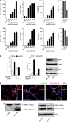
Figure 1
Figure 1
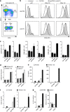
RLH activation induces secretion of proinflammatory cytokines and induction of apoptosis in murine pancreatic cancer cells. (a) Panc02 cells were stimulated with indicated amounts of ppp-RNA, poly(I:C) or left untreated. OH-RNA served as transfection control. IFN-β levels were analyzed with qRT-PCR relative to HPRT and secretion of CXCL10 or IL-6 was measured with ELISA; (b) Panc02 cells were stimulated with RNA (24 h for poly(I:C) and 48 h for ppp-RNA) and viability was assessed by FACS analysis using annexin V/PI staining; (c) Panc02 cells were incubated with siRNA specific for RIG-I or MDA5 for 24 h and subsequently stimulated with ppp-RNA or poly(I:C). Induction of apoptosis was measured by annexin V/PI staining. Silencing efficacy, as assessed by western blot, is shown; (d) activated caspase-9 (green) was visualized using green FLICA caspase-9 assay kit. Cell membranes were costained with cholera toxin B subunit (red) and nuclei with DAPI (blue); (e and f) Panc02 cells were treated as indicated for 48 h. Full length PARP-1 (116 kDa) and the cleaved large fragment of PARP-1 (89 kDa) (e) as well as the autophagy markers LC3B-I and LC3B-II (f) were analyzed by western blot. Results are representative of at least three independent experiments
Figure 2
Figure 2
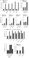
RLH activation induces characteristics of immunogenic cell death and sensitizes tumor cells towards Fas- and CTL-mediated killing. (ae) Panc02 cells were treated with RLH ligands for 24 h or left untreated. Surface expression of MHC-I (a), Fas (b) and calreticulin (c) was assessed with FACS analysis; (d and e) release of HMGB1 and Hsp70 in supernatants of RNA-treated tumor cells was measured with ELISA after 48 h; (f) tumor cells were stimulated for 12 h with RNA as indicated (50, 100, 200 ng/ml for poly(I:C) and 500, 1000, 2000 ng/ml for ppp-RNA) and subsequently stimulated with anti-Fas mAb (clone Jo2; 1 μg/ml) for another 24 h. Viability was assessed by annexin V/PI staining as double negative cell fraction; (g) lytic activity of OT-I T cells cocultured with RNA-treated PancOVA tumor cells for 6 h was assessed with an LDH-based cytotoxicity assay. As control, LDH release in the absence of T cells was measured. Representative results out of three independent experiments are shown
Figure 3
Figure 3
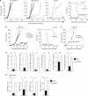
RLH-treated tumor cells induce DC activation. Panc02 cells were left untreated or stimulated with RNA as indicated before coculture with freshly isolated CD11c+ splenic DCs. (a) Representative FACS analysis of isolated DC populations, depicting CD11chigh CD8α+ and CD8α conventional DCs as well as CD11c+ B220+ pDCs; (b) CD86 surface expression of different DC subsets after 12 h exposure to RLH-treated tumor cells; (c) expression levels of CD69, CD80 and CD86 on CD8α+ DCs; (d) expression levels of CD86 on CD8α+ DCs isolated from wild-type, MDA5- or IRF-3/7-deficient mice exposed to poly(I:C)-treated tumor cells; (e and f) secretion of IL-6 and CXCL10 in supernatants of tumor cells and tumor cell/DC cocultures; (g) intracellular staining of IL-6 and CXCL10 in DC subsets. The results are representative of two (d, f, g) or three independent experiments (ac, e)
Figure 4
Figure 4
DC activation after exposure to RLH-treated tumor cells is mediated by a soluble tumor-derived factor. (a) Panc02 cells were treated as indicated to induce apoptosis and subsequently cocultured with CD8α+ DCs. CD86 surface expression of DCs was analyzed by flow cytometry; (b and c) tumor cell supernatant from RNA-treated Panc02 cells was collected and handled as indicated before addition to CD8α+ DC cultures; (b) influence of high -speed centrifugation at 15 000 g for 10 min, Benzonase, DNAse, RNAse A and H treatment of supernatants; (c) influence of boiling of the tumor cell supernatants; (d) influence of tumor cell incubation with cycloheximide (CHX) during RNA stimulation. The results are representative of three independent experiments
Figure 5
Figure 5
DC activation in response to RLH-activated tumor cells is mediated by type I IFN, but independent of TLR, RAGE or inflammasome signaling. (ac) Expression levels of CD86 on CD8α+ DCs isolated from wild-type mice incubated with supernatant from poly(I:C)-treated tumor cells were compared with DCs of (a) MyD88-, TRIF-, TLR4-, TLR2/4-deficient, (b) RAGE-deficient, (c) NLRP3-, P2X7-, ASC- and IL-18-deficient mice; (d) tumor cells were treated with RNA as indicated and supernatants were collected. Exogenous IL-6 (10 ng/ml), IFN-α (1000 U/ml), IFN-β (1000 U/ml) or a combination of IL-6 and IFN were added to the supernatants and transferred to CD8α+ DC cultures. CD86 expression of DCs was analyzed; (e) DCs were incubated either with a type IFN receptor (IFNAR) neutralizing or the corresponding control IgG antibody for 2 h before the addition of tumor cell supernatants; (f) CD86 expression of CD24high CD11blow CD45RA DCs (CD8α cDCs equivalents) generated from bone marrow cultures in the presence of rmFlt3L from IFNAR-deficient and respective wild-type mice after exposure to tumor cell supernatant. The results are representative of three independent experiments
Figure 6
Figure 6
CD8α+ DCs internalize and cross-present tumor antigen from RLH-activated tumor cells to CD8+ T cells. (ac) Panc02 cells were labeled with CFSE and treated with RNA as indicated. Total splenic DCs were added to the tumor cell culture for 3 h and antigen uptake by different DC populations was analyzed with flow cytometry (CFSE positivity); (b) influence of ice cooling or cytochalasin D on antigen uptake by CD8α+ DCs; (c) influence of different modes of cell death on antigen uptake by CD8α+ DCs; (d) quantification of SIINFEKL peptide (OVA257–264) bound to H-2Kb (MHC-I) on CD8α and CD8α+ DCs after coculture with RNA-treated OVA-expressing Panc02 tumor cells; (e) the ability of CD8α and CD8α+ DC populations cocultured with RNA-treated PancOVA tumor cells to induce proliferation of naive CFSE-labeled OT-I T cells was analyzed by flow cytometry after three days; (f) PancOVA cells were treated as indicated and CD8α+ DCs were added for 3 h. DCs were harvested and cocultured with naive, CFSE-labeled OT-I T cells for 3 days. OT-I proliferation was analyzed by flow cytometry. The results are representative of two or three independent experiments
Figure 7
Figure 7
RLH-based immunotherapy mediates prophylactic and therapeutic antitumor immunity in vivo. (a) Prophylactic vaccination: Panc02 tumor cells were treated with staurosporine or transfected with poly(I:C) for 24 h in vitro. Nonadherent cells were harvested and counted. A total of 5 × 10⁵ apoptotic tumor cells were injected s.c. into the left flank of C57BL/6 mice. Seven days later, mice where challenged with 5 × 10⁵ viable Panc02 cells injected into the right flank. Tumor growth and survival was monitored for 60 days (n=8 mice per group); (b and c) tumor therapy: established subcutaneous Panc02 (b) or T110299 (c) tumors (average size 50 mm2) were locally injected with 25 μg of OH-RNA or poly(I:C) complexed to in vivo-JetPEI. Injections were repeated twice weekly (arrows) for a total of up to six injections. Tumor growth was monitored daily and mice were killed when tumor size exceeded 150 mm2 or tumor showed ulcerations (n=8 mice per group); (d) Panc02 tumors were removed 18 h after local treatment, homogenized and mRNA expression profiles were obtained by qRT-PCR (n=3 mice per group); (e) surface marker expression of CD8α+ CD11c+ DCs in spleen of treated mice was analyzed by flow cytometry (n=3 mice per group). *P<0.05

Similar articles

Cited by

References

    1. Siegel R, Naishadham D, Jemal A. Cancer statistics, 2013. CA Cancer J Clin. 2013;63:11–30. - PubMed
    1. Fatima J, Schnelldorfer T, Barton J, Wood CM, Wiste HJ, Smyrk TC, et al. Pancreatoduodenectomy for ductal adenocarcinoma: implications of positive margin on survival. Arch Surg. 2010;145:167–172. - PubMed
    1. Zheng L, Xue J, Jaffee EM, Habtezion A. Role of immune cells and immune-based therapies in pancreatitis and pancreatic ductal adenocarcinoma. Gastroenterology. 2013;144:1230–1240. - PMC - PubMed
    1. Rabinovich GA, Gabrilovich D, Sotomayor EM. Immunosuppressive strategies that are mediated by tumor cells. Annu Rev Immunol. 2007;25:267–296. - PMC - PubMed
    1. Gabitass RF, Annels NE, Stocken DD, Pandha HA, Middleton GW. Elevated myeloid-derived suppressor cells in pancreatic, esophageal and gastric cancer are an independent prognostic factor and are associated with significant elevation of the Th2 cytokine interleukin-13. Cancer Immunol Immunother. 2011;60:1419–1430. - PMC - PubMed

Publication types

MeSH terms