

PARPi-FL--a fluorescent PARP1 inhibitor for glioblastoma imaging
- PMID: 24970386
- PMCID: PMC4198695
- DOI: 10.1016/j.neo.2014.05.005
PARPi-FL--a fluorescent PARP1 inhibitor for glioblastoma imaging
Abstract
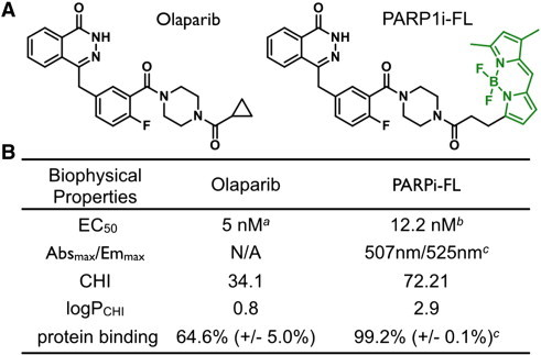
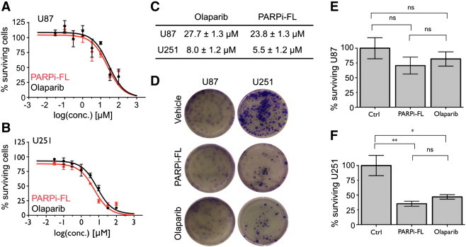
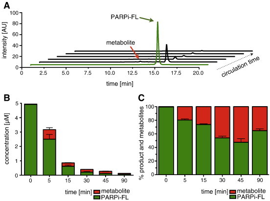
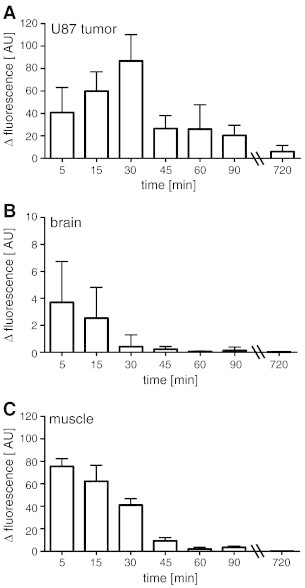
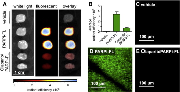
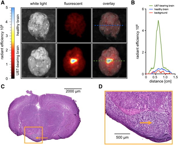
New intravital optical imaging technologies have revolutionized our understanding of mammalian biology and continue to evolve rapidly. However, there are only a limited number of imaging probes available to date. In this study, we investigated in mouse models of glioblastoma whether a fluorescent small molecule inhibitor of the DNA repair enzyme PARP1, PARPi-FL, can be used as an imaging agent to detect glioblastomas in vivo. We demonstrated that PARPi-FL has appropriate biophysical properties, low toxicity at concentrations used for imaging, high stability in vivo, and accumulates selectively in glioblastomas due to high PARP1 expression. Importantly, subcutaneous and orthotopic glioblastoma xenografts were imaged with high contrast clearly defining tumor tissue from normal surrounding tissue. This research represents a step toward exploring and developing PARPi-FL as an optical intraoperative imaging agent for PARP1 in the clinic.
Copyright © 2014 Neoplasia Press, Inc. Published by Elsevier Inc. All rights reserved.
Figures

Similar articles
-
Dual-Modality Optical/PET Imaging of PARP1 in Glioblastoma.Mol Imaging Biol. 2015 Dec;17(6):848-55. doi: 10.1007/s11307-015-0858-0. Mol Imaging Biol. 2015. PMID: 25895168 Free PMC article.
-
Restoration of Temozolomide Sensitivity by PARP Inhibitors in Mismatch Repair Deficient Glioblastoma is Independent of Base Excision Repair.Clin Cancer Res. 2020 Apr 1;26(7):1690-1699. doi: 10.1158/1078-0432.CCR-19-2000. Epub 2020 Jan 3. Clin Cancer Res. 2020. PMID: 31900275 Free PMC article.
-
Non-invasive PET Imaging of PARP1 Expression in Glioblastoma Models.Mol Imaging Biol. 2016 Jun;18(3):386-92. doi: 10.1007/s11307-015-0904-y. Mol Imaging Biol. 2016. PMID: 26493053 Free PMC article.
-
Radioiodinated PARP1 tracers for glioblastoma imaging.EJNMMI Res. 2015 Dec;5(1):123. doi: 10.1186/s13550-015-0123-1. Epub 2015 Sep 4. EJNMMI Res. 2015. PMID: 26337803 Free PMC article.
-
[PARP1 inhibitors: contemporary attempts at their use in anticancer therapy and future perspective].Postepy Hig Med Dosw (Online). 2016 Apr 13;70:280-94. doi: 10.5604/17322693.1199303. Postepy Hig Med Dosw (Online). 2016. PMID: 27117104 Review. Polish.
Cited by
-
Refining Glioblastoma Surgery through the Use of Intra-Operative Fluorescence Imaging Agents.Pharmaceuticals (Basel). 2022 Apr 29;15(5):550. doi: 10.3390/ph15050550. Pharmaceuticals (Basel). 2022. PMID: 35631376 Free PMC article. Review.
-
Detection and delineation of oral cancer with a PARP1 targeted optical imaging agent.Sci Rep. 2016 Feb 22;6:21371. doi: 10.1038/srep21371. Sci Rep. 2016. PMID: 26900125 Free PMC article.
-
A Radiotracer Strategy to Quantify PARP-1 Expression In Vivo Provides a Biomarker That Can Enable Patient Selection for PARP Inhibitor Therapy.Cancer Res. 2016 Aug 1;76(15):4516-24. doi: 10.1158/0008-5472.CAN-16-0416. Epub 2016 Jun 3. Cancer Res. 2016. PMID: 27261505 Free PMC article.
-
Approaches to PET Imaging of Glioblastoma.Molecules. 2020 Jan 28;25(3):568. doi: 10.3390/molecules25030568. Molecules. 2020. PMID: 32012954 Free PMC article. Review.
-
Molecular Imaging of PARP.J Nucl Med. 2017 Jul;58(7):1025-1030. doi: 10.2967/jnumed.117.189936. Epub 2017 May 4. J Nucl Med. 2017. PMID: 28473593 Free PMC article. Review.
References
-
- Hassa PO, Hottiger MO. The diverse biological roles of mammalian PARPS, a small but powerful family of poly-ADP-ribose polymerases. Front Biosci. 2008;12:3046–3082. - PubMed
Publication types
MeSH terms
Substances
Grants and funding
LinkOut - more resources
Full Text Sources
Other Literature Sources
Medical
Research Materials
Miscellaneous
