. 2014 Jul;141(13):2691-701.
doi: 10.1242/dev.108944. Epub 2014 Jun 12.

FGF signaling activates a Sox9-Sox10 pathway for the formation and branching morphogenesis of mouse ocular glands

Affiliations

FGF signaling activates a Sox9-Sox10 pathway for the formation and branching morphogenesis of mouse ocular glands

Ziyan Chen et al. Development. 2014 Jul.

Abstract

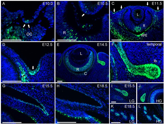
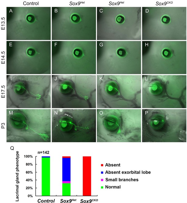
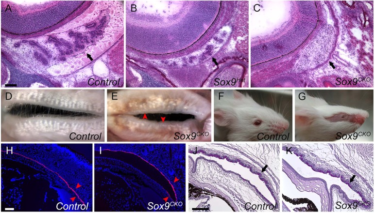
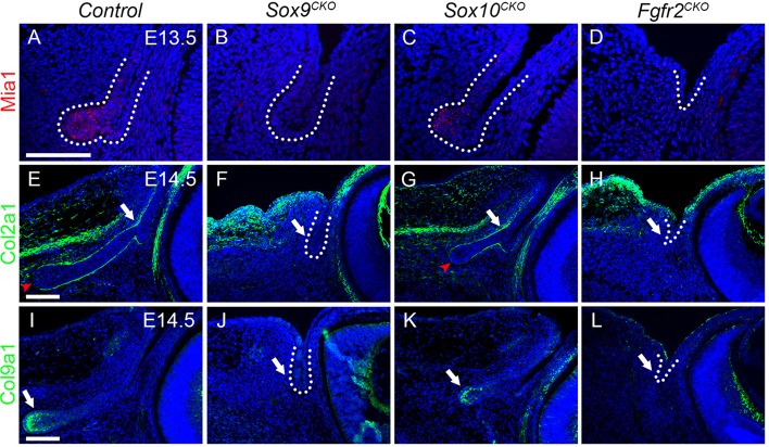
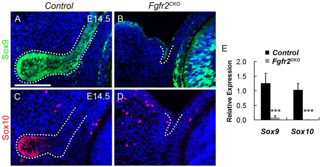
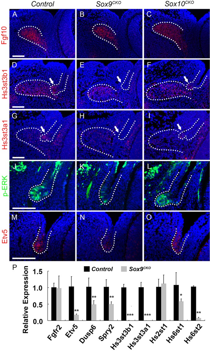
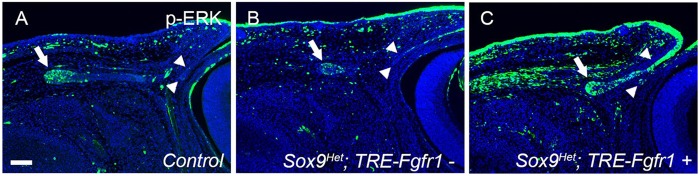
Murine lacrimal, harderian and meibomian glands develop from the prospective conjunctival and eyelid epithelia and produce secretions that lubricate and protect the ocular surface. Sox9 expression localizes to the presumptive conjunctival epithelium as early as E11.5 and is detected in the lacrimal and harderian glands as they form. Conditional deletion showed that Sox9 is required for the development of the lacrimal and harderian glands and contributes to the formation of the meibomian glands. Sox9 regulates the expression of Sox10 to promote the formation of secretory acinar lobes in the lacrimal gland. Sox9 and FGF signaling were required for the expression of cartilage-associated extracellular matrix components during early stage lacrimal gland development. Fgfr2 deletion in the ocular surface epithelium reduced Sox9 and eliminated Sox10 expression. Sox9 deletion from the ectoderm did not affect Fgf10 expression in the adjacent mesenchyme or Fgfr2 expression in the epithelium, but appeared to reduce FGF signaling. Sox9 heterozygotes showed a haploinsufficient phenotype, in which the exorbital branch of the lacrimal gland was absent in most cases. However, enhancement of epithelial FGF signaling by expression of a constitutively active FGF receptor only partially rescued the lacrimal gland defects in Sox9 heterozygotes, suggesting a crucial role of Sox9, downstream of FGF signaling, in regulating lacrimal gland branching and differentiation.

Keywords: FGF signaling; Harderian gland; Lacrimal gland; Sox10; Sox9.


Figures

Fig. 1.
Fig. 1.
Sox9 expression during development of the ocular surface epithelia. (A-C) Sox9 expression was first visible in a few lens pit cells at E10.0-10.5 (A,B, arrows). Expression ceased in the lens and spread throughout the ocular surface epithelium at E11.5, with evidence of concentration in the future conjunctival region (C, arrows). (D-F) Sox9 expression became restricted to the prospective conjunctival epithelium at E12.5 (D, arrow) and was expressed in the primary lacrimal bud epithelium at E14.5 (E,F). (G-L) Sox9 expression decreased in the conjunctival epithelium after E15.5 (G) and was hardly detected at E18.5 (H). However, Sox9 continued to be abundantly expressed in the epithelium of the lacrimal and harderian glands (I-L). lb, lacrimal bud; LG, lacrimal gland; HG, harderian gland; OC, optic cup; R, retina; L, lens; C, choroid; RPE, retinal pigmented epithelium. Scale bars: 100 μm.
Fig. 2.
Fig. 2.
Sox9 is required for the development of the lacrimal gland. (A-H) Lacrimal gland phenotypes were visualized by GFP expression from the Le-Cre transgene. The primary lacrimal bud formed at E13.5 and elongated at E14.5 (A,E). By contrast, lacrimal buds of Sox9Het embryos showed delayed budding and extension (B,C,F,G) and Sox9CKO embryos showed no bud formation (D,H). (I-P) Between E17.5 and P3, the intraorbital and exorbital lobes of the lacrimal glands formed and branched extensively in wild-type mice (I,M), whereas Sox9Het lacrimal glands were poorly branched (J,N) and, in some cases, lacked exorbital lobes (K,O), and Sox9CKO animals failed to develop lacrimal glands (L,P). (Q) Summary of lacrimal gland phenotypes in wild-type, Sox9Het and Sox9CKO P3 newborns (n=142 lacrimal glands). Sox9+/+; Le-Cre+/− embryos were used as controls.
Fig. 3.
Fig. 3.
Harderian and meibomian gland phenotypes and normal conjunctiva formation in Sox9CKO mice. (A-C) H&E staining of selected serial sections of the harderian gland at P3 showed that the harderian gland epithelium was present in wild type (A, arrow), reduced in Sox9Het mice (B, arrow) and absent in Sox9CKO mice, although the mesenchyme remained (C, arrow). (D,E) View of the inner surface of eyelids from adult wild-type (D) and Sox9CKO (E) mice showing reduced numbers of meibomian glands in both eyelids in the Sox9 mutant mice. (F,G) A Sox9CKO mouse at 3 weeks of age showing absence of hair on the eyelids and facial skin where the Le-Cre transgene was expressed. (H,I) Fluorescent RNA in situ hybridization (FISH) for keratin 4 (Krt4) in conjunctival epithelium from wild-type (H) and Sox9CKO (I) animals at P3. Krt4 expression in Sox9CKO pups (I, arrowheads) was similar to that in wild type (H, arrowheads). (J,K) Normal PAS staining pattern for conjunctiva goblet cells (arrow) in 3-month-old Sox9CKO mice (K) relative to the wild-type mice (J). Scale bars: 100 μm.
Fig. 4.
Fig. 4.
Sox9 is required for the expression of Sox10 during lacrimal gland formation. (A,B) Deletion of Sox9 resulted in the loss of Sox10 staining in the presumptive bud epithelium at E13.5 (dotted line). (C,D) Sox10 deletion maintained Sox9 expression in the poorly developed bud epithelium at E14.5 (dotted line). (E) qPCR analysis of relative Sox10 mRNA expression in E14.5 wild-type (control) and Sox9CKO temporal conjunctival epithelium. n=3; ***P<0.0001, Student's t-test; error bars represent s.d. Scale bar: 100 μm.
Fig. 5.
Fig. 5.
Sox10 is required for the formation of secretory acini in lacrimal and harderian glands. (A-F) Lacrimal gland phenotypes in Sox10Het and Sox10CKO mice. At E14.5, the primary lacrimal bud was present but shorter in Sox10CKO embryos (B, arrow). Sox10CKO glands had short, poorly branched lacrimal ducts at E17.5 (D, arrow) and P3 (F, arrow). (G-J) No evidence of secretory acini was detected in Sox10CKO lacrimal (H) or harderian (J) glands compared with wild type at P3 (G,I). (K,L) Sox10Het lacrimal glands at P21 had numerous myoepithelial cells stained for α-smooth muscle actin surrounding clusters of acinar cells. No myoepithelial cells were detectable in the Sox10CKO glands – just a few medium-size blood vessels. (M,N) H&E staining showed clusters of acini in P21 Sox10Het lacrimal glands, whereas Sox10CKO glands had no apparent acini – only blood vessels and what appeared to be degenerated ducts. Scale bars: 100 μm.
Fig. 6.
Fig. 6.
The expression of cartilage-associated factors in Sox9, Sox10 and Fgfr2 mutant early stage lacrimal glands. (A-D) FISH for Mia1 in wild-type (A), Sox9CKO (B), Sox10CKO (C) and Fgfr2CKO (D) embryos at E13.5. Mia1 transcripts were detected in wild-type bud tip cells (A, dotted line), but not in presumptive bud epithelium after Sox9 and Fgfr2 deletion (B,D, dotted line) and were still present in Sox10CKO embryos (C, dotted line). (E-H) Immunostaining for Col2a1 in wild-type (E), Sox9CKO (F), Sox10CKO (G) and Fgfr2CKO (H) embryos at E14.5. Col2a1 surrounded the conjunctiva and lacrimal bud epithelium (E, arrow), except at the bud tip (E, arrowhead). Col2a1 expression was lost in Sox9CKO (F, dotted line, arrow) and Fgfr2CKO (H, dotted line, arrow) mutants but was maintained in the normal pattern in Sox10CKO buds (G, arrow, arrowhead). (I-L) Immunostaining for Col9a1 in wild-type (I), Sox9CKO (J), Sox10CKO (K) and Fgfr2CKO (L) embryos at E14.5. Col9a1 protein was mainly localized within and at the basal surface of the bud tip cells (I, arrow). Col9a1 expression was lost in Sox9CKO (J, dotted line, arrow) and Fgfr2CKO (L, dotted line, arrow) embryos but was present in Sox10CKO buds (K, arrow). Scale bars: 100 μm.
Fig. 7.
Fig. 7.
FGF signaling is required for the expression of Sox9 and Sox10. (A,B) Sox9 expression decreases in the temporal conjunctival epithelium in the Fgfr2CKO mutant (B, dotted line) compared with the wild type (A, dotted line). (C,D) The Fgfr2 mutant showed no detectable Sox10 staining in the presumptive conjunctival epithelium (D, dotted line). (E) qPCR analysis of Sox9 and Sox10 expression in E14.5 temporal conjunctival epithelium, as normalized to Actb in Fgfr2CKO relative to the control mice. n=3; ***P<0.0001, Student's t-test; error bars represent s.d. Scale bar: 100 μm.
Fig. 8.
Fig. 8.
FGF signaling appears to be reduced after Sox9 deletion. (A-C) RNA FISH for Fgf10 in periocular mesenchyme surrounding the lacrimal bud in E13.5 wild-type (A), Sox9CKO (B) and Sox10CKO (C) mice. Both Sox9CKO (B) and Sox10CKO (C) embryos had similar expression of Fgf10 mRNA to the control (A). (D-I) RNA FISH for Hs3st3b1 (D-F) and Hs3st3a1 (G-I) in E14.5 wild-type (D,G), Sox9CKO(E,H) and Sox10CKO (F,I) embryos. In wild-type mice, Hs3st3b1 and Hs3st3a1 mRNAs were detected both in lacrimal bud tip cells (D,G, arrow, dotted line) and the adjacent mesenchyme (D,G, dotted line). However, after Sox9 deletion, Hs3st3b1 and Hs3st3a1 were expressed only in the adjacent mesenchyme (E,H, dotted line) but not in the presumptive bud epithelium (E,H, arrow, dotted line). Sox10 deletion did not affect Hs3st3b1 and Hs3st3a1 expression in lacrimal bud tip cells (F,I, arrow, dotted line) or the adjacent mesenchyme (F,I, dotted line). (J-L) Immunostaining for phospho-ERK in wild-type (J), Sox9CKO (K) and Sox10CKO (L) mice. In wild-type mice, phospho-ERK was prominent in the invaginating bud epithelium (J). In Sox9CKO conjunctival epithelium, levels of phospho-ERK decreased markedly (K), whereas the Sox10CKO bud (L) exhibited a similar level of phospho-ERK to the wild-type bud epithelium (J). (M-O) RNA FISH for Etv5 in E14.5 wild-type (M), Sox9CKO (N) and Sox10CKO (O) embryos. In wild-type mice, Etv5 transcripts were readily detected in the primary bud epithelium (M). In Sox9CKO conjunctival epithelium, Etv5 mRNA levels were greatly reduced (N); however, Sox10CKO buds expressed Etv5 mRNA at a similar level to controls (O). (P) Transcripts for key members or targets of the FGF signaling pathways were quantified by qPCR analysis at E14.5 for control and Sox9CKO temporal conjunctival epithelium. There was no significant difference in the expression levels of Fgfr2 between control and Sox9CKO epithelia. The transcript level for Etv5, Dusp6 and Spry2 decreased significantly in Sox9CKO embryos. Hs3st3b1 and Hs3st3a1 also showed a significant reduction, confirming the RNA FISH results. However, not all heparin-synthesizing enzymes were significantly decreased. n=3; *P<0.05, **P<0.001, ***P<0.0001, Student's t-test; error bars represent s.d. Scale bars: 100 μm.
Fig. 9.
Fig. 9.
Increased FGF signaling in the ocular surface epithelium moderately improves lacrimal gland defects in Sox9Het eyes. Activation of FGF signaling in the ocular surface epithelium increases the levels of phospho-ERK at E15.5. In wild-type (control) mice, phosphorylation of ERK was apparent in the lacrimal bud tip (A, arrow) and was decreased in Sox9Het; TRE-Fgfr1 mice (B, arrow). However, phospho-ERK was not detected in the conjunctival epithelium of control (A, arrowheads) or Sox9Het; TRE-Fgfr1 (B, arrowheads) mice. In Sox9Het; TRE-Fgfr1+ mice, ERK was intensely phosphorylated in the lacrimal bud and conjunctival epithelium (C, arrow and arrowheads), compared with the control and with Sox9Het; TRE-Fgfr1 mice. Scale bar: 100 μm.
Fig. 10.
Fig. 10.
Model of Sox9, Sox10 and FGF function during lacrimal gland branching morphogenesis. (A) Sox9 controls the initial budding and the elongation of the lacrimal bud; Sox10 is activated by Sox9 and continues the elongation and branching of the lacrimal bud and controls the formation of secretory acini. (B) Fgf10 regulates the expression of Sox9 and Sox10. In turn, Sox9 regulates the expression of heparan sulfate-synthesizing enzymes (HSSE), which are required for the synthesis and function of heparan sulfate (HS), to promote FGF signaling. Sox9 and FGF signaling regulate ECM components, which may contribute to the formation and elongation of the lacrimal bud. Identifying the Sox10-specific pathways in the lacrimal buds will require further exploration.

Similar articles

Cited by

References

    1. Abler L. L., Mansour S. L., Sun X. (2009). Conditional gene inactivation reveals roles for Fgf10 and Fgfr2 in establishing a normal pattern of epithelial branching in the mouse lung. Dev. Dyn. 238, 1999-2013 10.1002/dvdy.22032 - DOI - PMC - PubMed
    1. Akiyama H., Chaboissier M.-C., Martin J. F., Schedl A., de Crombrugghe B. (2002). The transcription factor Sox9 has essential roles in successive steps of the chondrocyte differentiation pathway and is required for expression of Sox5 and Sox6. Genes Dev. 16, 2813-2828 10.1101/gad.1017802 - DOI - PMC - PubMed
    1. Ashery-Padan R., Marquardt T., Zhou X., Gruss P. (2000). Pax6 activity in the lens primordium is required for lens formation and for correct placement of a single retina in the eye. Genes Dev. 14, 2701-2711 10.1101/gad.184000 - DOI - PMC - PubMed
    1. Bagheri-Fam S., Sim H., Bernard P., Jayakody I., Taketo M. M., Scherer G., Harley V. R. (2008). Loss of Fgfr2 leads to partial XY sex reversal. Dev. Biol. 314, 71-83 10.1016/j.ydbio.2007.11.010 - DOI - PubMed
    1. Belteki G., Haigh J., Kabacs N., Haigh K., Sison K., Costantini F., Whitsett J., Quaggin S. E., Nagy A. (2005). Conditional and inducible transgene expression in mice through the combinatorial use of Cre-mediated recombination and tetracycline induction. Nucleic Acids Res. 33, e51 10.1093/nar/gni051 - DOI - PMC - PubMed

Publication types

MeSH terms