. 2015 Apr 16;34(16):2138-2144.
doi: 10.1038/onc.2014.156. Epub 2014 Jun 9.

TRAIL-R2-specific antibodies and recombinant TRAIL can synergise to kill cancer cells

Affiliations

TRAIL-R2-specific antibodies and recombinant TRAIL can synergise to kill cancer cells

Mark H Tuthill et al. Oncogene. .

Abstract

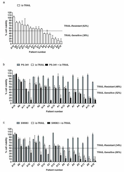
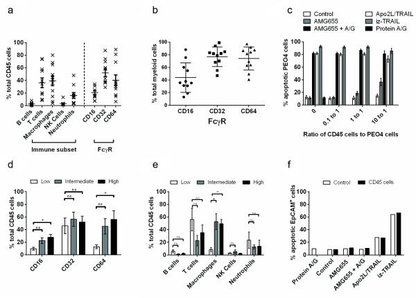
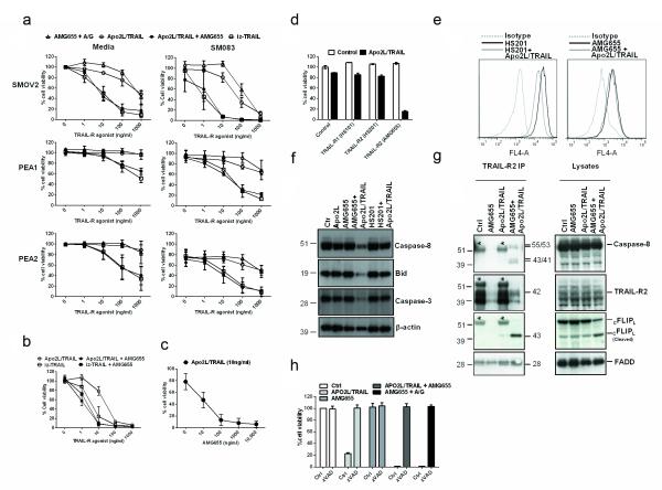
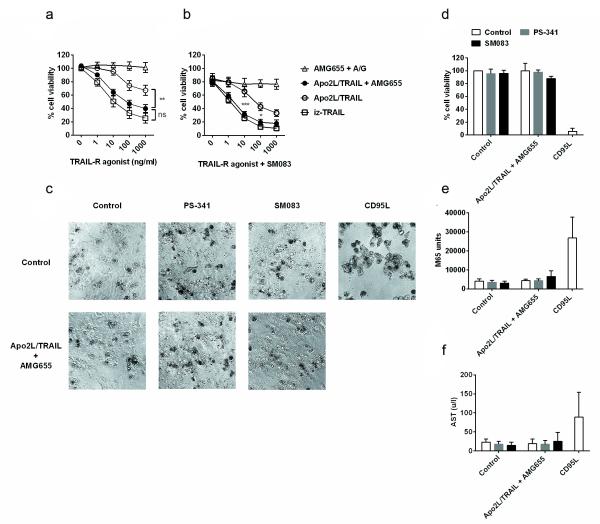
Tumour necrosis factor-related apoptosis-inducing ligand (TRAIL) induces apoptosis in cancer cells while sparing normal tissues. Despite promising preclinical results, few patients responded to treatment with recombinant TRAIL (Apo2L/Dulanermin) or TRAIL-R2-specific antibodies, such as conatumumab (AMG655). It is unknown whether this was due to intrinsic TRAIL resistance within primary human cancers or insufficient agonistic activity of the TRAIL-receptor (TRAIL-R)-targeting drugs. Fcγ receptors (FcγR)-mediated crosslinking increases the cancer-cell-killing activity of TRAIL-R2-specific antibodies in vivo. We tested this phenomenon using FcγR-expressing immune cells from patients with ovarian cancer. However, even in the presence of high numbers of FcγR-expressing immune cells, as found in ovarian cancer ascites, AMG655-induced apoptosis was not enabled to any significant degree, indicating that this concept may not translate into clinical use. On the basis of these results, we next set out to determine whether AMG655 possibly interferes with apoptosis induction by endogenous TRAIL, which could be expressed by immune cells. To do so, we tested how AMG655 affected apoptosis induction by recombinant TRAIL. This, however, resulted in the surprising discovery of a striking synergy between AMG655 and non-tagged TRAIL (Apo2L/TRAIL) in killing cancer cells. This combination was as effective in killing cancer cells as highly active recombinant isoleucine-zipper-tagged TRAIL (iz-TRAIL). The increased killing efficiency was due to enhanced formation of the TRAIL death-inducing signalling complex, enabled by concomitant binding of Apo2L/TRAIL and AMG655 to TRAIL-R2. The synergy of AMG655 with Apo2L/TRAIL extended to primary ovarian cancer cells and was further enhanced by combination with the proteasome inhibitor bortezomib or a second mitochondrial-derived activator of caspases (SMAC) mimetic. Importantly, primary human hepatocytes were not killed by the AMG655-Apo2L/TRAIL combination, also not when further combined with bortezomib or a SMAC mimetic. We therefore propose that clinical-grade non-tagged recombinant forms of TRAIL, such as dulanermin, could be combined with antibodies such as AMG655 to introduce a highly active TRAIL-R2-agonistic therapy into the cancer clinic.


Figures

Figure 1
Figure 1
Treatment of primary ovarian cancer cells with bortezomib or SMAC mimetics leads to enhanced iz-TRAIL-induced cell death. Primary ovarian cancer cells were isolated from 18 patients with advanced ovarian cancer (Supplementary Table S1 and Supplementary Figure S1). (a) Primary ovarian cancer cells were cultured in 50% RPMI and 50% ascites which was 0.22 μm sterile filtered and supplemented with 1% penicillin/streptomycin/gentamycin/glutamine at 37°C in a humidified incubator with 5% CO2 and subsequently treated with iz-TRAIL [100 ng/ml] in the absence or presence of bortezomib/PS-341 [20 nM] (Selleck Chemicals) (b), or the SMAC mimetic SM083 [100 nM] (29) (c). Cell viability was measured after 48 hours using the CellTiter-Glo® Luminescent Cell Viability Assay (Promega, Southampton, UK) according to the manufacturer’s instructions. Data represent the mean of 3 biological replicates (± S.D.) using cells from each donor.
Figure 2
Figure 2
Primary ascites-derived human CD45-positive cells are inefficient enablers of FcγR-dependent TRAIL-R2-mediated apoptosis. (a) Flow-cytometric analysis (FACS) of FcγRIIIA (CD16), FcγRIIA (CD32) and FcγRIA (CD64) on the surface of CD45-positive immune cells obtained from ascites of 11 patients with ovarian cancer. Flow cytometry was performed on a BD FACSCalibur and analysed using FlowJo software from Tree star. FcγR expression was determined by staining cells for CD45, CD16, CD32, and CD64; B-cells were identified via CD45, CD3, CD19; macrophages via CD45, CD68, CD14; T-cells via CD45 and CD3; NK cells via CD45, CD16, CD56; neutrophils via CD45, CD66. All antibodies were purchased from BioLegend, (London, UK). (b) A forward and side scatter gate was used to identify macrophages (CD45, CD68, CD14) and neutrophils (CD45, CD66) within ovarian cancer ascites (Supplementary Figure S2). FcγR expression was determined by staining cells for CD45 and CD16, CD32, and CD64. (c) Ascites-derived primary human CD45 immune cells were co-cultured with PEO4 cells and treated with AMG655 [500 ng/ml], AMG655 and recombinant protein A/G, Apo2L/TRAIL [500 ng/ml], or iz-TRAIL [500 ng/ml]. Primary CD45 cells were co-cultured with PEO4 cells in RPMI supplemented with 10% FCS and 1% penicillin/streptomycin/gentamycin/glutamine. Tumour-cell-specific death was quantified from a mixed population of tumour and immune cells 24 hours after TRAIL treatment and determined by detection of cleaved CK18 (M30 CytoDeath, Peviva, Bromma, Sweden), a specific marker of epithelial cell death by FACS. Data shown are mean (± S.D.) of 11 independent experiments using CD45 cells from 11 patients. (d) The relative expression of CD16, CD32 and CD64 on immune cells between patients with low (<10%), intermediate (>10% and <25%), and high (>25%) induction of AMG655-induced apoptosis in PEO4 cells (within the 10:1 ratio treated group, Supplementary Figure S3). (e) Differences between the immune cell profiles of patients with low, intermediate, and high enablers of FcγR-dependent AMG655-induced apoptosis (within the 10:1 ratio-treated group). Data were analysed using GraphPad Prism 5 software (GraphPad Software). Statistical analysis was performed using a two-tailed Student’s t-test (n.s = p>0.05, *=p<0.05, **=p<0.01, ***=p<0.001). (f) Primary ovarian cancer cells were cultured with patient-derived immune cells that had been determined to be capable of enabling AMG655-induced apoptosis in PEO4 cells (Supplementary Figure S5), at a ratio of 10 immune cells to 1 ovarian cancer cell, in the absence or presence of AMG655 [500 ng/ml], AMG655 and protein A/G, Apo2L/TRAIL [500 ng/ml], or iz-TRAIL [500 ng/ml]. Specific death of cancer cells was determined flow-cytometrically after 24 hours by release of cleaved cytokeratin 18. Primary CD45 cells and EpCAM+ cells were cultured in 50% RPMI and 50% ascites which was 0.22 μm sterile filtered and supplemented with 1% penicillin/streptomycin/ gentamycin/glutamine at 37°C in humidified incubator with 5% CO2. Data are representative of 2 independent experiments performed using cells from the same patient and experiments performed using samples from other patients.
Figure 3
Figure 3
AMG655 and Apo2L/TRAIL synergise to potently kill ovarian cancer cells via TRAIL-R2. (a) All ovarian cancer cell lines were cultured in RPMI 1640 supplemented with 10% Foetal Calf Serum (FCS) and 1% penicillin/streptomycin/glutamine. SMOV2, PEA1, and PEA2 cells were treated with AMG655 and recombinant protein A/G, Apo2L/TRAIL, iz-TRAIL, or AMG655 [10 μg/ml] and Apo2L/TRAIL with (right panel) and without (left panel) SM083 at the indicated concentrations. Cell viability was measured after 24 hours using the CellTiter-Glo® Luminescent Cell Viability Assay (Promega) according to the manufacturer’s instructions. (n=3 ± S.D.). The production and purification of iz-TRAIL was as described previously (4). Production and purification of Apo2L/TRAIL was performed as previously described by Ashkenazi et al. (2). (b) PEO4 cells were pre-incubated in the absence or presence of AMG655 [10 μg/ml] for 1 hour and then treated with Apo2L/TRAIL or iz-TRAIL at increasing concentrations. Cell viability was measured after 24 hours (n=5 ± S.D.). (c) PEO4 cells were treated with Apo2L/TRAIL [10 ng/ml] and increasing concentrations of AMG655. Cell viability was measured after 24 hours. (d) PEO4 cells were pre-treated with or without different TRAIL-R1- or TRAIL-R2-specific antibodies [10 μg/ml], and then treated with Apo2L/TRAIL [10 ng/ml]. The TRAIL-R1- and -R2-specific antibodies HS101 and HS201 are available from AdipoGen. (e) PEO4 cells were incubated with Apo2L/TRAIL [1 μg/ml] for 1 hour on ice, and then were washed 3 times, and incubated with HS201 [10 μg/ml] or AMG655 [10 μg/ml] or isotype control for 1 hour on ice. The cells were washed, and the binding of HS201 and AMG655 to TRAIL-R2 was determined by flow cytometry. (f) PEO4 cells were stimulated with control Apo2L/TRAIL [1 μg/ml], AMG-655 [10 μg/ml], AMG-655 [10 μg/ml] + Apo2L/TRAIL [1 μg/ml], HS201 [10 μg/ml], or HS201 [10 μg/ml] + Apo2L/TRAIL [1 μg/ml]. After 14 hours the cells were lysed, and equal amounts of proteins from lysates, as determined by the bicinchoninic acid (BCA) protein assay (Pierce), were separated by SDS-PAGE (NuPAGE) on 4-12% Bis-Tris-NuPAGE gels under reducing conditions (Invitrogen). Proteins were transferred onto nitrocellulose membranes (Amersham Pharmacia). Membranes were subjected to immunoprobing with antibodies against caspase-8 (clone C15, Enzo, Exeter, UK), Bid (R&D Systems, Abingdon, UK), caspase-3 (R&D Systems) and β-actin (Sigma, Gillingham, UK). Proteins were visualised using the Western Lightning® Plus-ECL, Enhanced Chemiluminescence Substrate ECL Western Blotting Detection Plus (GE Healthcare). (g) PEO4 cells were treated with either AMG655 [10 μg/ml], Apo2L/TRAIL [100 ng/ml], or Apo2L/TRAIL and AMG655 for 1 hour and then lysed. Either AMG655 [10 μg/ml] or Apo2L/TRAIL [100 ng/ml] was added to the lysates of cells stimulated in the absence of the respective substance before immunoprecipitation of TRAIL-R2 to ensure the observed changes in the TRAIL-R2 DISC were dependent on TRAIL-R2 stimulation (IP, immunoprecipitation. Protein concentrations of each lysate were determined as above. TRAIL-R2 was immunoprecipitated via AMG655, which was added to lysates if not used for stimulation, and uncoupled Protein G Sepharose (GE Healthcare). The beads were then washed and proteins were eluted from the beads with LDS buffer (NuPAGE, Invitrogen) containing DTT. Proteins were separated by SDS-PAGE as above and analysed by Western blotting as above. Membranes were subjected to immunoprobing with antibodies against TRAIL-R2 (Cell Signaling, Danvers, MA, USA), FADD (clone 1F7, Enzo), cFLIP (clone NF6, is available from Enzo) and Caspase-8 (clone C15, Enzo). *, unspecific bands representing the IgG1 heavy chain from the addition of exogenous AMG655 to the lysates. (h) PEO4 cells were treated for one hour with the pan-caspase inhibitor zVAD-fmk [20 μM] and subsequently with Apo2L/TRAIL [100 ng/ml], AMG655 [10 μg/ml] and Apo2L/TRAIL [100 ng/ml], AMG655 [10 μg/ml], or AMG655 and protein A/G. Cell viability was measured after 24 hours.
Figure 4
Figure 4
AMG655 and Apo2L/TRAIL synergise to kill primary ovarian cancer cells but not primary human hepatocytes (PHH). (a) Primary EpCAM+ ovarian cancer cells, isolated from 6 patients with advanced ovarian cancer, were treated with iz-TRAIL, AMG655 and recombinant protein A/G (A/G), Apo2L/TRAIL, or Apo2L/TRAIL and AMG655 [10 μg/ml], either alone (a) or in the presence of SM083 [100 nM] (b). Cell viability was measured after 48 hours (n = 6 ± S.D for (a) and n = 6 ± S.D. for (b). Data were analysed using GraphPad Prism 5 software (GraphPad Software) and two way ANOVA (non-significant (n.s) = p> 0.05, *=p<0.05, **=p<0.01, ***=p<0.001). Individual patient data are shown in Supplementary Figure S9. (c) PHH from 3 donors and PHH culture reagents were purchased from GIBCO® Life technologies. After thawing, the PHH were seeded as per the manufacturer’s recommendations and cultured in hepatocyte maintenance medium for 3 days. PHH were then treated with Apo2L/TRAIL [10 μg/ml] and AMG655 [10 μg/ml] with or without SM083 [100 nM] or bortezomib/PS-341 [20 nM]. Fc-CD95L [1 μg/ml] was used as a positive control. Fc-CD95L was produced and purified from HEK293T supernatant as previously described (4). (c) Phase contrast microscopy (10 × original magnification) of treated PHH from a representative donor (donor 3). (d) Viability of PHH was assessed 24 hours after the onset of treatment as in (a) (n=3 ± S.D.) using the CellTiter-Glo® Luminescent Cell Viability Assay (Promega) according to the manufacturer’s instructions. (e) PHH death was determined 24 hours after onset of treatment as in (a) by measuring release of soluble keratin 18 (K18) using the M65® ELISA (Peviva) according to the manufacturer’s instructions (n=3 ± S.D.). (f) Presence of the liver enzyme AST in PHH culture supernatants was measured 24 hours after onset of treatment. Release of intracellular hepatocyte-specific AST liver enzymes was measured using the Reflovet plus instrument (Roche, Burgess Hill, UK) according to the manufacturer’s instructions. Individual donor data are shown in Supplementary Figure S10.

Similar articles

Cited by

References

    1. Walczak H, Miller RE, Ariail K, Gliniak B, Griffith TS, Kubin M, et al. Tumoricidal activity of tumor necrosis factor-related apoptosis-inducing ligand in vivo. Nat Med. 1999;5(2):157–63. Epub 1999/02/04. - PubMed
    1. Ashkenazi A, Pai RC, Fong S, Leung S, Lawrence DA, Marsters SA, et al. Safety and antitumor activity of recombinant soluble Apo2 ligand. The Journal of clinical investigation. 1999;104(2):155–62. Epub 1999/07/20. - PMC - PubMed
    1. Lawrence D, Shahrokh Z, Marsters S, Achilles K, Shih D, Mounho B, et al. Differential hepatocyte toxicity of recombinant Apo2L/TRAIL versions. Nat Med. United States. 2001:383–5. - PubMed
    1. Ganten TM, Koschny R, Sykora J, Schulze-Bergkamen H, Buchler P, Haas TL, et al. Preclinical differentiation between apparently safe and potentially hepatotoxic applications of TRAIL either alone or in combination with chemotherapeutic drugs. Clinical cancer research: an official journal of the American Association for Cancer Research. 2006;12(8):2640–6. Epub 2006/04/28. - PubMed
    1. Herbst RS, Eckhardt SG, Kurzrock R, Ebbinghaus S, O’Dwyer PJ, Gordon MS, et al. Phase I dose-escalation study of recombinant human Apo2L/TRAIL, a dual proapoptotic receptor agonist, in patients with advanced cancer. Journal of clinical oncology: official journal of the American Society of Clinical Oncology. 2010;28(17):2839–46. Epub 2010/05/12. - PubMed

Publication types

MeSH terms