. 2014 Jun 5;510(7503):115-20.
doi: 10.1038/nature13413.

miR-34/449 miRNAs are required for motile ciliogenesis by repressing cp110

Affiliations

miR-34/449 miRNAs are required for motile ciliogenesis by repressing cp110

Rui Song et al. Nature. .

Abstract

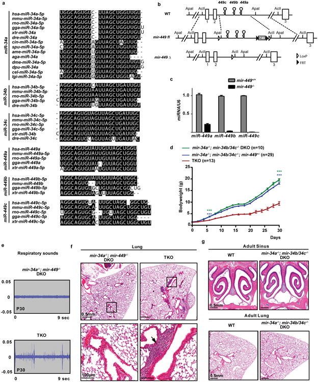
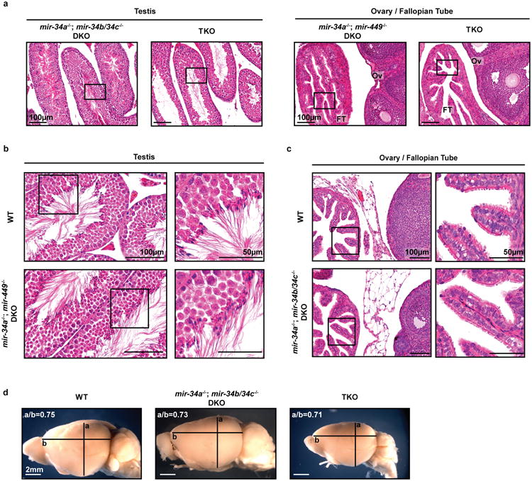
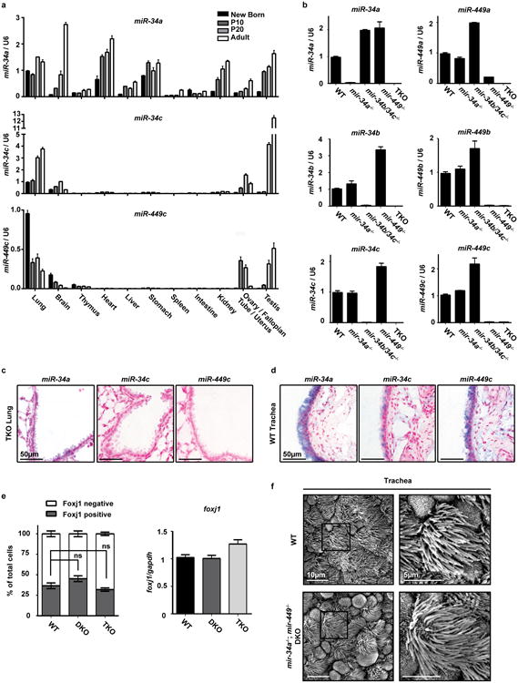
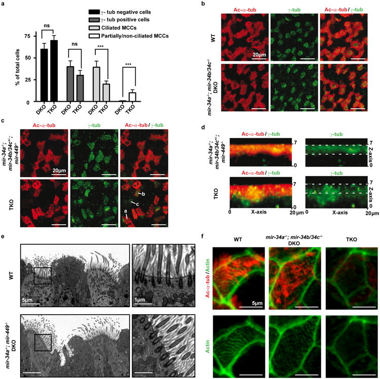
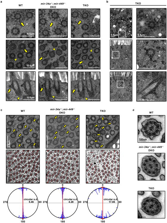
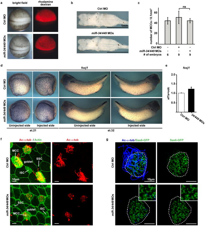
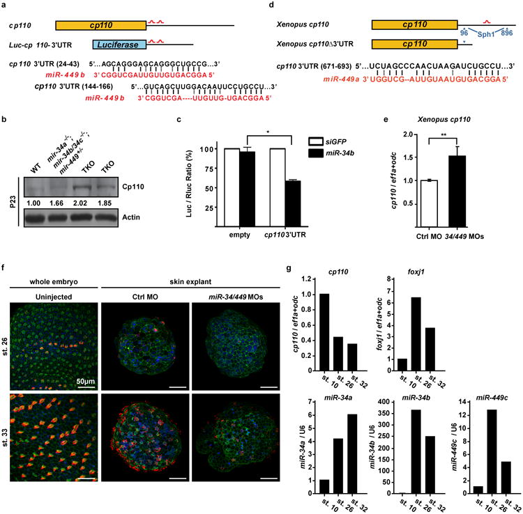
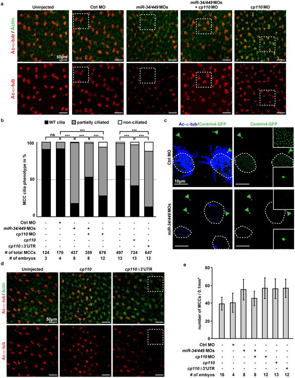
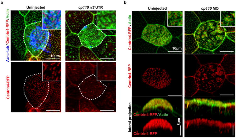
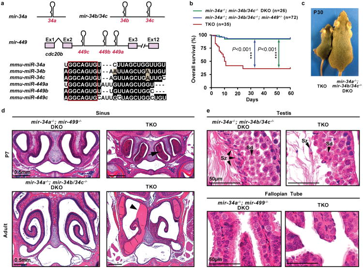
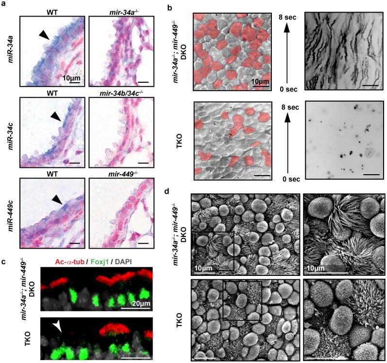
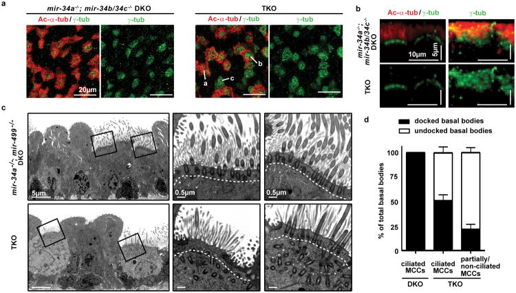
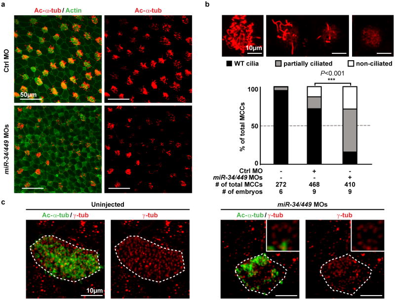
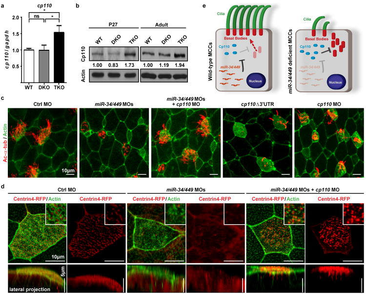
The mir-34/449 family consists of six homologous miRNAs at three genomic loci. Redundancy of miR-34/449 miRNAs and their dominant expression in multiciliated epithelia suggest a functional significance in ciliogenesis. Here we report that mice deficient for all miR-34/449 miRNAs exhibited postnatal mortality, infertility and strong respiratory dysfunction caused by defective mucociliary clearance. In both mouse and Xenopus, miR-34/449-deficient multiciliated cells (MCCs) exhibited a significant decrease in cilia length and number, due to defective basal body maturation and apical docking. The effect of miR-34/449 on ciliogenesis was mediated, at least in part, by post-transcriptional repression of Cp110, a centriolar protein suppressing cilia assembly. Consistent with this, cp110 knockdown in miR-34/449-deficient MCCs restored ciliogenesis by rescuing basal body maturation and docking. Altogether, our findings elucidate conserved cellular and molecular mechanisms through which miR-34/449 regulate motile ciliogenesis.


Figures

Extended Data Figure 1
Extended Data Figure 1. The generation and phenotypic characterization of miR-34/449 TKO mice
a. miR-34/449 miRNAs are evolutionarily conserved with extensive sequence homology across many species. miR-34a has a more ancient evolutionary history compared to the rest of miR-34/449 miRNAs. miR-34a is conserved in Deuterostome, Ecdysozoa and Lophotrochozoa, yet the rest of miR-34/449 miRNAs have only vertebrate homologues. b. Diagrams of the targeted deletion strategy to generate mir-449 knockout mice. Since all mir-449 miRNAs are within intron 2 of their host gene, cdc20b, we deleted mir-449 with a minimally predicted impact on cdc20b. c. miR-449 expression is absent in mir-449-/- knockout animals, as demonstrated by real time PCR analyses in lung tissues from littermate-controlled wild-type and mir-449-/- mice at postnatal day 35. n=3. miR-449a real time PCR primers exhibit a modest cross-reaction with miR-34 miRNAs. d. miR-34/449 TKO mice have a significant postnatal attenuation in body weight. Littermate-controlled mir-34a-/-; mir-34b/34c-/-, mir-34a-/-; mir-34b/34c-/-; mir-449+/- and TKO mice were monitored for their body weight every other day for 30 days after birth. Paired t-test, *** P<0.001. e. Surviving miR-34/449 TKO mice exhibit coughing/sneezing-like phenotype. The respiratory noise of littermate-controlled mir-34a-/-; mir-449-/- DKO and TKO mice was shown by sound wave analysis at postnatal day 30. n=14. f. Pulmonary inflammation occurs in a subset of miR-34/449 TKO mice. A representative H&E analysis of lung tissues from an adult TKO mouse indicates an increased infiltration by inflammatory cells. 3 out of 15 TKO mice examined exhibit lung infection. g. mir-34a-/-; mir-34b/34c-/- DKO mice resemble wild-type mice, exhibiting no obvious respiratory defects in paranasal sinus or lung. n=3. All error bars represent s.e.m.
Extended Data Figure 2
Extended Data Figure 2. Phenotypic characterization of reproductive organs and brain in miR-34/449 TKO mice
a. Adult male and female miR-34/449 TKO mice are infertile. Male (left) and female (right) reproductive organs from littermate-controlled DKO and TKO mice were subjected to H&E staining. n=3. Boxes indicate areas depicted in Figure 1e. b. Adult mir-34a-/-; mir-449-/- DKO male mice exhibit no defects in spermatogenesis. c. Adult mir-34a-/-; mir-34b/34c-/- DKO female mice display no defects in reproductive organs. d. The adult mir-34/449 TKO brains do not exhibit hydrocephalus, yet they are smaller in size than wild-type and DKO controls. a/b: the coronal to horizontal ratios. n=3 for b, c and d.
Extended Data Figure 3
Extended Data Figure 3. miR-34/449 miRNAs are enriched in airway MCCs
a. Most miR-34/449 miRNAs are enriched in tissues with motile cilia. Using real time PCR, the expression of miR-34a, miR-34c, and miR-449c were measured in multiple tissues from newborn, P10, P20, and adult wild-type mice. Both miR-34c and miR-449c are exclusively expressed in tissues with motile cilia, while miR-34a exhibits a broader expression pattern. n=3. b. The real time PCR assay for each miR-34/449 miRNA specifically detects the corresponding miRNA. The specificity of each miRNA real time PCR assay was validated using testis RNA from wild-type (WT), mir-34a-/-, mir-34b/34c-/-, mir-449-/-, and TKO mice at postnatal day 35. The miR-449a assay shows a slight cross reaction with homologous miRNAs. n=3. c. In situ hybridization of each miR-34/449 miRNA exhibits specific detection. No measurable miR-34/449 in situ signal is detected in TKO lung sections at postnatal day 25. n=2. d. miR-34/449 miRNAs are enriched in tracheal MCCs. In situ hybridization analyses demonstrate that miR-34c and miR-449c are specifically expressed in the tracheal MCCs, while miR-34a is expressed in both tracheal MCCs and the surrounding cell types. n=2. e. miR-34/449 TKO mice do not exhibit significant alterations in Foxj1 expression. Quantification of Foxj1 positive cells (left, n=3) and foxj1 mRNA (right, n=4) was performed for well controlled wild-type, DKO and TKO tracheas, using immunofluorescence and real time PCR, respectively. Paired t-test, ns: P > 0.05. f. mir-34a-/-; mir-449-/- DKO tracheal epithelia are morphologically indistinguishable from wild-type controls in scanning electron microscopy (SEM) analyses. n=3. All error bars represent s.e.m.
Extended Data Figure 4
Extended Data Figure 4. miR-34/449 deficiency causes defective ciliation and basal body docking in mouse airway MCCs
a. miR-34/449 TKO trachea exhibit reduced MCC ciliation. Quantification of fully ciliated MCCs (γ-tub and Ac-α-tub double-positive) and partially/non-ciliated MCCs, Ac-α-tub weak/negative) was performed in littermate controlled DKO and TKO mouse tracheas, using data from all three experiments in Figure 3a. The number of cells with MCC identity (γ-tub positive) is unaffected in TKO tracheas, yet one third of the TKO MCCs display aberrant Ac-α-tub staining, indicating ciliation defects. Paired t-test, ns: P > 0.05, *** P < 0.001. b. The mir-34a-/-; mir-34b/34c-/- DKO mice exhibit normal ciliogenesis in tracheal MCCs. Whole tracheas from age matched adult wild-type and mir-34a-/-; mir-34b/34c−/−- DKO mice were analyzed by immunofluorescence staining for Ac-α-tub (cilia) and γ-tubulin (basal bodies). n=3 c. miR-34/449 TKO primary tracheal epithelial cells exhibit ciliation defects in air liquid interface (ALI) culture. ALI culture of MCCs were derived from tracheas of littermate-controlled mir-34a-/-; mir-34b/34c+/-; mir-449-/- and TKO mice, and subjected to immunofluorescence staining for Ac-α-tub (cilia) and γ-tub (basal bodies). In TKO and control ALI culture, comparable levels of γ-tub positive cells are observed, yet a large portion of TKO γ-tub positive cells displayed a partial or complete loss of Ac-α-tub staining. a: fully, b: partially, c: non ciliated MCCs; n=2. d. Basal bodies fail to dock to the apical membrane of miR-34/449 TKO MCCs in ALI culture. Lateral projections of confocal micrographs described in (c) show impaired apical localization of γ-tub staining in TKO MCCs from ALI cultures, suggesting a defective basal body docking to the apical membrane. e. mir-34a-/-; mir-449-/- DKO trachea exhibit no defects in basal body docking using transmission electron microscopy (TEM) analyses. n=3. f. TKO tracheal MCCs exhibit a defective subapical Actin network. Whole tracheas from adult wild-type, mir-34a-/-; mir-34b/34c-/- DKO and TKO mice were analyzed by immunofluorescence staining for Ac-α-tub (cilia) and phalloidin-488 (Actin). n=2. All error bars represent s.e.m.
Extended Data Figure 5
Extended Data Figure 5. Major basal body structural components are intact in miR-34/449 TKO MCCs revealed by transmission electron microscopy
Apically docked (a) and undocked (b) basal bodies in miR-34/449 TKO MCCs have intact structural components. Basal body transition fibers (top), basal feet (middle) and striated rootlets (bottom) have comparable morphology among WT, DKO and TKO MCCs. Top panel: arrow, a representative transverse view of transition fibers. Middle panel: arrow, a representative transverse view of nine microtubule triplets with basal feet; arrowhead, a representative transverse view collected from a different height of a basal body, containing nine microtubule triplets without basal feet. Bottom panel: arrow, the longitudinal view of basal feet; arrowhead, the striated rootlet structure. c. Directionality of basal bodies (top) and axonemes (middle) is moderately affected in miR-34/449 TKO MCCs. Top panel: arrows point to the directions indicated by basal foot. Middle panel: red lines connecting the central pair of axonemes indicate the rotational polarity of each ciliary axoneme. Bottom panel: the angles of the axoneme directionality were statistically analyzed as bidirectional circular data. The average angel was set from 0° to 180° axis. miR-34/449 TKO ciliary axonemes have moderately un-coordinated directionality compared to WT and DKO controls. d. miR-34/449 TKO axonemes exhibit intact structures, including nine outer microtubule doublets, two central microtubule singlets, and dynein arms. n=3.
Extended Data Figure 6
Extended Data Figure 6. miR-34/449 deficiency in frog MCCs causes defective ciliogenesis without affecting cell fate specification
a. Injection of Ctrl or miR-34/449 MOs does not affect general embryonic development or neural tube closure. Xenopus laevis embryos were injected unilaterally with MOs at the 2-4 cell stage and analyzed at neurula stages (18-20). Targeting of the skin ectoderm was confirmed by coinjection of fluorescent rhodamine dextran. b. Frog miR-34/449 morphants do not exhibit hydrocephalus. Embryos were injected animally with control or miR-34/449 MOs into both dorsal blastomeres at the 4 cell stage to target the neural tube and brain regions. Subsequently, the whole brains were dissected and analyzed at stage 45/46. The lack of hydrocephalus in miR-34/449 morphants argues against a role of miR-34/449 in ependymal ciliation. c. Quantification of fully ciliated, partially ciliated or non-ciliated MCCs reveals no significant change in total number of MCC-fated cells in miR-34/449 morphants. Error bars represent s.d. Wilcoxon Two Sample test, n.s., P > 0.05. d, e. foxj1 expression and specification of MCC fate is unaltered in miR-34/449 deficient embryos. d. Embryos were unilaterally injected with Ctrl or miR-34/449 MOs to the right side at 2-4 cell stage, cultured until stage 21 or 32 and processed for in situ hybridization to monitor foxj1 expression in the mucociliary epithelium of the skin. No change in foxj1 expression can be detected. e. Real time PCR analysis in Ctrl or miR34/449 MOs injected skin explants at stage 26 (onset of ciliation) does not indicate reduced expression levels of foxj1. f. miR-34/449 deficient frog embryos exhibit normal development of the mucociliary ectodermal epithelium. Detailed analysis of the embryonic skin at stage 30-32 reveals the presence (specification and intercalation) of all cell types in miR-34/449 morphants, including large goblet cells, small secretory cells (SSC), Ac-α-tub positive ciliated cells (MCC) and non-tubulin enriched ion secreting cells (ISC). g. miR-34/449 morphant MCCs exhibit an uneven distribution of basal bodies. Sas6-gfp mRNA was injected at the 2-4 cell stage to visualize basal bodies at stage 30-32. In control embryos Sas6-GFP foci are evenly distributed in fully ciliated MCCs, while miR-34/449 morphant MCCs are characterized by an uneven distribution and aggregation of basal bodies, which frequently fail to grow cilia (Ac-α-tub staining). Such phenotype is characteristic for basal body docking defects. Embryos/cells analyzed: Ctrl MO (4/7), miR-34/440MOs (6/10).
Extended Data Figure 7
Extended Data Figure 7. cp110 is a direct target of miR-34/449 miRNAs
a. A schematic representation of two predicted miR-34/449 binding sites in the mouse cp110 3′UTR and in the luciferase reporter construct that contains cp110 3′UTR. b. Cp110 protein levels at postnatal day 23 are elevated in miR-34/449 TKO tracheal epithelia. c. The expression of Luc-cp110-3′UTR exhibits miR-34b-dependent repression in NIH/3T3 cells. Error bars represent s.e.m., n=3. Paired t-test, * P < 0.05. d. A schematic representation of one predicted miR-34/449 binding site in the frog cp110 3′UTR. A truncated cp110 construct, cp110Δ3′UTR, was made to generate a cp110 cDNA without the miR-34/449 target site. e. Real time PCR monitoring cp110 reveals elevated mRNA expression levels of cp110 in miR-34/440 morphant frog skin explants as compared to Ctrl MO injected specimens. f. Timeline of MCC ciliation and recapitulation of ciliation defects in skin explants (animal caps). Representative confocal images from staged whole embryos and skin explants injected with either Ctrl MO or miR-34/449 MOs show the onset of ciliation at stage 26 and fully ciliated skin ectoderm at stage 32 in whole embryos and Ctrl MO injected skin explants. miR-34/449 MOs injected skin explants develop MCC ciliogenesis defects comparable to whole embryo treatment. Cilia: Ac-α-Tub (red), Actin: Phalloidin-488 (green) and nuclei: DAPI (blue). g. Expression of cp110, foxj1 and miR-34/449 RNAs during time course of ciliation in skin explants. Explants at stage 10 represent unciliated MCC precursors, explants at stage 26 represent MCCs at the onset of ciliation, and stage 32 explants represent fully ciliated ectodermal epithelium. cp110 mRNA levels decrease over the time course of ciliation, with the strongest decrease between stage 10 and 26, while foxj1 mRNA levels rapidly increase during this time. miR-34a, -34b and -449c levels strongly increase between stage 10 and stage 26; and only a moderate increase or even decrease can be observed between stage 26 and 32, similar to foxj1 expression levels. Error bars represent s.e.m. n=2, technical replicates on pools of 30 skin explants for each time point.
Extended Data Figure 8
Extended Data Figure 8. miR-34/449 miRNAs promote ciliogenesis by repressing cp110
a. Representative examples of confocal images used for quantification of MCC ciliation in (b). Embryos were stained for Ac-α-tub (cilia) and phalloidin-488 (Actin). White boxes indicate areas depicted in Figure 5c. b. Quantification of MCC ciliation in (a), (d) and Figure 5c. χ2-test, ns P > 0.05, *** P < 0.001. c. Centrin4-GFP incorporation into basal bodies is affected in miR-34/449 deficient embryos. centrin4-gfp mRNA was injected at 2-4 cell stage to visualize basal bodies in MCCs at stage 32, and centrosomes in neighboring epithelial cells. In Ctrl morphant embryos, Centrin4-GFP staining in basal bodies (smaller foci in ciliated cells) and centrosomes (bigger foci in non-ciliated cells, green arrowheads) are equally strong. In contrast, Centrin4-GFP staining in basal bodies is greatly reduced in miR-34/449 morphants, without alteration of fluorescent intensity in centrosomes of neighboring cells. Embryos/cells analyzed: Ctrl MO (6/17), miR-34/449 MOs (7/23). d. Representative examples of confocal images from cp110 overexpression experiments used for quantification of MCC ciliation in (b). White boxes indicate areas depicted in Figure 5c. e. The number of MCC-fated cells in miR-34/449 or cp110 morphants, and embryos injected with cp110 DNA constructs is not reduced. Quantification of total MCC numbers (fully ciliated, partially ciliated or non-ciliated MCCs) is shown for frog embryos injected with various MOs/DNAs (corresponding to (a), (b), (d) and Figure 5c). Error bars represent s.d.
Extended Data Figure 9
Extended Data Figure 9. Gain and loss of cp110 affects MCC basal bodies, but not the subapical Actin meshwork
a. cp110 overexpression phenocopies miR-34/449 knockdown. Centrin4-RFP enrichment is strongly reduced in cp110Δ3′UTR overexpressing MCCs. It is noteworthy, that while ciliation and incorporation of Centrin4 are strongly affected in cp110Δ3′UTR injected embryos, formation of the subapical Actin meshwork appears largely unaffected. Together with the lack of cp110 knockdown to rescue the subapical Actin meshwork in miR-34/449 morphants, these data indicate an additional effect of miR-34/449 miRNAs on Actin formation/organization, which is cp110 independent. Cilia: Ac-α-tub, basal bodies: Centrin4-RFP, Actin: phalloidin-488. Embryos/cells analyzed: Uninjected (4/7), cp110Δ3′URT (6/10). b. The cellular basis for ciliation defects in cp110 morphants is likely due to the atypical failure of basal bodies to separate from each other, thus they appear to be aggreageted in clusters of cp110-deficient MCCs. Nevertheless, Centrin4 incorporation or apical localization of basal bodies is not affected in cp110 morphants. Basal bodies: Centrin4-RFP, Actin: phalloidin-488. Embryos/cells analyzed: Uninjected (2/3), cp110 MO (5/8). Embryos were derived from at least two females and independent fertilizations per Xenopus experiment.
Figure 1
Figure 1. miR-34/449 TKO mice exhibit defective mucociliary airway clearance and infertility
a. Gene structure (top) and sequence alignment (bottom) of mouse miR-34/449 miRNAs. Red box: seed sequences. b. TKO mice exhibit frequent postnatal mortality. Log-rank test. c. Surviving TKO mice display postnatal growth retardation. d. Excessive mucus accumulation and infection in paranasal sinuses of dying TKO at P7 (top) and surviving adult TKO mice (bottom). Arrow: infection; arrowhead: mucus accumulation; n=15. e. Adult TKO males and females are infertile. Although early spermatids (Sd) are developed, few intact spermatozoa (Sz) are generated (top, n=3). A significant MCC reduction is observed in TKO fallopian tubes (bottom, n=3).
Figure 2
Figure 2. miR-34/449 deficiency causes ciliogenesis defects in respiratory MCCs
a. miR-34/449 are strongly enriched in MCCs of respiratory epithelia (arrowheads), shown by in situ hybridization. n=2. b. TKO tracheal epithelia exhibit defective mucociliary clearance demonstrated by live imaging of fluorescent bead transport. Red: visibly ciliated MCCs; n=4. c. Cell-fate specification of MCCs is unaffected in TKO tracheas. Immunofluorescence staining for Foxj1 (a MCC marker) is unaltered in TKO tracheas, yet a large number of Foxj1-positive MCCs (arrowhead) have decreased staining for Ac-α-tub (a cilia marker). n=3. d. TKO tracheal MCCs have a significant reduction in cilia number and length, revealed by scanning electron microscopy. n=3.
Figure 3
Figure 3. miR-34/449 deficiency causes defective basal body docking in mouse airway MCCs
a. TKO tracheas exhibit ciliation defects, shown by immunofluorescence staining for Ac-α-tub (cilia) and γ-tubulin (basal bodies). a: fully, b: partially, c: non ciliated MCC; n=3. b, c, d. Basal bodies fail to dock to the apical membrane of TKO MCCs. b. Lateral projections of confocal micrographs shown in (a) c. Transmission electron microscopy (TEM) confirms basal body docking defects in TKO MCCs. n=3. d. Quantification of basal body docking based on TEM studies in (c). Docked and undocked basal bodies exhibit a distance ≤ 0.3 μm and > 0.3 μm to the apical membrane, respectively. Error bar, s.e.m.
Figure 4
Figure 4. miR-34/449 deficiency causes defective ciliogenesis in the Xenopus embryonic epidermis
a. MCCs in miR-34/449 morphants show reduced cilia length and number, demonstrated by immunofluorescence for Ac-α-tub (cilia) and phalloidin-488 (Actin). b. Quantification of MCC ciliation in (a). χ2-test. c. Co-staining of Ac-α-tub (cilia) and γ-tub (basal bodies) in miR-34/449 morphants reveals uneven/aggregated distribution of basal bodies, which frequently fail to form cilia. Embryos/cells analyzed: Uninjected (4/14), miR-34/449 MOs (5/30). Embryos were derived from at least two females and independent fertilizations per experiment. Error bar, s.e.m.
Figure 5
Figure 5. miR-34/449 miRNAs are required for ciliogenesis by repressing cp110
a. cp110 mRNA is derepressed in TKO tracheal epithelia (n=4). Paired t-test, ns P>0.05, * P<0.05; error bars, s.e.m. b. Cp110 protein is elevated in TKO tracheal epithelia. n=3.c, d. miR-34/449 represses cp110 to regulate MCC ciliation and basal body maturation and docking. c. Co-injection of miR-34/449 and cp110 MOs rescues MCC ciliation, whereas cp110Δ3′UTR overexpression phenocopies miR-34/449 morphants. cp110 MO alone also induced ciliation defects. Ac-α-tub: cilia, phalloidin-488: Actin. (Quantification: Extended Data Figure 8b.) d. Basal body maturation/docking is reestablished in miR-34/449 morphants upon cp110 knockdown. Basal bodies: Centrin4-RFP, Actin: phalloidin-488; Insets: subapical Actin meshwork. Embryos/cells analyzed: Ctrl MO (2/3), miR-34/440MOs (5/10), miR-34/449 MOs + cp110 MO (4/7). Embryos were derived from at least two females and independent fertilizations per Xenopus experiment. e. Proposed Model of regulatory role of miR34/449 during MCC ciliogenesis through direct repression of cp110.

Comment in

Similar articles

Cited by

References

    1. Ambros V. The functions of animal microRNAs. Nature. 2004;431:350–355. - PubMed
    1. He L, Hannon GJ. MicroRNAs: small RNAs with a big role in gene regulation. Nat Rev Genet. 2004;5:522–31. - PubMed
    1. Kim VN. Small RNAs: classification, biogenesis, and function. Mol Cells. 2005;19:1–15. - PubMed
    1. Du T, Zamore PD. microPrimer: the biogenesis and function of microRNA. Development. 2005;132:4645–4652. - PubMed
    1. Lee RC, Feinbaum RL, Ambros V. The C. elegans heterochronic gene lin-4 encodes small RNAs with antisense complementarity to lin-14. Cell. 1993;75:843–54. - PubMed

Publication types

MeSH terms