. 2014 May;26(5):2168-2183.
doi: 10.1105/tpc.113.120535. Epub 2014 May 30.

In Planta Recognition of a Double-Stranded RNA Synthesis Protein Complex by a Potexviral RNA Silencing Suppressor

Affiliations

In Planta Recognition of a Double-Stranded RNA Synthesis Protein Complex by a Potexviral RNA Silencing Suppressor

Yukari Okano et al. Plant Cell. 2014 May.

Abstract

RNA silencing plays an important antiviral role in plants and invertebrates. To counteract antiviral RNA silencing, most plant viruses have evolved viral suppressors of RNA silencing (VSRs). TRIPLE GENE BLOCK PROTEIN1 (TGBp1) of potexviruses is a well-characterized VSR, but the detailed mechanism by which it suppresses RNA silencing remains unclear. We demonstrate that transgenic expression of TGBp1 of plantago asiatica mosaic virus (PlAMV) induced developmental abnormalities in Arabidopsis thaliana similar to those observed in mutants of SUPPRESSOR OF GENE SILENCING3 (SGS3) and RNA-DEPENDENT RNA POLYMERASE6 (RDR6) required for the trans-acting small interfering RNA synthesis pathway. PlAMV-TGBp1 inhibits SGS3/RDR6-dependent double-stranded RNA synthesis in the trans-acting small interfering RNA pathway. TGBp1 interacts with SGS3 and RDR6 and coaggregates with SGS3/RDR6 bodies, which are normally dispersed in the cytoplasm. In addition, TGBp1 forms homooligomers, whose formation coincides with TGBp1 aggregation with SGS3/RDR6 bodies. These results reveal the detailed molecular function of TGBp1 as a VSR and shed new light on the SGS3/RDR6-dependent double-stranded RNA synthesis pathway as another general target of VSRs.


Figures

Figure 1.
Figure 1.
TGBp1 Induces Developmental Defects Resembling Those Observed in tasiRNA-Deficient Mutants and Significantly Reduces tasiRNA Accumulation. (A) Photographs of 4-week-old TGBp1 transgenic lines (1F, 7A, 10D, and 7D), tasiRNA-deficient mutants (sgs3, rdr6, dcl4, and ago7), TBSV p19, CMV 2b, and GUS transformants, and a wild-type Col-0 plant. (B) Immunoblot analysis with anti-TGBp1 polyclonal antibody to detect TGBp1 protein levels in ∼5-week-old TGBp1 transgenic lines. Coomassie Brilliant Blue (CBB) staining is shown as a loading control. (C) RNA gel blot analysis of TAS2 and TAS3 tasiRNAs, miR173, miR390, and miR171. Total RNA was prepared from the indicated transformants or mutants. The numbers below each lane show accumulation levels relative to wild-type Col-0, after normalization against ethidium bromide–stained low-molecular-weight (LMW) RNAs. (D) Quantitative real-time RT-PCR analysis of PPR and ARF3 mRNA levels in plants at the reproductive stage. Relative expression values (means ± sd, n = 3) were normalized against ACTIN2 mRNA levels. [See online article for color version of this figure.]
Figure 2.
Figure 2.
TGBp1 Does Not Affect miRNA-Directed Cleavage of the TAS Transcripts but Represses dsRNA Synthesis. (A) Schematic representation of detection of the 3′ cleavage products (thick lines) of the primary TAS transcripts using RLM-5′ RACE PCR. In the TAS2 pathway, a 313-bp product generated from the TAS2 primary transcript by the AGO1/miR173 complex could be detected. In the TAS3 pathway, a 77-bp product generated from the TAS3 primary transcript by the AGO7/miR390 complex, as well as a 110-bp product generated by the AGO1/TAS3 tasiRNA [5′D2(–)] complex, could be detected. (B) RLM-5′ RACE PCR analysis of the 3′ cleavage product of the primary TAS2 and TAS3 transcripts in wild-type Col-0 plants, GUS transformants, the TGBp1 transgenic lines, the p19 transformants, and the rdr6 and dcl4 mutants at the reproductive stage. Black and white arrowheads indicate the bands corresponding to the 3′ cleavage product generated from the TAS primary transcript by the AGO/miRNA complex and that generated from the TAS3 primary transcript by the AGO1/TAS3 tasiRNA [5′D2(–)] complex, respectively. ACTIN2 was used as a control. (C) Detection of double-stranded TAS2 and TAS3 RNAs using the RNase protection assay. Total RNAs were treated with DNase I and subsequently treated with 0, 1, and 5 units (U) of RNase I, which digests single-stranded RNA and leaves dsRNA intact, followed by RT-PCR amplification. (D) RNA gel blot analysis performed on 20 μg of a high-molecular-weight RNA fraction from plants at the reproductive stage to detect complementary RNA derived from the TAS2 transcript. Relative gel loadings are shown by ethidium bromide staining of rRNA.
Figure 3.
Figure 3.
TGBp1 Interacts with RDR6 and SGS3 in Planta. (A) Coimmunoprecipitation immunoblot analyses. Dual combinations of SGS3, RDR6, and GUS tagged with Flag epitope and those tagged with triple c-myc epitope were coexpressed in N. benthamiana leaves. Coimmunoprecipitation analyses were performed by using anti-Flag antibody, and the input and immunoprecipitated (IP) proteins were analyzed by immunoblot analysis using anti-Flag (α-Flag) and anti-myc (α-myc) antibodies. Coomassie Brilliant Blue (CBB) staining is shown as a loading control. (B) BiFC assays between TGBp1 and SGS3. TGBp1-YFPN and SGS3-YFPC were coexpressed by agroinfiltration in leaf epidermal cells of N. benthamiana plants. The left panel shows a YFP fluorescence image showing the generation of the intracellular fluorescent aggregate (arrowhead). The middle panel shows an overlay of a bright-field image and the left panel. The right panel shows a higher magnification view of the left panel, showing the aggregate composed of minute vesicles. Bars in the left and middle panels = 25 μm; bar in the right panel = 5 μm. (C) Agrobacterium-mediated RNA silencing suppression assay of TGBp1 mutants. Wild-type N. benthamiana leaves were coinfiltrated with Agrobacterium mixtures containing a vector expressing GFP and GUS (top left patches of each panel), wild-type TGBp1 (bottom left patches), or TGBp1 mutant (TGBp1AKT, TGBp1E82A, TGBp1P110L, and TGBp1T192A; right patches) expression vectors. GFP fluorescence was visualized under UV light at 4 d after inoculation. (D) BiFC assay to detect the interaction between TGBp1 mutants and SGS3. SGS3-YFPN and TGBp1AKT-YFPC, TGBp1E82A-YFPC, TGBp1P110L-YFPC, or TGBp1T192A-YFPC were coexpressed in leaf epidermal cells of N. benthamiana plants. Arrowheads indicate the intracellular fluorescent aggregates. Bars = 25 μm.
Figure 4.
Figure 4.
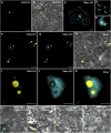
TGBp1 Alters the Subcellular Localization of SGS3. (A) to (E) Confocal sections of N. benthamiana leaves expressing SGS3-YFP alone ([A] and [B]) or TGBp1-CFP alone ([C] to [E]). (B) shows an overlay of a bright-field image and the fluorescence image of (A). (D) shows a higher magnification view of lower right boxed region in (C). The arrowheads in (D) indicate puncta embedded in cell walls. (E) shows a higher magnification view of upper left boxed region in (C). Arrows indicate aggregates adjacent to the nucleus. (F) to (K) Confocal sections of N. benthamiana leaves coexpressing SGS3-YFP and TGBp1-CFP. In (F) to (H), the yellow and cyan signals of the same plane are presented in (F) and (G), and both signals are merged and overlaid with the bright-field image in (H). Arrowheads show the region where TGBp1-CFP coaggregates with SGS3-YFP bodies. (I) to (K) show higher magnification views of the boxed regions in (F) to (H), respectively. (L) to (O) Confocal sections of N. benthamiana leaves coexpressing SGS3-YFP and each TGBp1-CFP mutant, TGBp1AKT (L), TGBp1E82A (M), TGBp1P110L (N), and TGBp1T192A (O). The panels show overlays of bright-field and fluorescence images. Bars in (A) to (C), (F) to (H), and (L) to (O) = 25 μm; bars in (D) and (E), (I) to (K), and insets in (L) to (O) = 5 μm.
Figure 5.
Figure 5.
TGBp1 Forms Homooligomers. (A) Immunoblot analysis of fractionated proteins extracted from N. benthamiana leaves transiently expressing TGBp1. Total proteins extracted from agroinfiltrated leaves at 3 d after inoculation were subjected to ultracentrifugation at 30,000g for 30 min to obtain the insoluble protein fraction (P30) and the soluble protein fraction (S30) and were analyzed by immunoblotting using anti-TGBp1 antibody. Coomassie Brilliant Blue (CBB) staining is shown as a loading control. (B) Immunoblot analysis of TGBp1 mutants. Total proteins extracted from agroinfiltrated leaves were analyzed by immunoblotting using anti-TGBp1 antibody. Coomassie blue staining is shown as a loading control. (C) Quantification of each band representing monomers (25 kD), dimers (50 kD), and trimers (75 kD) in (B) by ImageJ software version 1.40. The bars show accumulation levels relative to the monomer of each TGBp1 mutant.
Figure 6.
Figure 6.
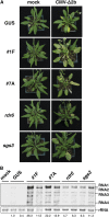
TGBp1 Increases the Accumulation Level of the 2b Deletion Mutant of CMV. (A) Symptoms of Arabidopsis plants (GUS transformants, the TGBp1 transgenic lines 1F and 7A, and the rdr6 and sgs3 mutants) infected with CMV-Δ2b. Plants were mechanically inoculated using sap from CMV-Δ2b–infected N. benthamiana leaves and photographed 3 weeks after inoculation. (B) RNA gel blot analysis of CMV-Δ2b RNAs in the plants shown in (A) using a DIG-labeled RNA probe specific for the 3′ untranslated region conserved in all four CMV RNAs. rRNA was used as the loading control. The numbers below each lane show average signal intensities of RNA1, RNA2, and RNA3 relative to the GUS transformants.
Figure 7.
Figure 7.
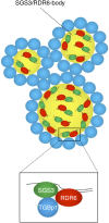
A Model Explaining the Mechanism of RNA Silencing Inhibition Mediated by TGBp1. TGBp1 inhibits dsRNA synthesis by interacting with RDR6 and SGS3, which are localized to SGS3/RDR6 bodies dispersed in the cytoplasm, and by forming the aggregates with SGS3/RDR6 bodies as a result of its ability to form homooligomers.

Similar articles

Cited by

References

    1. Adenot X., Elmayan T., Lauressergues D., Boutet S., Bouché N., Gasciolli V., Vaucheret H. (2006). DRB4-dependent TAS3 trans-acting siRNAs control leaf morphology through AGO7. Curr. Biol. 16: 927–932 - PubMed
    1. Allen E., Howell M.D. (2010). miRNAs in the biogenesis of trans-acting siRNAs in higher plants. Semin. Cell Dev. Biol. 21: 798–804 - PubMed
    1. Allen E., Xie Z., Gustafson A.M., Carrington J.C. (2005). MicroRNA-directed phasing during trans-acting siRNA biogenesis in plants. Cell 121: 207–221 - PubMed
    1. Amari K., Lerich A., Schmitt-Keichinger C., Dolja V.V., Ritzenthaler C. (2011). Tubule-guided cell-to-cell movement of a plant virus requires class XI myosin motors. PLoS Pathog. 7: e1002327. - PMC - PubMed
    1. Azevedo J., Garcia D., Pontier D., Ohnesorge S., Yu A., Garcia S., Braun L., Bergdoll M., Hakimi M.A., Lagrange T., Voinnet O. (2010). Argonaute quenching and global changes in Dicer homeostasis caused by a pathogen-encoded GW repeat protein. Genes Dev. 24: 904–915 - PMC - PubMed

LinkOut - more resources