Comparative Study
. 2014 Jul;3(7):867-78.
doi: 10.5966/sctm.2013-0154. Epub 2014 May 22.

Directed differentiation of human induced pluripotent stem cells toward bone and cartilage: in vitro versus in vivo assays

Affiliations
Comparative Study

Directed differentiation of human induced pluripotent stem cells toward bone and cartilage: in vitro versus in vivo assays

Matthew D Phillips et al. Stem Cells Transl Med. 2014 Jul.

Abstract

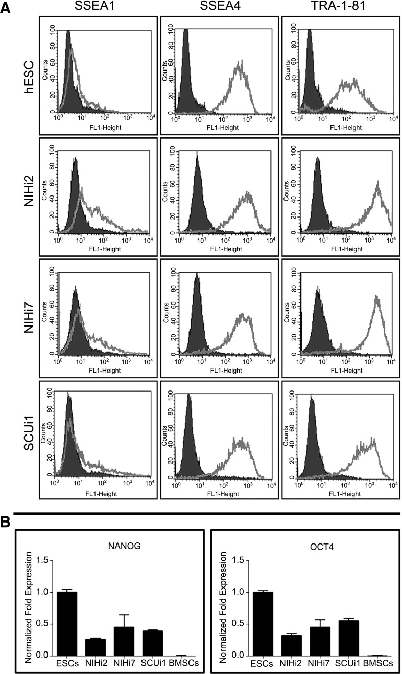
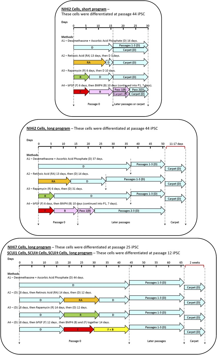
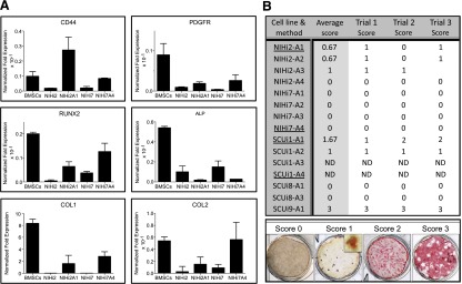
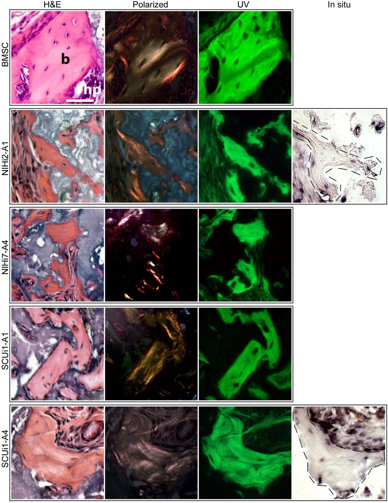
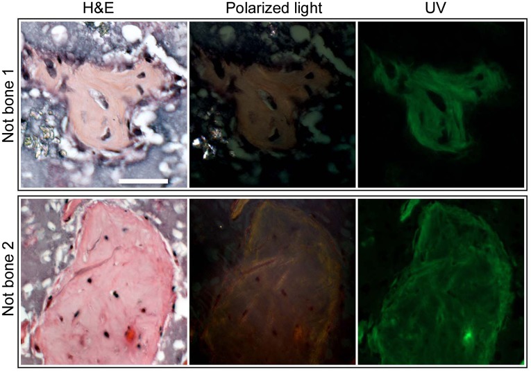
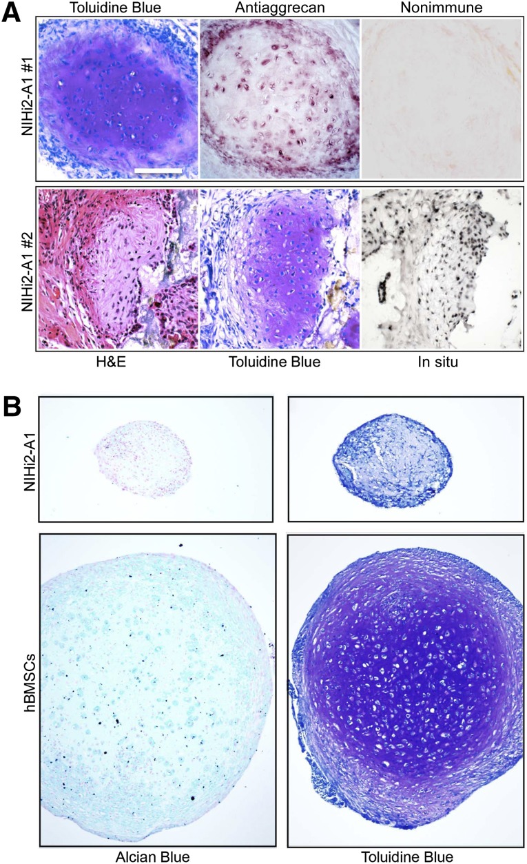
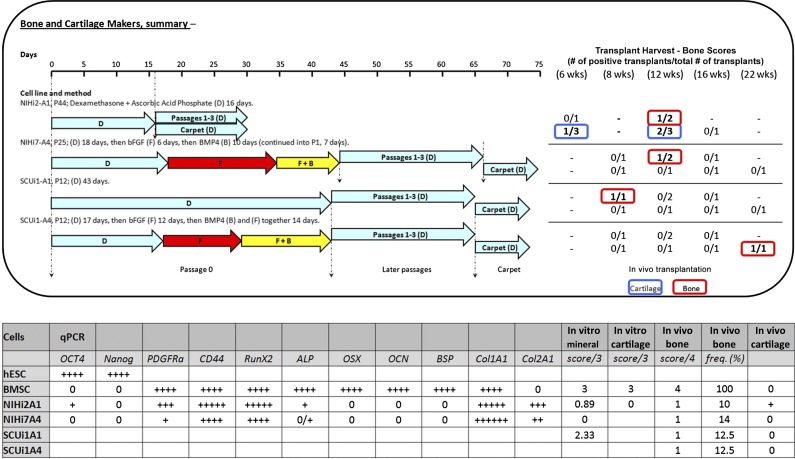
The ability to differentiate induced pluripotent stem cells (iPSCs) into committed skeletal progenitors could allow for an unlimited autologous supply of such cells for therapeutic uses; therefore, we attempted to create novel bone-forming cells from human iPSCs using lines from two distinct tissue sources and methods of differentiation that we previously devised for osteogenic differentiation of human embryonic stem cells, and as suggested by other publications. The resulting cells were assayed using in vitro methods, and the results were compared with those obtained from in vivo transplantation assays. Our results show that true bone was formed in vivo by derivatives of several iPSC lines, but that the successful cell lines and differentiation methodologies were not predicted by the results of the in vitro assays. In addition, bone was formed equally well from iPSCs originating from skin or bone marrow stromal cells (also known as bone marrow-derived mesenchymal stem cells), suggesting that the iPSCs did not retain a "memory" of their previous life. Furthermore, one of the iPSC-derived cell lines formed verifiable cartilage in vivo, which likewise was not predicted by in vitro assays.

Keywords: Bone; Chondrogenesis; Induced pluripotent stem cells; Osteoblast; Transplantation.


Figures

Figure 1.
Figure 1.
Evidence for the reprogramming of induced pluripotent stem cell lines. (A): Expression of characteristic surface markers as determined by flow cytometry is displayed for NIHi2, NIHi7, and SCUi1 compared with that of a hESC control line. The isotype control antibody for each antibody tested is in black, and the gray line represents the specified antibody results. (B): Normalized fold expression of Nanog and Oct4 mRNA levels is displayed for NIHi2, NIHi7, and SCUi1 compared with the level found in human embryonic stem cells (ESC column). Virtually identical results were found for SCUi8 and SCUi9 by fluorescence-activated cell sorting and by quantitative polymerase chain reaction (data not shown). Abbreviations: BMSCs, bone marrow stromal stem cells; ESC, embryonic stem cell; hESC, human embryonic stem cell.
Figure 2.
Figure 2.
Representation of the methods used to differentiate iPSC lines into a bone fate. Each of the three sections represents all the differentiation methods used. At the top of each section is a differentiation timeline. Underneath the timeline, the lengths and positions of the colored arrows represent the amount of time the cells in a particular program were exposed to each condition. The color of each arrow indicates that different additives were used as shown (e.g., RA [orange], R [green], F [red], B [pink], and F + B [yellow]). Light blue arrows always indicate the presence of D in the medium. Where carpet culture arrows are not shown, carpets were not made. Dashed lines indicate significant events in each program (such as differentiation, passaging, and transplant). Abbreviations: B, bone morphogenetic protein; bFGF, basic fibroblast growth factor; BMP4, bone morphogenetic protein; D, dexamethasone and ascorbic acid phosphate; F, basic fibroblast growth factor; iPSC, induced pluripotent stem cell; Pass, passage; R, rapamycin; RA, retinoic acid.
Figure 3.
Figure 3.
In vitro assays. (A): Quantitative PCR evidence for differentiation of induced pluripotent stem cell lines to a bone or cartilage fate. When available, three independent biological samples from cell lines and methods that successfully made bone in vivo were used to perform qPCR on mRNA harvested prior to cell transplantation. The results were compared with bone marrow stromal cell controls, which form bone in vivo and cartilage in vitro. (B): Results from in vitro mineralization assays. The in vitro mineralization assay was performed as described on all the cell lines and methods listed in the summary table, when available, in triplicate. After alizarin red S staining, each well was assigned a numerical score based on the amount of red staining visible, as shown in the scored samples across the bottom of the figure. For a score of 0, the well must have no significant red staining even under high magnification. For a score of 1, red staining must be visible only with magnification (inset), and wells scored as 2 must have red staining visible to the eye, but less than half the plate. To achieve a score of 3, the majority of the well must be stained. In the summary table, underlined cell lines and methods were those that made bone in vivo. The average score is the mathematical average of the three trial scores shown. Abbreviations: BMSCs, bone marrow stromal stem cells; ND, not determined because of a lack of cells (in vivo transplantation was the priority); PCR, polymerase chain reaction; qPCR, quantitative PCR.
Figure 4.
Figure 4.
Evidence that true bone was formed in certain in vivo transplants. For each cell line listed on the left, H&E, polarized light, and fluorescent light views of a single transplant section are shown compared with transplants of BMSCs, which form abundant areas of bone (b) and support hematopoiesis (hp). For H&E staining, the bone appears red and the hydroxyapatite carrier particles stain purple. ALU-positive cells (osteocytes) are observed in lacunae. When illuminated with polarized light, the collagen bundles in the bone matrix become visible as orange or pink striata, and the hydroxyapatite remains dark. Under fluorescent conditions, the pieces of bone are clear and bright green, and the surrounding tissue and carrier particles remain dark. For NIHi2-A1 and SCUi1-A4 transplants, an additional picture of an adjacent serial section is shown, in which human-specific ALU in situ hybridization was performed. Few intact nuclei were present for in situ hybridization because these are small pieces of bone, but enough are present to indicate that the bone and surrounding fibrous tissues are of human origin. All panels are at the same magnification (scale bar = 50 μm). Human bone (positive control) and mouse bone (negative control) are shown in supplemental online Figure 3. Abbreviation: BMSC, bone marrow stromal cell.
Figure 5.
Figure 5.
H&E-stained histological sections are not reliable indicators of true bone. When stained with H&E, several promising formations appeared red, which is characteristic of bone matrix when stained with H&E. Note also the appearance of widely spaced nuclei contained in apparent lacunae. However, when the same sections were examined under polarized light, no organized collagen bundles were apparent. Likewise, under fluorescent light, the bone-like formations remained dark with little autofluorescence, again indicating the lack of organized collagen that indicates true bone. These areas also did not stain with Toluidine Blue. All panels are at the same magnification (scale bar = 50 μm). Abbreviations: Not bone 1, NIHi2-A3; Not bone 2, SCUi9-A1.
Figure 6.
Figure 6.
Differentiated NIHi2-A1 cells formed cartilage. (A): Serial sections from two pieces of cartilage are shown, one in each row. Results of H&E and Toluidine Blue staining indicate the presence of characteristic cartilage matrix containing proteoglycans and glycosaminoglycans. In addition, the last two panels in row 1 show the result of antiaggrecan immunohistochemistry (including nonimmune control). Positive aggrecan staining strongly indicates the presence of cartilage. In row 2, the final panel shows the result of an in situ hybridization directed against human-specific ALU repeats. (B): NIHi2-A1 was tested in an in vitro pellet assay (protocol 1 is shown here) for the ability to form cartilage in comparison with bone marrow stromal cells (BMSCs). The resulting pellets were sectioned and stained using either Alcian blue with Nuclear Fast Red or Toluidine Blue. Chondrocytes are seen surrounded with Alcian blue positive matrix in BMSC pellets, but not in NIHi2-A1 pellets. However, note that staining of matrix in the BMSC pellet with Toluidine Blue that becomes purple (metachromasia) is far more definitive. One can see bona fide chondrocytes lying in lacunae surrounded by purple-stained matrix. At the same time, pellets made with NIHi2-A1 cells did not stain prominently with Alcian blue and did not display the characteristic metachromatic (purple) color associated with binding to sulfated glycosaminoglycans but remained a dull blue, with no histological evidence of cartilage formation visible. All panels are at the same magnification (scale bar = 50 μm). Abbreviation: hBMSCs, human bone marrow stromal stem cells.
Figure 7.
Figure 7.
Data summary. In the top panel, the cell lines and method used to successfully differentiate induced pluripotent stem cells into bone in vivo are shown on the left, with the results on the right. Both regular and carpet culture transplant results are shown. The results of in vivo transplantation are displayed on the right in table form. In the results table, blue signifies that cartilage was found in a transplant, and red indicates that true bone was found. The results are stated as the number of bone- or cartilage-producing transplants over the total number of transplants created. In the bottom panel, a summary table shows qPCR, in vitro mineralization scores, in vitro cartilage formation scores, in vivo bone formation scores, in vivo bone formation frequency as a percentage of all transplants made, and an indicator for the presence of in vivo cartilage. In vitro mineralization scores are averaged as described in Figure 4. For in vivo bone scores, the highest score achieved in any transplant for the corresponding cell type is shown. In vivo bone frequency refers to the number of times true bone appears in a transplant, calculated as a percentage of all transplants attempted for the corresponding cell type. In vivo cartilage is represented as present (+) or not present (0). Abbreviations: B, bone morphogenetic protein; bFGF, basic fibroblast growth factor; BMP4, bone morphogenetic protein; BMSC, bone marrow stromal stem cell; D, dexamethasone and ascorbic acid phosphate; F, basic fibroblast growth factor; hESC, human embryonic stem cell; qPCR, quantitative polymerase chain reaction; R, rapamycin; RA, retinoic acid.

Similar articles

Cited by

References

    1. Buttery LD, Bourne S, Xynos JD, et al. Differentiation of osteoblasts and in vitro bone formation from murine embryonic stem cells. Tissue Eng. 2001;7:89–99. - PubMed
    1. Ahn SE, Kim S, Park KH, et al. Primary bone-derived cells induce osteogenic differentiation without exogenous factors in human embryonic stem cells. Biochem Biophys Res Commun. 2006;340:403–408. - PubMed
    1. Kärner E, Unger C, Sloan AJ, et al. Bone matrix formation in osteogenic cultures derived from human embryonic stem cells in vitro. Stem Cells Dev. 2007;16:39–52. - PubMed
    1. Toh WS, Yang Z, Liu H, et al. Effects of culture conditions and bone morphogenetic protein 2 on extent of chondrogenesis from human embryonic stem cells. Stem Cells. 2007;25:950–960. - PubMed
    1. Kim S, Kim SS, Lee SH, et al. In vivo bone formation from human embryonic stem cell-derived osteogenic cells in poly(d,l-lactic-co-glycolic acid)/hydroxyapatite composite scaffolds. Biomaterials. 2008;29:1043–1053. - PubMed

Publication types

MeSH terms

LinkOut - more resources