Review
. 2014 Oct;71(19):3711-47.
doi: 10.1007/s00018-014-1638-8. Epub 2014 May 21.

Moving towards a paradigm: common mechanisms of chemotactic signaling in Dictyostelium and mammalian leukocytes

Affiliations
Review

Moving towards a paradigm: common mechanisms of chemotactic signaling in Dictyostelium and mammalian leukocytes

Yulia Artemenko et al. Cell Mol Life Sci. 2014 Oct.

Abstract

Chemotaxis, or directed migration of cells along a chemical gradient, is a highly coordinated process that involves gradient sensing, motility, and polarity. Most of our understanding of chemotaxis comes from studies of cells undergoing amoeboid-type migration, in particular the social amoeba Dictyostelium discoideum and leukocytes. In these amoeboid cells the molecular events leading to directed migration can be conceptually divided into four interacting networks: receptor/G protein, signal transduction, cytoskeleton, and polarity. The signal transduction network occupies a central position in this scheme as it receives direct input from the receptor/G protein network, as well as feedback from the cytoskeletal and polarity networks. Multiple overlapping modules within the signal transduction network transmit the signals to the actin cytoskeleton network leading to biased pseudopod protrusion in the direction of the gradient. The overall architecture of the networks, as well as the individual signaling modules, is remarkably conserved between Dictyostelium and mammalian leukocytes, and the similarities and differences between the two systems are the subject of this review.


Figures
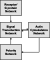
Fig. 1
Fig. 1
Overview of the networks contributing to chemotaxis. The four proposed networks required for amoeboid chemotaxis with arrows representing the interactions between them
Fig. 2
Fig. 2
Spatiotemporal regulation of “front” and “back” proteins with chemoattractant stimulation. Many proteins involved in chemotaxis are differentially localized to the front or back of migrating cells and this helps establish the balance between protrusion and retraction that leads to directed cellular migration. Thus in a cell exposed to a chemoattractant gradient the “front” proteins (shown in red) localize to the pseudopods, which are oriented toward the gradient, whereas the “back” proteins (shown in blue) line the back and sides of the cell. In a round cell with a disrupted cytoskeleton, for example, via actin-depolymerizing drugs, the “front” proteins localize to the high side of the gradient in a “crescent”, whereas the “back” proteins have opposing localization. In a resting cell or in a cell with a disrupted cytoskeleton in the absence of a chemoattractant the “back” proteins are localized uniformly along the membrane or cortex, whereas the front proteins are in the cytosol. When a cell makes a protrusion, the back proteins dissociate from that region, and the front proteins associate with that extending region of the cell periphery [396, 397]. With a uniform stimulation, the “front” proteins transiently re-localize to the entire membrane and “back” proteins transiently dissociate. These protein translocations after chemoattractant stimulation occur within 10–30 s and then return to basal state after about 30-60 s [19, 85, 397]. The precise kinetics of specific signaling components in different contexts will be addressed further in this review. Chemoattractants also trigger activation of some proteins at the front or the back of a cell without affecting localization of those proteins. In this case, fluorescently-tagged biosensors that recognize activated versions of the protein or their enzymatic products act as “front” or “back” proteins
Fig. 3
Fig. 3
Topology of the signal transduction network in Dictyostelium and mammalian leukocytes. The signal transduction network is placed between the receptor/G protein and the actin cytoskeleton networks, while the polarity network is omitted from this figure. The individual modules within the Signal Transduction Network are represented by a specific color and these colors will be used to represent the modules in subsequent figures. The arrows depict interactions between the modules that are strongly supported in the literature
Fig. 4
Fig. 4
Ras GTPase module in Dictyostelium and mammalian leukocytes. The arrow and bar lines represent positive and inhibitory links, respectively, and the lines shown depict interactions that are strongly supported in the literature
Fig. 5
Fig. 5
PI3K/PIP3 module in Dictyostelium and mammalian leukocytes. The arrow and bar lines represent positive and inhibitory links, respectively, and the lines shown depict interactions that are strongly supported in the literature
Fig. 6
Fig. 6
TORC2/PKB module in Dictyostelium and mammalian leukocytes. The arrow and bar lines represent positive and inhibitory links, respectively, and the lines shown depict interactions that are strongly supported in the literature
Fig. 7
Fig. 7
Rap1 and KrsB/Mst1 pathways in Dictyostelium and mammalian leukocytes. The arrow and bar lines represent positive and inhibitory links, respectively, and the lines shown depict interactions that are strongly supported in the literature
Fig. 8
Fig. 8
Dictyostelium cGMP/Myosin II and leukocyte RhoA/Myosin II pathways. The arrow and bar lines represent positive and inhibitory links, respectively, and the lines shown depict interactions that are strongly supported in the literature
Fig. 9
Fig. 9
Non-specific and specific modes of cell adhesion during migration. To be able to migrate cells must generate traction, which is most commonly achieved by cell adhesion to the substrate. A. Dictyostelium cells possess the ability to navigate over numerous different substrates found in their environment. Their migration is independent of canonical integrin-mediated focal adhesions. Integrins are transmembrane adhesion receptors that comprise a family of 18 α and 8 β subunits in mammals a can associate to form 24 different heterodimers that bind to a variety of ligands [398]. A transmembrane protein similar to integrin beta (SibA) is found in Dictyostelium and has some similarity to integrin β-chains, including the ability to bind talin, but they do not possess the homologous genes for focal adhesion kinase (FAK) or integrin-α chains [305]. The interaction between SibA and talinA is required for proper substrate adhesion and motility in vegetative Dictyostelium cells [–401]. Although cells lacking SibA have impaired adhesion, they have no defects in cell migration possibly due to the presence of redundant family members [400, 402]. In contrast, lack of talin A/B results in reduced adhesion, as well as migration of vegetative cells; however, it remains unclear which receptor, if any, is necessary for these effects [399]. Two nine transmembrane domain-containing proteins, Phg1 and SadA, positively regulate adhesion to non-specific substrates in Dictyostelium [403, 404]. Recent evidence indicates that an important mechanism for cell-substrate attachment in Dictyostelium is via van der Waals interactions, which allow the cells to adhere to a variety of surfaces within minutes [401]. Although the molecular mechanism of Dictyostelium attachment to the substrate is largely unknown, several signaling pathways that alter cell spreading and adhesion have been identified and are discussed in this review. B. There are focal adhesion-associated proteins, including talin, paxillin, and vinculin, that make transient foci on the basal surface of migrating cells, which may be homologous to mammalian focal adhesions. TIRF microscopy analyzing the basal layer of the cells shows paxillin localizing to these foci [–407]. C. In 2D environments, leukocytes specifically attach to the tissues via selectins, addressins, and to the extracellular matrix via integrins. L-selectin and P-selectin are cell–cell adhesion molecules that bind glycoproteins, and are utilized by leukocytes and endothelial cells, respectively, to regulate adhesion of leukocytes to specific tissues [408, 409]. Selectins are associated with numerous intracellular signaling proteins to modify cell behavior. Endothelial target localized addressins (such as MAdCAM-1) are bound by leukocyte homing receptors (for example, CD34) to help leukocytes adhere to their targets [410]. Although leukocytes express a number of different integrins, α4β1 (very late antigen-4, VLA-4) and β2-containing integrins αLβ2 (lymphocyte function-associated antigen 1, LFA-1, CD11a/CD18) and αMβ2 (Mac-1, macrophage antigen-1, CD11b/CD18) are particularly important for leukocyte chemotaxis [411, 412]. D. Unlike integrins found in mesenchymal cells, leukocyte integrins do not form well-defined focal adhesions, even though they associate with many proteins typically found in these structures, including talin, paxillin, and focal adhesion kinase (FAK). Paxillin localization in foci at the bottom of a leukocyte under TIRF microscopy is shown [407]. The reason for the absence of well-defined focal adhesions in leukocytes is likely the requirement for very rapid turnover of the cell attachments to the extracellular matrix to allow for the fast migration rates of these cells. Integrins also bind cell adhesion molecules (CAMs), such as VCAM, to aid in targeting appropriate vasculature adhesion [413]. Interestingly, while important for migration on 2D surfaces, in 3D environments, leukocytes switch to integrin-independent migration, where confinement by the extracellular matrix generates enough traction to allow forward propulsion [8]

Similar articles

Cited by

References

    1. Theveneau E, Mayor R (2012) Neural crest delamination and migration: From epithelium-to-mesenchyme transition to collective cell migration. Developmental Biology 366 (1):34-54. doi:10.1016/j.ydbio.20112.041 - DOI - PubMed
    1. Richardson BE, Lehmann R (2010) Mechanisms guiding primordial germ cell migration: strategies from different organisms. Nat Rev Mol Cell Biol 11 (1):37-49. doi:http://www.nature.com/nrm/journal/v11/n1/suppinfo/nrm2815_S1.html - PMC - PubMed
    1. Sadik CD, Luster AD. Lipid-cytokine-chemokine cascades orchestrate leukocyte recruitment in inflammation. J Leukoc Biol. 2012;91(2):207–215. - PMC - PubMed
    1. Kolaczkowska E, Kubes P. Neutrophil recruitment and function in health and inflammation. Nat Rev Immunol. 2013;13(3):159–175. - PubMed
    1. Sixt M (2011) Interstitial locomotion of leukocytes. Immunology Letters 138 (1):32-34. doi:10.1016/j.imlet.2011.02.013 - DOI - PubMed

Publication types

MeSH terms

Substances

LinkOut - more resources