. 2014 May 15;9(5):e97580.
doi: 10.1371/journal.pone.0097580. eCollection 2014.

Interactions between exosomes from breast cancer cells and primary mammary epithelial cells leads to generation of reactive oxygen species which induce DNA damage response, stabilization of p53 and autophagy in epithelial cells

Affiliations

Interactions between exosomes from breast cancer cells and primary mammary epithelial cells leads to generation of reactive oxygen species which induce DNA damage response, stabilization of p53 and autophagy in epithelial cells

Sujoy Dutta et al. PLoS One. .

Abstract

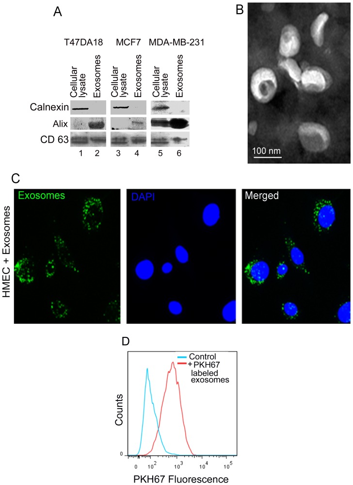
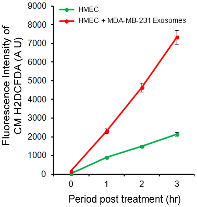
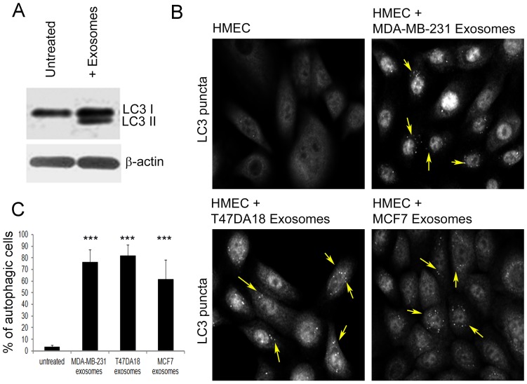
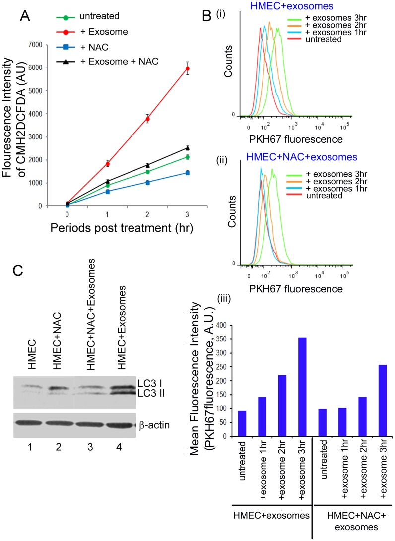
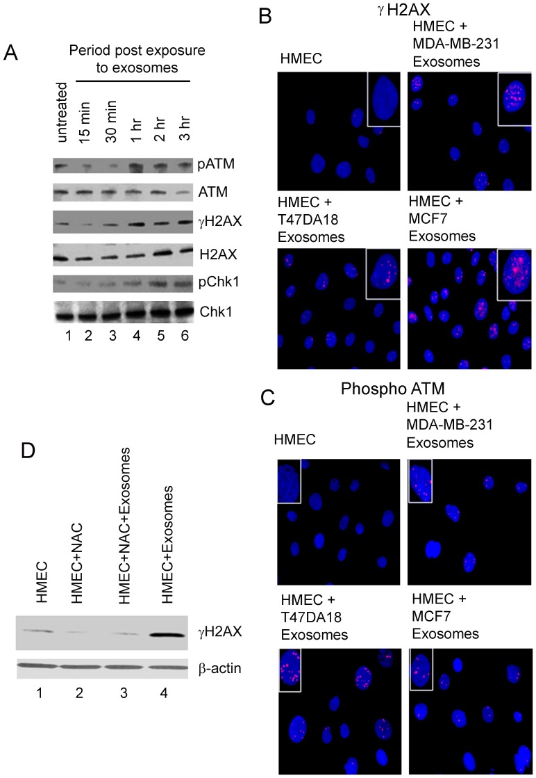
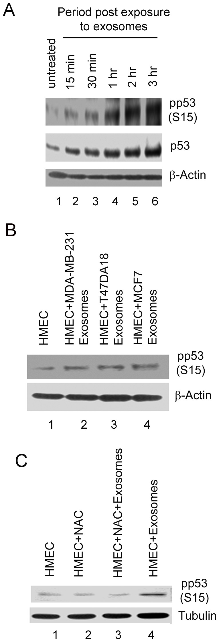
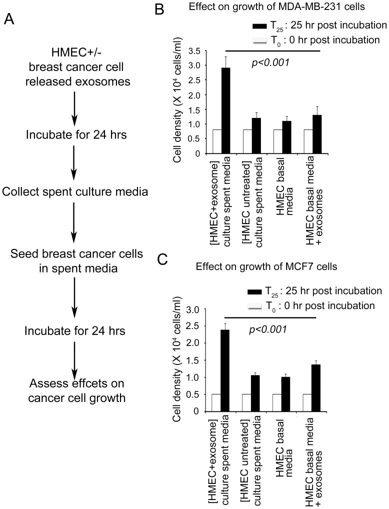
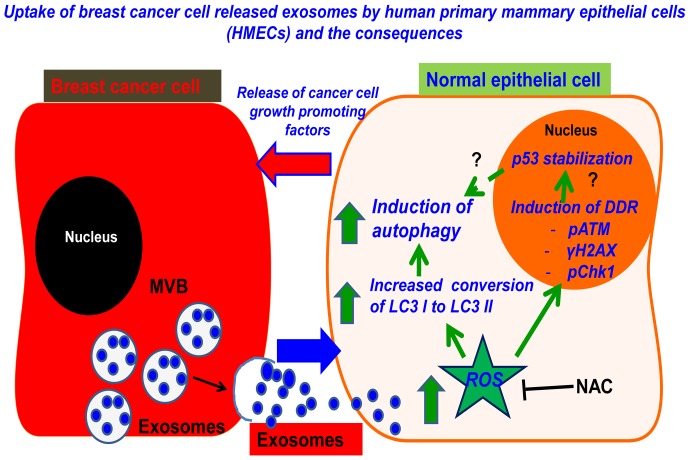
Exosomes are nanovesicles originating from multivesicular bodies and are released by all cell types. They contain proteins, lipids, microRNAs, mRNAs and DNA fragments, which act as mediators of intercellular communications by inducing phenotypic changes in recipient cells. Tumor-derived exosomes have been shown to play critical roles in different stages of tumor development and metastasis of almost all types of cancer. One of the ways by which exosomes affect tumorigenesis is to manipulate the tumor microenvironments to create tumor permissive "niches". Whether breast cancer cell secreted exosomes manipulate epithelial cells of the mammary duct to facilitate tumor development is not known. To address whether and how breast cancer cell secreted exosomes manipulate ductal epithelial cells we studied the interactions between exosomes isolated from conditioned media of 3 different breast cancer cell lines (MDA-MB-231, T47DA18 and MCF7), representing three different types of breast carcinomas, and normal human primary mammary epithelial cells (HMECs). Our studies show that exosomes released by breast cancer cell lines are taken up by HMECs, resulting in the induction of reactive oxygen species (ROS) and autophagy. Inhibition of ROS by N-acetyl-L-cysteine (NAC) led to abrogation of autophagy. HMEC-exosome interactions also induced the phosphorylation of ATM, H2AX and Chk1 indicating the induction of DNA damage repair (DDR) responses. Under these conditions, phosphorylation of p53 at serine 15 was also observed. Both DDR responses and phosphorylation of p53 induced by HMEC-exosome interactions were also inhibited by NAC. Furthermore, exosome induced autophagic HMECs were found to release breast cancer cell growth promoting factors. Taken together, our results suggest novel mechanisms by which breast cancer cell secreted exosomes manipulate HMECs to create a tumor permissive microenvironment.


Conflict of interest statement

Competing Interests: The authors have declared that no competing interests exist.

Figures

Figure 1
Figure 1. Characterization of exosomes secreted by breast cancer cells and exosome uptake by HMECs.
Exosomes were isolated from conditioned media of 3 different breast cancer cell lines, T47DA18, MCF7 and MDA-MB-231 and characterized by (A) detection of exosome specific proteins by western blotting and (B) electron microscopy. (A) Western blotting for endoplasmic reticulum specific protein calnexin and exosome marker proteins Alix and CD63 in total cellular lysates (lanes 1, 3 and 5) and exosome preparations (2, 4 and 6). 10 µg of protein was analyzed for each sample. (B) Characterization of exosomes from MDA-MB-231 cells by transmission electron microscopy (TEM). Exosomes isolated by multi-step centrifugation were fixed, negatively stained using phosphotungstic acid and observed by TEM. (C) Exosomes isolated from conditioned media of MDA-MB-231 cells were labeled with fluorescent dye PHK67. 10 µg protein equivalent of labeled exosomes were incubated with 5×10⁴ HMECs for 24 h. HMECs were washed extensively with PBS, fixed in PFA and observed using an epifluorescence microscope. Green fluorescent “specs” represent PKH-67 labeled exosomes taken up by the HMECs. (D) Flow cytometric analysis of HMECs exposed to PKH-67 labeled exosomes as described in (C).
Figure 2
Figure 2. Detection of ROS production during exosome-HMEC interactions.
Semi-confluent layers of 5×10⁴ HMECs were incubated with 10 µg protein equivalent of exosomes from MDA-MB-231 cells and ROS detection agent 10 µM CMH2DCFDA in a total volume of 300 µl of epithelial cell basal growth media for up to 3 h. Fluorescence of oxidized CMH2DCFDA was assessed fluorimetrically at the indicated time points to detect ROS production during exosome-HMEC interactions.
Figure 3
Figure 3. Induction of autophagy in HMECs following uptake of breast cancer cell released exosomes.
(A) Western blot analysis for detection of proteins LC3 I and II in cellular lysates of untreated HMECs and those incubated with exosomes from MDA-MB-231 cells for 24 h. Equal protein concentrations of whole cell lysates were analyzed by western blots. β- actin was used as an equal loading control. (B) IFA of LC3 “puncta” formation in HMECs untreated or incubated with exosomes from either MDA-MB-231, T47DA18 or MCF7 cells for 24 h. Cells were washed, fixed with paraformaldehyde, permeabilized with saponin, blocked with normal donkey sera and reacted with rabbit polyclonal anti-LC3 antibodies. LC3 expression was detected using donkey anti-rabbit IgG secondary antibodies labeled with Alexa 594 fluorophore. White arrows indicate LC3 “puncta” characteristic of autophagy. (C) Quantitation of cells with LC3 puncta in cultures incubated with or without exosomes. A minimum of 10 independent fields of view/50 cells were chosen for colocalization analysis. Error bars indicate SEM values.***: p<0.001.
Figure 4
Figure 4. Effects of NAC on ROS production, exosome uptake and induction of autophagy during exosome-HMEC interactions.
(A) HMECs were treated with or without NAC were incubated with or without exosomes from MDA-MB-231 cells for up to 3 h. ROS production was detected fluorimetrically using CMH2DCFDA at the indicated times. (B) Flow cytometry analysis of the effects of NAC on uptake of exosomes from MDA-MB-231 cells. HMECs were incubated with exosomes labeled with PKH-67 dye for different time periods and exosome uptake was assessed by flow cytometry (i). (ii) HMECs were treated with µM NAC for 1 hr and then incubated with PKH-67 labeled exosomes in the presence of NAC for different time periods and analyzed by flow cytometry. (iii) Comparisons of mean fluorescence intensities of HMECs under conditions described in (i) and (ii). (C) Western blot analysis for detection of autophagy protein LC3 I and II in cellular lysates of HMECs that were treated with or without NAC and incubated with or without exosomes from MDA-MB-231 cells for 3 h. Equal protein concentrations of cellular lysates were analyzed by western blots. β- actin was used as an equal loading control.
Figure 5
Figure 5. Detection of DNA damage response in HMECs incubated with exosomes and its abrogation by NAC.
(A) Western blot analysis for expression of phosphorylated ATM (pATM), H2AX (γH2AX), and Chk1 (pChk1) in untreated HMECs and those incubated with exosomes from MDA-MB-231 cells for up to 3 h. Equal protein concentrations of cellular lysates were analyzed by western blots for phosphorylated and total protein levels. (B) IFA of phosphorylated H2AX (γH2AX) “micronuclei” formation in HMECs untreated or incubated with exosomes from either MDA-MB-231, T47DA18 and MCF7 cells for 24 h. Cells were washed, fixed with paraformaldehyde, permeabilized with saponin, blocked with normal donkey sera and reacted with mouse polyclonal anti-phospho H2AX antibodies. γH2AX expression was detected using donkey anti-rabbit IgG secondary antibodies labeled with Alexa 594 fluorophore. Nuclei were stained with DAPI. (C) IFA of phospho ATM in HMECs untreated or incubated with exosomes from either MDA-MB-231, T47DA18 and MCF7 cells for 24 h. Cells processed as described in (B) and reacted with rabbit polyclonal anti-phospho ATM antibodies. phospho ATM expression was detected using donkey anti-rabbit IgG secondary antibodies labeled with Alexa 594 fluorophore. Nuclei were stained with DAPI. (D) Western blot analysis for expression of phosphorylated H2AX (γH2AX) in HMECs that were untreated, treated with NAC alone, treated with NAC and incubated with exosomes, or left untreated but incubated with exosomes from MDA-MB-231 cells for 3 h. Equal protein concentrations of cellular lysates were analyzed. β- actin was used as an equal loading control.
Figure 6
Figure 6. Detection of phosphorylation of p53 in HMECs incubated with exosomes and its abrogation by NAC.
(A) Western blotting for detection of phosphorylation of p53 at serine 15 (pp53 S15) in HMECs incubated with exosomes from MDA-MB-231 cells for up to 3 h. Equal protein concentrations of cellular lysates were analyzed by western blots for pp53 S15 and total levels of p53. β- actin was used as an equal loading control. (B) Western blot analysis for pp53 S15 in cellular lysates of HMECs untreated and those treated with exosomes from 3 different breast cancer cell lines, MDA-MB-231, T47DA18 and MCF7 respectively for 24 h. β- actin was used as an equal loading control. (C) Western blot analysis for pp53 S15 in cellular lysates of HMECs untreated, treated with NAC alone, treated with NAC and incubated with exosomes, in untreated but incubated with exosomes from MDA-MB-231 cells for 3 h. Tubulin was used as an equal loading control.
Figure 7
Figure 7. Effects of conditioned media from HMECs incubated with exosomes on growth of breast cancer cells.
(A) Schematics of experimental design. HMECs were untreated or incubated with exosomes from MDA-MB-231 and MCF7 cells respectively in human epithelial cell basal culture media for 24 h. Spent media from HMEC cultures exposed to exosomes was collected and filtered using a 0.22 µm sterile filter and used as culture media to grow breast cancer cell lines for 24 h as described in materials and methods. (B) Growth of MDA-MB-231 cells in spent media from HMECs incubated with exosomes from MDA-MB-231 cells and controls, spent culture media from untreated HMECs, HMEC basal growth media and HMEC basal growth media supplemented with exosomes from MDA-MB-231 cells. (C) Growth of MCF7 cells in spent culture media from HMECs incubated with exosomes from MCF7 cells and controls, spent culture media from untreated HMECs, HMEC basal growth media and HMEC basal growth media supplemented with exosomes from MCF7 cells.
Figure 8
Figure 8. Proposed model for breast cancer cell and HMEC crosstalk.
Exosomes released from breast cancer cells interact and are taken up by HMECs. Exosome-HMEC interactions induce ROS, which further induces autophagy, phosphorylation of ATM, H2AX and Chk1 (DDR) and stabilization of p53. Inhibition of ROS by NAC abrogates autophagy, DDR and stabilization of p53. Exosome induced autophagic HMECs release breast cancer cell growth promoting factors.

Similar articles

Cited by

References

    1. Howlader N, Noone AM, Krapcho M, Neyman N, Aminou R, et al... (eds) (2012) SEER Cancer Statistics Review, 1975–2009 (Vintage 2009 Populations). National Cancer Institute. Bethesda, MD, 2012.
    1. Leonard GD, Swain SM (2004) Ductal carcinoma in situ, complexities and challenges. J Natl Cancer Inst 96: 906–920. - PubMed
    1. Cowell CF, Weigelt B, Sakr RA, Ng CK, Hicks J, et al. (2013) Progression from ductal carcinoma in situ to invasive breast cancer: revisited. Mol Oncol 7: 859–869. - PMC - PubMed
    1. Bissell MJ, Radisky D (2001) Putting tumours in context. Nat Rev Cancer 1: 46–54. - PMC - PubMed
    1. Egeblad M, Littlepage LE, Werb Z (2005) The fibroblastic coconspirator in cancer progression. Cold Spring Harb Symp Quant Biol 70: 383–388. - PMC - PubMed

Publication types

Grants and funding

This work was supported by H. M. Bligh Cancer Research support to BC and RFUMS (CMS)-ALGH pilot research grant to BC and SD. The funders had no role in study design, data collection and analysis, decision to publish, or preparation of the manuscript.