. 2014 May 8;157(4):922-934.
doi: 10.1016/j.cell.2014.03.038.

GroEL/ES chaperonin modulates the mechanism and accelerates the rate of TIM-barrel domain folding

Affiliations

GroEL/ES chaperonin modulates the mechanism and accelerates the rate of TIM-barrel domain folding

Florian Georgescauld et al. Cell. .

Abstract

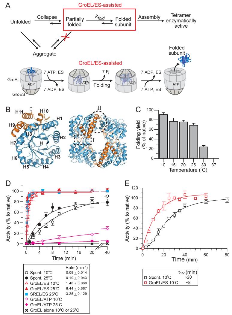
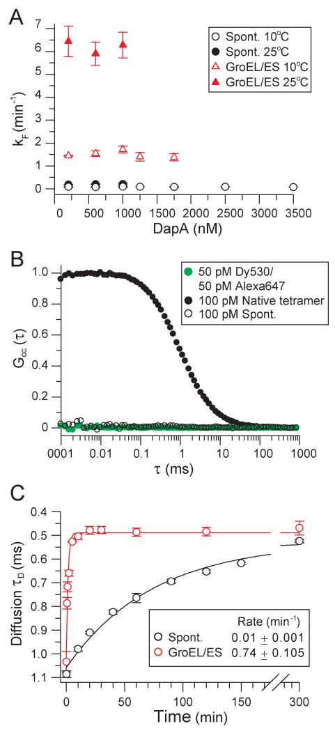
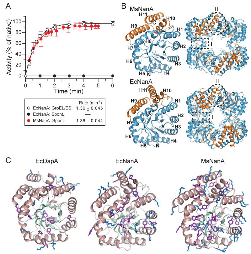
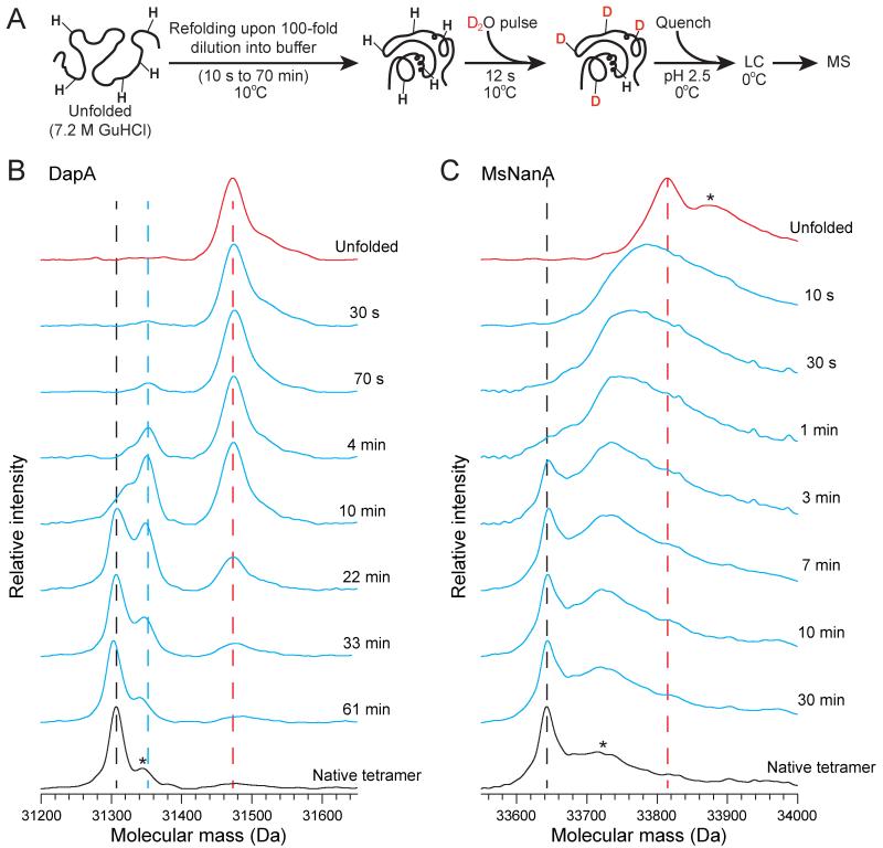
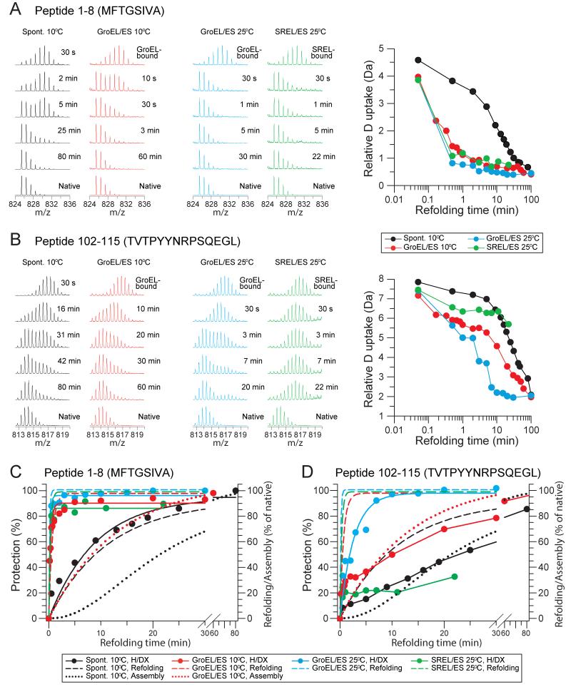
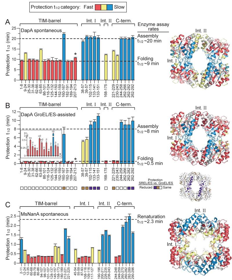
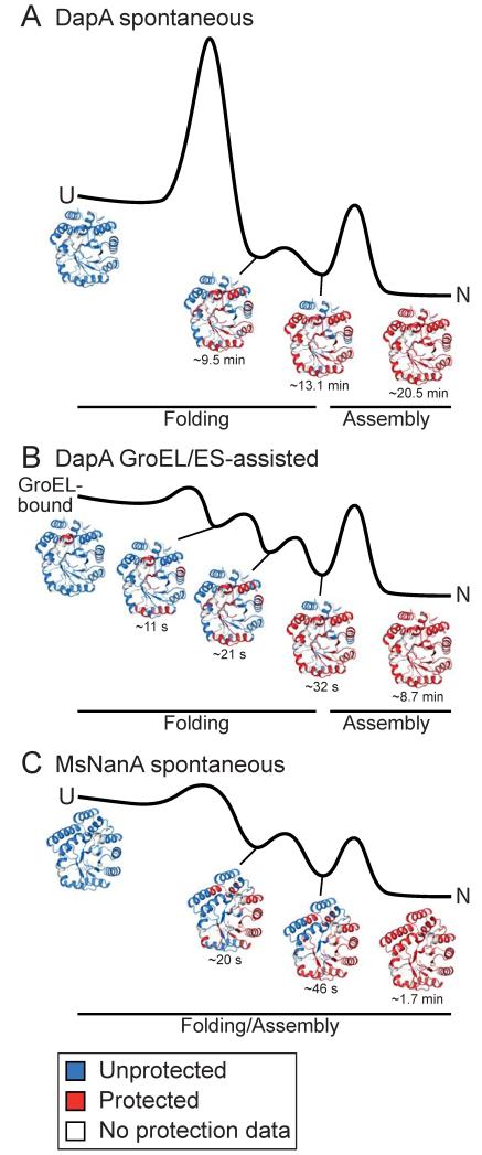
The GroEL/ES chaperonin system functions as a protein folding cage. Many obligate substrates of GroEL share the (βα)8 TIM-barrel fold, but how the chaperonin promotes folding of these proteins is not known. Here, we analyzed the folding of DapA at peptide resolution using hydrogen/deuterium exchange and mass spectrometry. During spontaneous folding, all elements of the DapA TIM barrel acquire structure simultaneously in a process associated with a long search time. In contrast, GroEL/ES accelerates folding more than 30-fold by catalyzing segmental structure formation in the TIM barrel. Segmental structure formation is also observed during the fast spontaneous folding of a structural homolog of DapA from a bacterium that lacks GroEL/ES. Thus, chaperonin independence correlates with folding properties otherwise enforced by protein confinement in the GroEL/ES cage. We suggest that folding catalysis by GroEL/ES is required by a set of proteins to reach native state at a biologically relevant timescale, avoiding aggregation or degradation.


Figures

Figure 1
Figure 1. Spontaneous and Chaperonin-assisted Refolding of DapA
(A) Schematic representation of DapA refolding/assembly. Top panel: Spontaneous refolding/assembly, highlighting the steps modulated by GroEL/ES in preventing off-pathway aggregation (red cross) and/or accelerating subunit folding (red box). Bottom panel: Model of the GroEL/ES mechanism of assisted refolding. (B) Structure of E. coli DapA monomer (left) and tetramer (right) in ribbon representation (PDB 1YXC). The (βα)8 TIM-barrel domain is shown in blue and the C-terminal domain in gold. Helices H1 to H11 and locations of the strong and weak interfaces of the tetramer (interface I and II, respectively) are indicated. (C) Yield of spontaneous DapA refolding at 10°C-37°C. Refolding was initiated by diluting GuHCl-denatured DapA into refolding buffer B to a final concentration of 200 nM monomer and yields analyzed by enzyme assay after 1.5 h (15°C-37°C) and after 16 h (10°C). Folding yields are plotted as DapA activities in % of native enzyme control incubated at the respective temperature. (D) Rates of spontaneous and chaperonin-assisted DapA subunit folding at 10°C and 25°C. Refolding was measured at 200 nM DapA by diluting the denatured protein into refolding buffer as in (C) or into buffer containing 2 μM GroEL or SREL and 4 μM GroES. Assisted refolding was initiated by addition of ATP. In the case of SREL, urea-denatured DapA and low salt refolding buffer C was used (see Experimental Procedures). Spontaneous refolding was stopped by addition of 0.8 μM GroEL D87K and GroEL/ES-assisted refolding with 50 mM CDTA. In the case of SREL/ES, 50 mM CDTA and 60 mM GuHCl were added to stop folding. Reactions were incubated for 1 h at 25°C to allow for complete assembly prior to enzyme assay. Single exponential rates are indicated. (E) Spontaneous and assisted assembly of DapA. Refolding reactions were performed as in (D) at 10°C with 2 μM DapA and 4 μM GroEL/8 μM GroES when indicated. The reactions were not stopped and enzyme activities were measured at the time points indicated. All standard deviations are from at least 3 independent experiments.
Figure 2
Figure 2. GroEL/ES Catalyzes DapA Refolding
(A) Rates of spontaneous and GroEL/ES-assisted refolding are concentration independent. Spontaneous and GroEL/ES-assisted subunit refolding was performed at 10°C and 25°C as in Figure 1D over a range of DapA concentrations. (B) Absence of inter-molecular association during refolding by FCCS. DapA-293C was labeled with either Dy530 or Alexa647 fluorophores. The labeled proteins were mixed, denatured and diluted into refolding buffer to a final concentration of 50 pM each. FCCS was recorded within the first 30 min of refolding. As a positive control, DapA-293C tetramer refolded and assembled from Dy530 and Alexa647 labeled subunits at 200 nM concentration was diluted to 100 pM. A 1:1 mixture of the free dyes at 50 pM each was used as a negative control. (C) Spontaneous and assisted refolding of DapA-293C labeled with Alexa647 was measured at a final concentration of 100 pM at 20°C. GroEL and GroES, when present, were 2 μM and 4 μM, respectively. Refolding was stopped either by addition of 2 μM GroEL (spontaneous refolding) or by addition of Apyrase (assisted refolding). The difference in diffusion rate between not-yet folded DapA bound to GroEL and folded DapA monomer free in solution was monitored by FCS, resulting in rates of subunit refolding. All standard deviations are from at least 3 independent experiments.
Figure 3
Figure 3. Chaperonin-Dependent and Independent TIM-Barrel Proteins
(A) Assisted refolding of EcNanA and spontaneous refolding of MsNanA occur at similar rates. Spontaneous and GroEL/ES assisted refolding of EcNanA were analyzed at 25°C (400 nM EcNanA) in buffer D essentially as described in Figure 1D. Spontaneous renaturation of MsNanA (400 nM final concentration) in buffer D was analyzed by direct enzyme assay at the time points indicated. The observed kinetics reflects both subunit folding and assembly. Standard deviations are from at least 3 independent experiments. B) Crystal structures of MsNanA (PDB 4N4P, this study) and EcNanA (PDB 2WO5) are shown as for DapA in Figure 1B. Left, monomer; right, tetramer. Helices H1 to H11 as well as the locations of the interfaces I and II of the tetramer are indicated. C) Amino acid compositional bias in MsNanA. The monomer structures of EcDapA, EcNanA and MsNanA are shown in ribbon representations with α-helices and β-strands indicated in salmon and pale green, respectively. The side-chains of lysines and aromatic residues (Phe and Tyr) are highlighted in blue and purple, respectively.
Figure 4
Figure 4. Different Properties of DapA and MsNanA Refolding by H/DX
(A) Schematic representation of the H/DX pulse experiment. After different times of spontaneous refolding, proteins are pulse-labeled with D2O buffer E for 12 s, followed by acid quenching of the H/DX reaction and LC-MS analysis. See Extended Experimental Procedures for details. (B and C) Mass spectra during spontaneous refolding of DapA (B) and MsNanA (C). The positions of the unfolded proteins and the folded tetramers in the mass spectra are indicated by red and black dotted lines, respectively. In the case of DapA, the blue dotted line marks the position of folded monomer. Asterisk in the native tetramer in B indicates a potassium adduct. Asterisks on the broad peaks of unfolded MsNanA and MsNanA tetramer in C represent the presence of potassium and sodium adducts (1 sodium, 1 potassium, 2 sodium and 2 potassium).
Figure 5
Figure 5. Deuterium Incorporation into Peptides of DapA During Spontaneous and Assisted Refolding
(A and B) Deuterium incorporation into P1-8 of the TIM-barrel domain (A) and P102-115 of interface I (B) of DapA. Left panels: Examples of mass spectra for DapA peptides P1-8 and P102-115 at different times during spontaneous, GroEL/ES-assisted and SREL/ES-assisted refolding at 10°C and 25°C, as indicated. Amino acid sequences of the peptides are indicated in single letter code. Right panels: Deuterium uptake in Da is plotted versus refolding time (see Figure S5 for full dataset). (C and D) Time courses of H/DX protection during refolding for P1-8 (C) and P102-115 (D). For comparison, subunit refolding and assembly based on enzymatic assay (Figures 1D and 1E) are also shown (dashed and dotted lines, respectively).
Figure 6
Figure 6. Comparison of DapA and MsNanA Refolding at Peptide Resolution
Apparent half-times of H/DX protection for peptides along the amino acid sequence during spontaneous (A) and GroEL/ES-assisted refolding/assembly (B) of DapA, and spontaneous renaturation of MsNanA (C) at 10°C are presented in the bar graphs and are mapped on the tetramer structures of DapA and MsNanA (right panels). Peptides are assigned either to TIM-barrel domain, C-terminal domain or interfaces I and II (when at least 25 % of the sequence is in the interface; see Figure S5). Half-times of protection are color-coded: Red bars indicate peptides with half-times as fast or faster as subunit refolding in enzymatic refolding assays (Figure 1D); blue bars denote peptides with half-times of protection as slow as assembly (Figure 1E), and yellow bars denote peptides with intermediate half-times of protection. Note that P207-213 (asterisk) in GroEL/ES-assisted folding is already fully protected in the GroEL-bound state. The insert in (B) highlights the differences in protection of TIM-barrel domain peptides (x-axis labeling as in main figure of panel B) and the secondary structure of the peptides is indicated: α, α-helix; β, β-strand; cα, coil and α-helix; cβ, coil and β-strand. Squares below the bar graph in (B) indicate protection properties of peptides upon refolding with SREL/ES in comparison to GroEL/ES at 25°C. Peptides with reduced protection with SREL/ES are highlighted in the tetramer structure. In the case of MsNanA, refolding and assembly are coupled and half-times of protection are colored from fast (red <0.5 min) to slow (blue >1.0 min).
Figure 7
Figure 7. Mechanisms of Spontaneous and GroEL/ES-Assisted TIM-Barrel Folding
Free energy diagrams, schematically summarizing the salient features of spontaneous (A) folding and GroEL/ES-assisted folding (B) of DapA and spontaneous folding of MsNanA (C). Intermediate states populated during folding with the approximate half-times indicated, as determined by H/DX-MS at peptide resolution, are tentatively assigned to different phases of the energy diagrams. Ribbon diagrams show acquisition of H/DX protection during folding in red. U, unfolded state; N, native tetramer. See Discussion for detail.

Similar articles

Cited by

References

    1. Apetri AC, Horwich AL. Chaperonin chamber accelerates protein folding through passive action of preventing aggregation. Proc. Natl. Acad. Sci. U.S.A. 2008;105:17351–17355. - PMC - PubMed
    1. Azia A, Unger R, Horovitz A. What distinguishes GroEL substrates from other Escherichia coli proteins? FEBS J. 2012;279:543–550. - PubMed
    1. Baumketner A, Jewett A, Shea JE. Effects of confinement in chaperonin assisted protein folding: Rate enhancement by decreasing the roughness of the folding energy landscape. J. Mol. Biol. 2003;332:701–713. - PubMed
    1. Bicout DJ, Szabo A. Entropic barriers, transition states, funnels, and exponential protein folding kinetics: A simple model. Protein Sci. 2000;9:452–465. - PMC - PubMed
    1. Brinker A, Pfeifer G, Kerner MJ, Naylor DJ, Hartl FU, Hayer-Hartl M. Dual function of protein confinement in chaperonin-assisted protein folding. Cell. 2001;107:223–233. - PubMed

Publication types

MeSH terms

LinkOut - more resources