Review
. 2014 May 7;82(3):522-36.
doi: 10.1016/j.neuron.2014.01.018.

Transmitting pain and itch messages: a contemporary view of the spinal cord circuits that generate gate control

Affiliations
Review

Transmitting pain and itch messages: a contemporary view of the spinal cord circuits that generate gate control

João Braz et al. Neuron. .

Abstract

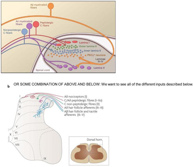
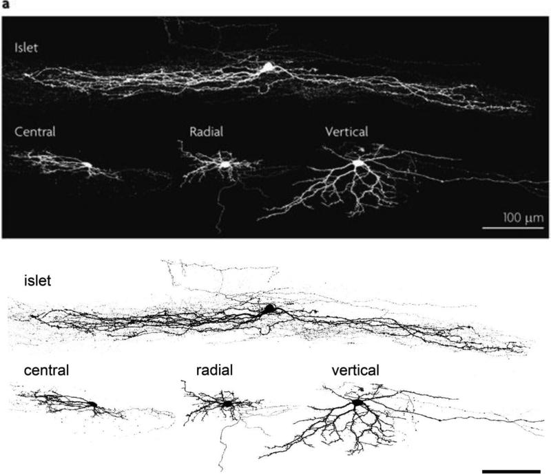
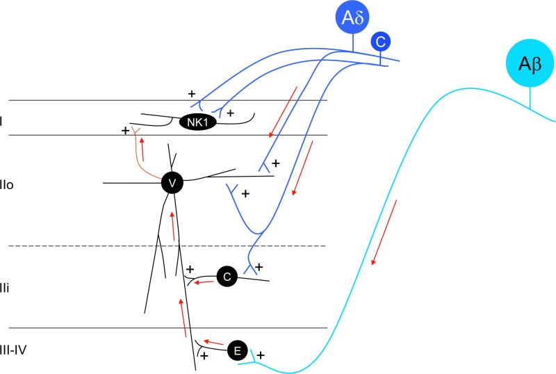
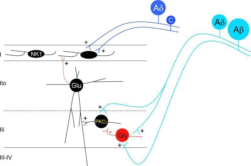
The original formulation of Gate Control Theory (GCT) proposed that the perception of pain produced by spinal cord signaling to the brain depends on a balance of activity generated in large (nonnociceptive)- and small (nociceptive)-diameter primary afferent fibers. The theory proposed that activation of the large-diameter afferent "closes" the gate by engaging a superficial dorsal horn interneuron that inhibits the firing of projection neurons. Activation of the nociceptors "opens" the gate through concomitant excitation of projection neurons and inhibition of the inhibitory interneurons. Sixty years after publication of the GCT, we are faced with an ever-growing list of morphologically and neurochemically distinct spinal cord interneurons. The present Review highlights the complexity of superficial dorsal horn circuitry and addresses the question whether the premises outlined in GCT still have relevance today. By examining the dorsal horn circuits that underlie the transmission of "pain" and "itch" messages, we also address the extent to which labeled lines can be incorporated into a contemporary view of GCT.


Figures

Figure 1
Figure 1
The original formulation of Gate Control Theory proposed that spinal cord signaling to the brain (the “Action System”) depended on the balance of activity of Large (L) and small (S) diameter primary afferent fibers. The large fibers not only can excite output/projection cells (transmission; T), but the large fibers also exert a feedforward inhibition of the T cells by concurrently activating inhibitory interneurons in the substantia gelatinosa (SG). The inhibition was presumed to involve presynaptic connections to the T cell, however, postsynaptic inhibition of the T cell by SG interneurons was not ruled out. The net effect of the large diameter afferents is to “close” the Gate. Critical to GCT, however, was the activity of small diameter (presumptive nociceptors), which not only directly excite the T cells and thus the Action System, but also engage inhibitory circuits that reduce the activity of the SG inhibitory interneurons. This presumably multisynaptic inhibitory mechanism “opens” the Gate. Finally, the GCT schematic included an illustration of the engagement of supraspinal control systems (the Central Control Trigger) that can regulate the output of the spinal cord dorsal horn.
Figure 2
Figure 2
The different populations of primary afferent fibers target different regions of the dorsal horn of the spinal cord, with the input from C and Aδ nociceptors concentrated in the superficial dorsal horn (laminae I and II). The peptidergic, TRPV1-expressing subset of nociceptors targets laminae I and outer II; the nonpeptidergic subset, many of which expresses Mrgpr GPCRs, targets inner lamina II. Aδ nociceptors target both laminae I and V. This figure also includes the PKCγ-expressing population of interneurons that have been implicated in nerve injury-evoked mechanical allodynia provoked by stimulation of large diameter (Aβ) afferents (See also Figure 5).
Figure 3
Figure 3
Four morphologically distinct subsets of interneurons predominate in lamina II of the superficial dorsal horn. Islet cells have the longest dendritic arborization, extending long axons and dendrites in the rostrocaudal plane, largely within the lamina where their cell bodies reside. Central cells also have rostrocaudally directed intralaminar arborizations, but the extent of the arborization is less than that of the islet cells. Vertical cells have complex and ramified dendritic arbors that extend largely in the dorsoventral plane, crossing multiple laminae, where they can receive convergent inputs from different populations of primary afferents. Many of the vertical cell axons arborize dorsally in lamina I. Radial cells have a restricted dendritic arbor and a variably-directed axon. Finally, whereas islets cells are exclusively inhibitory, vertical and radial cells are predominantly glutamatergic and thus excitatory. Both inhibitory and excitatory central cells have been described. (Image provided by Dr. Andrew Todd).
Figure 4
Figure 4
Primary afferent-derived excitatory and inhibitory drive to the spinal cord dorsal horn. Ventral to dorsal excitatory activation circuits: Nociceptive C- and Aδ- fibers (dark blue) directly activate vertical cells (V) of lamina IIo and these, in turn, excite NK1-expressing projection neurons of lamina I (green axon). In addition, nociceptive C-fibers activate central cells (C) of lamina IIi, and the central cells directly contact, and likely excite vertical cells in lamina IIo. Of course, projection neurons of lamina I also receive direct nociceptive C and Aδ input. Finally, non-nociceptive Aβ fibers (light blue) project to deeper laminae (III-IV) where they establish monosynaptic connections with local excitatory interneurons (E). The latter directly activate vertical cells. These dorsally-directed circuits are the route through which both noxious and innocuous primary afferent input can engage the projection neurons of lamina I. As described in Figure 5, in the setting of nerve injury, additional routes of Aβ engagement of the projection neurons are uncovered. Feedforward inhibitory control of superficial dorsal horn circuits: These excitatory circuits are subject to profound inhibitory controls (red neurons). The majority of the inhibitory neurons (I) in lamina II are islet cells (I-i) and these can be directly engaged by input from low-threshold, mechanoreceptive C-fibers (light blue). The islet cells in turn establish monosynaptic inhibitory connections with both vertical cells of lamina IIo and NK1 receptor-expressing projection neurons of lamina I. Low threshold Aδ (D-hair; light blue) and Aβ fibers also directly engage inhibitory interneurons in laminae IIi-IV. The latter in turn exert inhibitory control of a variety of excitatory interneurons, including vertical (V) and central (C) cells. The Aβ to inhibitory cell circuit presumably underlies the circuit through which the “Gate” of Gate Control Theory can be closed. Figure 5 illustrates the neurochemical identity of critical elements in this circuit.
Figure 4
Figure 4
Primary afferent-derived excitatory and inhibitory drive to the spinal cord dorsal horn. Ventral to dorsal excitatory activation circuits: Nociceptive C- and Aδ- fibers (dark blue) directly activate vertical cells (V) of lamina IIo and these, in turn, excite NK1-expressing projection neurons of lamina I (green axon). In addition, nociceptive C-fibers activate central cells (C) of lamina IIi, and the central cells directly contact, and likely excite vertical cells in lamina IIo. Of course, projection neurons of lamina I also receive direct nociceptive C and Aδ input. Finally, non-nociceptive Aβ fibers (light blue) project to deeper laminae (III-IV) where they establish monosynaptic connections with local excitatory interneurons (E). The latter directly activate vertical cells. These dorsally-directed circuits are the route through which both noxious and innocuous primary afferent input can engage the projection neurons of lamina I. As described in Figure 5, in the setting of nerve injury, additional routes of Aβ engagement of the projection neurons are uncovered. Feedforward inhibitory control of superficial dorsal horn circuits: These excitatory circuits are subject to profound inhibitory controls (red neurons). The majority of the inhibitory neurons (I) in lamina II are islet cells (I-i) and these can be directly engaged by input from low-threshold, mechanoreceptive C-fibers (light blue). The islet cells in turn establish monosynaptic inhibitory connections with both vertical cells of lamina IIo and NK1 receptor-expressing projection neurons of lamina I. Low threshold Aδ (D-hair; light blue) and Aβ fibers also directly engage inhibitory interneurons in laminae IIi-IV. The latter in turn exert inhibitory control of a variety of excitatory interneurons, including vertical (V) and central (C) cells. The Aβ to inhibitory cell circuit presumably underlies the circuit through which the “Gate” of Gate Control Theory can be closed. Figure 5 illustrates the neurochemical identity of critical elements in this circuit.
Figure 5
Figure 5
Disruption of glycinergic inhibitory controls contributes to the development of nerve injury-induced pain hypersensitivity: Low threshold Aβ and possibly Aδ (light blue) inputs directly activate PKCγ-expressing interneurons of lamina II and these interneurons in turn activate projection neurons of lamina I, likely via polysynaptic pathways involving glutamatergic (Glu) excitatory vertical cells of lamina IIo. Under normal conditions, this excitatory drive is tonically inhibited by glycinergic interneurons, and the inhibition can be enhanced by activity of low threshold afferents. In conditions where this tonic glycinergic inhibitory control is disrupted (e.g. after peripheral nerve injury), primary afferent-derived innocuous inputs can gain access to the pain transmission circuitry of the superficial dorsal horn, leading to mechanical allodynia and thermal hyperalgesia.
Figure 6
Figure 6
Prostaglandins induce hypersensitivity in the setting of inflammation by regulating glycinergic inhibitory circuits: Peripheral inflammation evokes cyclooxygenase (Cox)-dependent release into the superficial dorsal horn of prostaglandin E2 (PGE2), likely from astrocytes and/or microglia. The PGE2 binds to neuronal EP2 receptors expressed by many interneurons, including those under inhibitory glycinergic control. Through a cAMP-dependent phosphorylation of the GlyR3 subunit of glycine receptors, the PGE2 reduces the effects of glycine on what are presumed to be excitatory interneurons that ultimately engage neurons in lamina I. The net result is that innocuous stimuli can provoke pain (allodynia) and noxious stimuli exacerbate pain (hyperalgesia) in the setting of tissue injury. In contrast to the effects of PGE2 on glycinergic signaling, there is no influence on GABAergic signaling, even though GABA and glycine are co-released from many superficial dorsal horn inhibitory interneurons.
Figure 7
Figure 7
Microglial-neuronal interactions disrupt GABAergic inhibitory control: Peripheral nerve injury activates spinal cord microglia, which in turn release numerous molecules that enhance the excitability of spinal corn neurons. Some studies indicate that chemokine ligand 2 (CCL2), which is upregulated in and released from injured primary sensory neurons, binds to the microglial chemokine receptor 2 (CCR2) to induce microglial activation. Nerve-injury-induced release of ATP, via an action on microglial P2X4 receptors, also activates the microglia. The latter release brain-derived neurotrophic factor (BDNF), which via an action on neuronal TrkB receptors, downregulates the neuronal potassium-chloride co-transporter KCC2. The net result is a reduced chloride gradient that alters the magnitude of any GABAergic inhibitory input to the neuron. In this setting there may be ongoing pain, mechanical allodynia and thermal hyperalgesia.
Figure 8
Figure 8
A labeled line for the transmission of itch messages: Several subpopulations of primary afferent fiber (pruritoceptors) transmit messages provoked by a host of itch-producing stimuli that engage the afferent directly or indirectly. To what extent itch and pain-producing afferents (nociceptors) overlap is unclear. Activation of the pruritoceptors evokes the release of natriuretic polypeptide B (NPPB) in the superficial dorsal horn. The receptor for NPPB, natriuretic peptide receptor A (NPRA) is expressed by presumptive excitatory interneurons of the superficial dorsal horn, and these cells in turn release gastrin-releasing peptide (GRP) and engage another interneuron population, namely one that expresses GRPR, the receptor for GRP. Whether GRPR-expressing cells project directly to the brain or signal locally to other projection neurons in the spinal cord is unclear, but our recent findings (Wang et al., 2013) suggest that the GRPR cells are interneurons. Another view holds that GRP, in fact, derives predominantly from pruritoceptors (Zhao et al., 2013), not from dorsal horn interneurons. Whether the “pain-responsive” NK1 receptor-expressing projection neurons also transmit itch messages to the brain (e.g. via inputs from GRPR cells) is also unclear. Finally, this figure illustrates that a subset of Bhlhb5-dependent inhibitory interneurons regulates the output of superficial dorsal horn itch circuits, as do primary afferent neurons that express the VGLUT2 subtype of vesicular glutamate transporter (Lagerstrom et al., 2010; Liu et al., 2010; Scherrer et al., 2010). Loss of either Bhlhb5 or of primary afferent-derived VGLUT2 generates mice with an exaggerated itch phenotype, but limited effects on pain processing.

Similar articles

Cited by

References

    1. Abbadie C, Lindia JA, Cumiskey AM, Peterson LB, Mudgett JS, Bayne EK, DeMartino JA, MacIntyre DE, Forrest MJ. Impaired neuropathic pain responses in mice lacking the chemokine receptor CCR2. Proc Natl Acad Sci (USA) 2003;100:7947–7952. - PMC - PubMed
    1. Abraira VE, Ginty DD. The sensory neurons of touch. Neuron. 2013;79:618–639. - PMC - PubMed
    1. Ahmadi S, Lippross S, Neuhuber WL, Zeilhofer HU. PGE(2) selectively blocks inhibitory glycinergic neurotransmission onto rat superficial dorsal horn neurons. Nat Neurosci. 2002;5:34–40. - PubMed
    1. Al-Khater KM, Todd AJ. Collateral projections of neurons in laminae I, III, and IV of rat spinal cord to thalamus, periaqueductal gray matter, and lateral parabrachial area. J Comp Neurol. 2009;515:629–646. - PMC - PubMed
    1. Albuquerque C, Lee CJ, Jackson AC, MacDermott AB. Subpopulations of GABAergic and non-GABAergic rat dorsal horn neurons express Ca2+-permeable AMPA receptors. Eur J Neurosci. 1999;11:2758–2766. - PubMed

Publication types